Taegeun
Lim
ab,
Eun-Geun
Kim
c,
Jungil
Choi
*d and
Sunghoon
Kwon
*ab
aQuantaMatrix Inc., Seoul National University Hospital CMI, Seoul, 03082, Republic of Korea
bDepartment of Electrical Engineering and Computer Science, Seoul National University, Seoul, 08826, Republic of Korea. E-mail: skwon@snu.ac.kr
cLowendtechnologies Co., Anyang, 14056, Republic of Korea
dSchool of Mechanical Engineering, Kookmin University, Seoul, 02707, Republic of Korea. E-mail: jchoi@kookmin.ac.kr
First published on 2nd November 2020
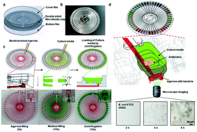
Antibiotic resistance is a global threat to modern society. Rapid determination of suitable antibiotics that inhibit bacterial growth can effectively reduce antibiotic resistance and improve clinical treatment. The conventional methods of antimicrobial susceptibility testing (AST) depend on optical density measurements, which require long-time incubation. Various kinds of rapid AST systems which utilize various technologies from the field of lab on a chip have promised a great reduction in measurement time, but cannot achieve high-throughput, user-friendly testing due to the complexity of the testing system. Here, we introduce a capillary and centrifuge-based rapid AST system that reduces the time of loading the sample and culture media while achieving a high-throughput testing capacity. The capability of the proposed system is validated in a systematic analysis that includes sample loading characteristics and AST trials with standard strains. The proposed system provides a useful tool for drug testing in cell-culture systems with user-friendly and high-throughput analysis.
In the past decade, various kinds of rapid AST have reduced the test times to a few hours.9 The detection principle can be phenotypic or genotypic. Phenotypic methods reduce the AST time by lowering the detection limit of bacterial growth using some techniques: microscopic imaging of bacteria,10–16 measuring the protein displacement profile,17 monitoring bacterial metabolism,18 detecting the pH of culture media,19 measuring fluorescence signals,20 laser light scattering,21 electrical resistance,22 Raman spectroscopy,23 droplet digital polymerase chain reaction,24 or detecting the bacteria-induced vibrations of a cantilever.25 Although these methods promise to reduce the AST time, they lack a user-friendly protocol and a high-throughput system for integration into clinical practice. To determine whether a bacterial strain is susceptible, intermediate or resistant, conventional AST systems test up to 20 clinically available antibiotics at 2–4 concentrations simultaneously.26 To meet this requirement, a high-throughput testing platform must handle approximately 50 testing conditions. A 96-well testing system satisfies these criteria, but the clinical environment demands a quick sample-loading system to handle multiple tests from multiple patients.
The most commonly used AST systems are Vitek2 and MicroScan, which can simultaneously test 30–100 samples. These systems employ their own processes for handling the samples and loading the culture media. However, new rapid AST systems either are incapable of high-throughput testing, or require cumbersome procedures (i.e., loading the samples and testing their antimicrobial susceptibilities). Specifically, both the bacterial sample and culture media must be loaded into each testing well, which doubles the pipetting time per well. Multiple pipetting also increases the possibility of misloading and contamination. To remove these difficulties, a simple system that loads a liquid sample into separate testing chambers is required.
A lab-on-a-disc system operated on a centrifuge platform is an attractive method for loading samples into separate testing chambers.27 A lab on a disc allows sequential sample tests through accurately designed microfabricated channels. Under the control of the centrifugal force induced by rotational acceleration, the system guides the direction of the fluid in the channel and the timing of the reaction. In addition, the small-scale capillary forces passively control the microfluidic flow.28 Recently, a fidget spinner introduced a new method for concentrating pathogens for AST that shows a centrifuge platform having broad applications including AST.29
Here, we introduce a capillary and centrifugal-based AST (C2-AST) system for quick loading of the sample and culture media. The C2-AST also includes a microscopic cell-growth detection protocol. The microfluidic channels realize a robust and accurate loading and culturing system. The loading performance of the C2-AST chip was tested in a systematic analysis. The accuracy of the system was then evaluated in comparison tests with the standard method. Tests were performed on standard strains in the presence of several antibiotics. This system can potentially reduce the burden of sample-loading and drug-testing in general cell-culture systems. The loading protocol does not require a complex pipetting system and it simplifies the overall form factor of the automated system for clinical applications. The reduced pipetting procedure will also reduce the chance of contamination of samples. The small microfluidic based testing chamber is favorable to high-throughput testing.
Capillary forces along the bottom channels drive the movement of the first fluid through a length of 22 mm. At this stage, the channel is 600 μm wide and 400 μm high. The fluid then enters a vertical hole of 500 μm diameter and 1.5 mm length. The vertical hole and the second vertical hole are separated but linked at the bottom of the incubation cell. As the second fluid is injected at a much higher volume than the first fluid, its path should run along the top surface. The top channels are 1.2 mm wide, 2.4 mm high, and 17.8 mm long, and each channel stores approximately 45 μl of the second fluid.
The ends of one paired top and the bottom channel are connected to an incubation cell, which functions as an independent diagnostic laboratory. In each incubation cell, the second fluid is introduced after the gelation of the first fluid, forming an interface which must be located near the imaging plane of the first (gelated) fluid. One incubation cell is composed of an imaging well, a reservoir well, and other components. The imaging plane appears in the imaging well filled with the first fluid, which flows through the bottom path. The reservoir well contains a pre-dried antimicrobial reagent, which dissolves easily in the second fluid in a few minutes. The second fluid completely fills the reservoir well and spills into the remainder of the incubation cell. To close the channels of each path, two films with inlet and outlet holes are attached at both sides of the chip. The holes are aligned with a film-attaching jig for ease and efficiency.
The culture media are pipetted into the top inlet hole, through which they fully fill the top channels under capillary forces. The excess media are removed by pulling back from the top buffer space. While pulling the excess liquid back, the beginning section of the top channels holds the liquid tightly, ensuring an equal amount of liquid in each channel. Finally, the chip is rotated at 1200 rpm, and the second liquid is introduced to the incubating cells. The second liquid, which is intended to simultaneously supply the microorganisms in the gelated matrix with nutrients and antimicrobials, completely covers the gelated first liquid.
Table 1 compares the estimated times of each step of the AST operation in the proposed and conventional methods. The loading processes of the first and second reagents in the C2-AST system are especially critical, as they convert the repeated time-consuming process into a single simple pipetting process. Broth microdilution (BMD) was chosen as the conventional method for it was considered as one of the gold-standard method. The BMD method is usually done with a 96-well rounded bottom plate which has individual cells in which individual pipetting is performed; therefore, it requires 48 times more pipetting than the C2-AST system does. The actual duration or workload difference was 12–18 times because the injection volume of the C2-AST is much larger, and the multi-dispensing function of electric pipettes is usually used for the BMD method. The rotation process was implemented only in the C2-AST system. The preparation time of the whole sequence in the C2-AST system was 25 s, approximately 8 times faster than in the conventional method (210 s).
Operating time | C2-AST (proposed method) | Broth microdilution (conventional method) |
---|---|---|
Equipment | C2-AST chip with a manual pipette | 96-Well rounded bottom plate with a multi-dispensing pipette |
First reagent loading (s) | 5 | 90 |
Second reagent loading (s) | 10 | 120 |
Rotation (s) | 10 | — |
Total (s) | 25 | 210 |
The concentration and temperature of the agarose solution obviously affected the agarose filling time. The concentration was the dominant changeable variable, as the temperature cannot be altered far beyond the range 35–45 °C. At these temperatures, the solution remains in the liquid phase without imparting a critical heat shock to the bacterial sample. However, optimizing the temperature of the chip might additionally improve the circumstances of the liquid loading by slowing the cooling speed of the liquid. Fig. 2(a) shows the liquid filling times in the systems with different agarose concentrations and C2-AST chip temperatures. The results were clearly related to the agarose concentration but showed no correlation with the chip temperature. Theoretically, the chip temperature should affect the movement of the agarose, but its effects were apparently concealed by experimental error. In contrast, the agarose concentration dominantly affected the liquid loading, although the results at 0.375% and 0.5% were quite similar. The chip material and the surface modification method also affected the filling time, confirming that the capillary force provided the main motive power. Changing the intensity of the vacuum plasma treatment applied to the C2-AST chip altered the surface energy of the chip substrate to different extents. Increasing the treatment intensity decreased the filling time, and (by implication) increased the loading speed of the liquid (see Fig. 2(b)). This result also suggests that the highest treatment conditions did not saturate the hydrophilic surface. The result after the treatment at 100 W for 30 seconds deviated from the above trend, probably because an error in the agarose preparation process affected the physical properties of agarose. At the same treatment intensity, the hydrophilicity and original surface energies depended on the materials used. General-purpose polystyrene provided the most powerful capillary force under every plasma condition, whereas polycarbonate provided the weakest force. The contact-angle differences before and after the treatment also depended on the material (see contact angle measurements in Fig. S3†). As shown in Fig. 2(c), the filling time increased with agarose concentrations above 0.5%, similar to that in Fig. 2(b). Finally, we selected an agarose concentration of 0.5%, a surface treatment of 200 W for two minutes, and a chip material of general-purpose polystyrene. The 0.5% agarose condition yielded firmer bacterial colonies than the 0.375% agarose condition, and the 200 W plasma treatment for two minutes was near the equipment limit in the manufacturer's guidelines. The only thermal process was maintaining the agarose solution at 45 °C before loading.
The medium loading was influenced by the capillary force as expected; moreover, the material and surface-treatment conditions determined in the agarose loading investigation were sufficient to complete the first step of medium loading, which (as mentioned above) was powered mainly by the capillary force. The medium injection ceased when the medium reached the capillary valve located between the channel and the incubation cell. The second step bursts the capillary valve using the centrifugal force generated by rotating the chip. For this purpose, the rotational frequency should exceed a critical value, which should be minimized to reduce turbulent flows in the incubating cell. To determine the minimum required rotational speed, we increased the rotational speed from 0 to 1200 rpm, and counted the number of wells in which the liquid was successfully transferred into the incubating cell from the channel. Because the densities and viscosities vary in biological applications, these tests were performed using water and glycerol solutions in addition to the culture medium.
As shown in Fig. 2(d), all fluids were successfully loaded at 1200 rpm, but the results widely differed among the fluid compositions at lower speeds. Water made the highest contact angle with the hydrophilic treated polystyrene surface, and it was the most easily loaded fluid. The 50 wt% glycerol aqueous solution was the least easily loaded fluid at any given rotation speed. These two sequential loading steps enable precise volume control of a simple and simultaneous medium loading, thereby shortening the time of the reagent loading sequence.
![]() | ||
Fig. 3 Time-lapse bright field and fluorescence images of the rhodamine solution slowly diffusing into agarose prepared at different concentrations: (a) 0.375% and (b) 0.5% (scale bar = 2 mm). |
The experimental images were translated into normalized values by the same method applied in our previous work.12Fig. 4(a) shows a matrix of images showing the gradual changes in the E. coli colonies during 0 to 4 hours of exposure to gentamicin at different concentrations (control, 0.12, 0.25, and 0.50 μg ml−1). The acquired images were converted into binary images, revealing the colonies as white pixels against a black background (Fig. 4(b)). From the binary images, the areas of each bacterial colony were easily calculated. The average values of the area of the 20 biggest colonies before being normalized at different antibiotic concentrations are plotted in Fig. 4(c). Under the control (no antibiotic), 0.12, and 0.25 μg ml−1 conditions, the E. coli colonies grew over time, but no growth was detected under the 0.5 μg ml−1 condition. The minimum inhibitory concentration (MIC) of gentamicin for E. coli was thus determined as 0.5 μg ml−1. According to the CLSI guidelines, most MICs of the E. coli/gentamicin combination are statistically within the 0.25–1.00 μg ml−1 range. As the broth microdilution (BMD) protocol of CLSI is the gold standard of AST evaluation, the above result demonstrates the suitability of the C2-AST system.
The MIC results of all the antibiotics are listed in Tables 2 and 3. All the results were within the range suggested in the CLSI guidelines. The BMD results, including those of the above-described E. coli/gentamicin combination, also appeared within the suitable range, although some of them showed a two-fold difference from the C2-AST results. In this test, the testing chip contains 4–5 concentrations per each antibiotic (Fig. S5†). To test broad spectrum antibiotics, the system can reduce the number of concentrations and increase the testing antibiotics. As the system is capable of testing 48 combinations of antibiotics and their concentrations, it can be applied for high-throughput analysis of AST (Fig. S6†).
Antibiotic | QC range | C2-AST | BMD test |
---|---|---|---|
Amikacin | 0.50–4.00 | 0.5 | 1 |
Cefotaxime | 0.03–0.12 | 0.12 | 0.06 |
Meropenem | 0.008–0.06 | 0.03 | 0.015 |
Gentamicin | 0.25–1.00 | 0.5 | 0.25 |
Ampicillin | 2.00–8.00 | 8 | 4 |
Cefepime | 0.015–0.12 | 0.06 | 0.06 |
Imipenem | 0.06–0.25 | 0.12 | 0.12 |
Colistin | 0.25–2.00 | 1 | 0.5 |
Ceftazidime | 0.06–0.50 | 0.25 | 0.25 |
Antibiotic | QC range | C2-AST | BMD |
---|---|---|---|
a The result of S. aureus/tetracycline was obtained after 6 hours of incubation. QC: quality control, BMD: broth microdilution. | |||
Penicillin | 0.25–2.00 | 0.25 | 0.5 |
Oxacillin | 0.12–0.50 | 0.5 | 0.25 |
Levofloxacin | 0.06–0.50 | 0.25 | 0.25 |
Vancomycin | 0.50–2.00 | 0.5 | 1 |
Clindamycin | 0.06–0.25 | 0.06 | 0.12 |
Tetracycline | 0.12–1.00 | 0.25a | 0.5 |
Rifampicin | 0.004–0.015 | 0.004 | 0.008 |
Erythromycin | 0.25–1.00 | 0.25 | 0.5 |
The C2-AST tended to give higher antibiotic resistances than the BMD protocol in the E. coli test; the opposite tendency was observed in the S. aureus test. This discordance might be caused by the different culturing methods in the C2-AST system and BMD method; in the former system, the bacteria grow and form colonies within the solid substrate, whereas in the BMD method, they grow in a liquid culture without forming colonies. The growth and antibiotic-resistance characteristics apparently differ between colonies formed from fixed cells in a solid substrate (agarose in this case) and freely growing cells. In the liquid medium, the bacteria can freely access the nutrients and metabolize at full speed, as they are homogeneously distributed before saturation. In agarose, only the outer cells of the colonies can access the nutrients; the interior cells slowly die, so the total colony mass multiplies in a linear fashion. Bacterial clumps can effectively resist chemical assaults by minimizing their surface-to-volume ratio. In addition, the agarose matrix physically (and occasionally chemically) changes the action mechanism and/or diffusion of the nutrients and drugs.
Finally, and perhaps most importantly, the culturing times before determination differ in the two methods. Some bacterial/drug combinations are strongly time-dependent, exhibiting fake, late, or slow growth, and yielding false positive or false negative results at certain antibiotic concentrations. The result of the slow-growing S. aureus/tetracycline combination was settled only in the 6-hourly data.
The spinning can be done with any rotating devices such as spin coaters or centrifuges. The C2-AST chip was preferred to be pinned by the rotating axis, which runs through the entrance of the chip. A spin coater was preferred for its flexible speed programming over a centrifuge, though we used a centrifuge for confirming the concept of rotation as shown in Fig. S3(c).† In this experiment, the spinning was applied with a spin coater (SF-100ND, RHABDOS, Seoul, Republic of Korea) with setting values of max speed and duration at the max speed. The duration was set as 10 s while the max speed was stepwise-increased from 0 to 1200 rpm. The spin coater has a vacuum chuck, which can hold the C2-AST chip while spinning. After each rotation, the remaining culture media were imaged for further analysis.
Each cultured bacterial strain was diluted to 0.5 McFarland using a nephelometer (DensiCHEK Plus, Biomerieux, Marcy-l'Étoile, France), mixed with liquid-state agarose prepared at ∼40 °C, and injected into the bottom of the chip through the inlet. The bacterial sample (100 μl, containing 1.5 × 10⁸ CFU ml−1) mixed with liquid state agarose (3 ml) contained 5 × 10⁶ CFU mL−1. As 5 μl of the mixture and 50 μl of the culture medium were filled to each chamber, the final inoculation concentration was 5 × 10⁵ CFU mL−1.
The C2-AST chip was incubated at 35 °C and time-lapse images were obtained after four hours using a microscope (Eclipse Ti, Nikon, Tokyo, Japan). All images, taken at 1-hour intervals, were processed by a binary conversion algorithm. To determine whether an antibiotic at a specific concentration blocked the colony growth, the areas occupied by colonies were calculated, and assessed whether the growth in the antibiotic wells (as a ratio of growth in the control medium) exceeded the given threshold. Here, the threshold growth ratio was set to 0.15 for all the antibiotics.
Footnote |
† Electronic supplementary information (ESI) available. See DOI: 10.1039/d0lc00753f |
This journal is © The Royal Society of Chemistry 2020 |