Andreas R. Baudy*a,
Monicah A. Otienob,
Philip Hewittc,
Jinping Gan
d,
Adrian Rothe,
Douglas Keller
f,
Radhakrishna Surag,
Terry R. Van Vleetg and
William R. Proctorh
aMerck & Co., Inc., Kenilworth, NJ, USA. E-mail: andreas.baudy@merck.com
bJanssen Pharmaceutical Research and Development, Spring House, PA, USA
cMerck KGaA, Darmstadt, Germany
dBristol-Myers Squibb, New York City, NY, USA
eRoche Innovation Centre, Basel, Switzerland
fSanofi, Bridgewater, NJ, USA
gAbbVie, Inc., Chicago, IL, USA
hGenentech, Inc., South San Francisco, CA, USA
First published on 20th November 2019
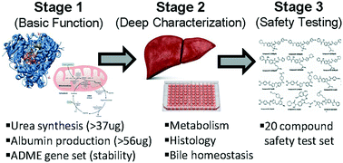
The liver is critical to consider during drug development because of its central role in the handling of xenobiotics, a process which often leads to localized and/or downstream tissue injury. Our ability to predict human clinical safety outcomes with animal testing is limited due to species differences in drug metabolism and disposition, while traditional human in vitro liver models often lack the necessary in vivo physiological fidelity. To address this, increasing numbers of liver microphysiological systems (MPS) are being developed, however the inconsistency in their optimization and characterization often leads to models that do not possess critical levels of baseline performance that is required for many pharmaceutical industry applications. Herein we provide a guidance on best approaches to benchmark liver MPS based on 3 stages of characterization that includes key performance metrics and a 20 compound safety test set. Additionally, we give an overview of frequently used liver injury safety assays, describe the ideal MPS model, and provide a perspective on currently best suited MPS contexts of use. This pharmaceutical industry guidance has been written to help MPS developers and end users identify what could be the most valuable models for safety risk assessment.
Despite progress in the development of novel in vitro and in vivo models, as well as biomarkers of DILI, prediction of the multiple facets of human DILI during preclinical drug development remains challenging. Immortalized cell lines and primary cell models have limitations and poor prediction potential of DILI in humans.6 These single cell type models lack the multicellular environment found in liver and while many complex 3D structural technologies have been developed, the fidelity of drug disposition, cellular adaptation, and biological response are lacking. Therefore, there is a clear need for improved physiologically relevant models that can better recapitulate human sensitivity to hepatotoxic drugs.
Human liver microphysiological systems (MPS) may offer solutions to the identified gaps through engineering and designs that better reproduce human liver physiology. We define liver MPS as going beyond traditional 2D sandwich culture and could include several of the following design aspects: having a multi-cellular environment incorporated within a biopolymer or tissue-derived matrix, a 3D structure, being exposed to media flow, utilizing primary or stem cell derived cells (both hepatocyte and non-parenchymal cells), and/or inclusion of immune system components.7
It is important for each emerging liver MPS model to have well-defined context(s) of use (CoU) and these should be considered in relation to simpler and established assays and technologies. A CoU is defined by the FDA-NIH BEST (Biomarkers, EndpointS, and other Tools) Resource as “A statement that fully and clearly describes the way the medical product development tool is to be used and the medical product development-related purpose of the use”.8 By identifying this for emerging liver MPS, developers and end users (e.g. pharma) can build a path for qualification since comprehensive data packages around specific CoUs will need to be generated in order for these tools to replace traditional processes in drug discovery and development. One of the intentions of this manuscript is to outline the biology that may define conditions under which liver MPS can address specific biological questions (e.g. CoUs) to enable broad industry and ultimately regulatory acceptance.
Albumin production requires hepatocellular transcription of prepro-albumin, translation, cleavage of the N-terminal peptide to form pro-albumin, subsequent release from the rough endoplasmic reticulum, and a final cleavage into albumin within the Golgi apparatus prior to exportation into serum.9,10 Urea synthesis requires viable mitochondria with appropriate metabolic activity to support the biochemical conversion of intracellular ammonia.11 Together, these biomarkers cover a range of hepatocellular organellular functionality and can offer a view into the overall health status of the hepatocyte.
Primary human hepatocytes plated in 2D rapidly lose physiological function as a consequence of separation from their native environment and this is exemplified by rapidly declining albumin production and urea synthesis over time.12 Likewise, lower functioning hepatocyte models (e.g. HepG2, poorly differentiated stem cell derived hepatocytes, hypoxic 3D tissues) also have lower production rates compared to higher functioning MPS. We have calculated the native human in vivo production range for albumin and urea (Table 1) and suspect that if these physiological rates could be achieved and sustained by MPS in vitro, then other normal hepatocyte functions may also be retained.
Parameter | Estimated human liver outputa | 2D primary plated hepatocytes (day 3)9 | 2D primary plated hepatocytes (day 7)9 |
---|---|---|---|
a Assuming a range of 1.1–3.2 × 10¹¹ hepatocytes/liver.73b Based on estimate of 160 mg albumin produced per kg per day.74c Assuming 10 mg urea per h kg.75 | |||
Albumin (μg per day per 1 million hepatocytes) | 37–105b | 2 | 1 |
Urea (μg per day per 1 million hepatocytes) | 56–159c | 140 | 35 |
Decreases in absorption, distribution, metabolism, and excretion (ADME) gene transcription is an early event related to the loss of metabolic hepatocyte functionality and so optimizing MPS conditions to maintain ADME genes will be important.13 Not only will this help ensure that adequate amounts of metabolite(s) can be generated during a duration of drug testing, but it will also add to the reliability of toxicity assay endpoints by reducing baseline system variability. Taken together, we therefore recommend that measures of albumin, urea, as well as gene expression of key drug metabolizing phase I/II enzymes and transporters are evaluated over a 14 day time course in the first stage (stage 1) of characterizing MPS model performance (Table 2).
Measure | Function assessed | Specifications |
---|---|---|
Albumin production | Liver transcription, translation, processing, and export function | • >37 μg per day per 1 million hepatocytes• Daily production rates should remain stable across a 14 day time frame∘ Less than a 50% change over a 14 day period with <30% C.V. of mean daily production rates |
Urea synthesis | Mitochondrial and biochemical synthesis | • >56 μg per day per 1 million hepatocytes• Daily production rates should remain stable across a 14 day time frame∘ Less than a 50% change over a 14 day period with <30% C.V. of mean daily production rates |
Baseline quantitative gene expression profiling | mRNA expression of ADME genes, stability over time, and levels in comparison to that of a cryopreserved hepatocyte in freshly prepared suspension or human liver sample | • Phase I CYP450 enzymes∘ CYP3A4, CYP2B6, CYP2C9, CYP2C19, CYP1A2, CYP2D6, CYP2C8, CYP2E1• Phase II enzymes∘ UGT1A1, GSTA1• Hepatocyte uptake transporters∘ SLCO1B1, SLCO1B3, SLC22A1• Hepatocyte efflux transporters∘ ABCC2, ABCG2, ABCB1, ABCB11 |
If the described stage 1 thresholds are successfully met, then a second stage (stage 2) evaluation involving more resource intensive and deeper characterization would be advised to gain further confidence in MPS function (Table 3). Stage 2 includes assessments of predominant drug metabolizing enzymes and transporter functions, morphology, cytokine stability (if Kupffer and/or stellate cells are present) and integrity of hepatobiliary networks. Results from such evaluation could then support or reject proceeding to a final stage of compound testing in the section below.
Measure | Function assessed | Specifications |
---|---|---|
Alanine aminotransferase (ALT), lactate dehydrogenase (LDH), miR122, cytokines | Indications of cell damage and MPS stability over time | • ≤30% C.V. for mean daily baseline release levels across a 14 day time frame |
Baseline and induced metabolic enzymes functional activity using a set of standard probe substrates | Liver phase I/II metabolizing enzymes capability (measure of CYP450 enzymatic capacity and induction) | • 3A4 (midazolam → 1′hydroxymidazolam); show elevated turnover when CYP is induced with rifampicin• 1A2 (phenacetin → APAP); show elevated turnover when CYP is induced with omeprazole• 2B6 (bupropion → hydroxyl bupropion); show elevated turnover when CYP is induced with phenobarbital• 2C9 (diclofenac → diclofenac 2′,3′ oxide or 4-OH diclofenac)• 2D6 (dextromethorphan → dextrorphan)• UGT1A1 (estradiol → estradiol 3-glucuronide; or 7-hydroxycoumarin → 7-hydroxycoumarin glucuronide)• GST (rilpivirine → glutathione conjugate; or dichloronitrobenzene → chloronitrobenzene glutathione) |
Benchmark levels specified for each enzyme compared to fresh hepatocytes and demonstrate <30% CV (as measure of stability of enzymatic activity rates over time) | ||
Transporter function and bile acid homeostasis: uptake, metabolism, and export | Measures of daily rates of transporter substrate and bile acid uptake, metabolism, conjugation, and export in media | • Assess transporter functionality using fluorescent probe substrates (e.g. cholyl-lysyl-fluorescein for OATP/MRP2/BSEP; tauro-nor-THCA-24-DBD for NTCP/BSEP/MRP2)• Assess bile acid flux using stable label biochemical (e.g. labelled GCDCA) with mass spectrometry for bile acid transport |
Histology of MPS | Allows comparison to that of normal human in vivo liver architecture and cellular morphology | • Immunohistochemical analysis of bile canaliculi (BSEP, MRP2, AQP1), Kupffer cells (CD68), and stellate cells (desmin). Electron microscopy of liver sinusoidal endothelial cells to show fenestrations• H&E staining for presence of polygonal, non-rounded, hematoxylin positive, polarized hepatocytes |
We also must stress the importance of developing animal MPS models of relevant preclinical tox species (e.g. rat, dog, cyno, rhesus). Comparisons of such MPS to animals using similar characterization approaches described herein would be beneficial to bolster the confidence the end user has in the platform's fidelity. Additionally, access to such animal MPS would also be advantageous for drug developers to support the currently FDA required animal safety toxicity testing studies.
We have compiled a DILI compound test set based on our combined IQ MPS affiliate industry experience, focusing on drugs that include the aforementioned DILI mechanisms, as well as some that have been challenging to de-risk with current available in vitro models (Table 4). We recommend testing human liver MPS models with these compounds to assess their ability to successfully separate diverse liver toxicants from safe drugs.
Tool liver toxicant | DILI presentation | Mechanism of toxicity | Appropriate less toxic comparator | DILI presentation | Comparator characteristic |
---|---|---|---|---|---|
Sitaxsentan | ALT elevations after 2 weeks | Reactive metabolites, mitochondrial toxicity, BSEP inhibition76–78 | Ambrisentan | Minimal ALT elevations79 | Targets the same receptor as sitaxsentan |
Clozapine | ALT elevations after 1 week | Reactive metabolite80–82 | Olanzapine | No DILI concerns l83 | Structurally similar to clozapine |
Diclofenac | ALT elevations within 1 month | Reactive metabolites, mitochondrial dysfunction, bile acid dysfunction84–87 | |||
Zileuton | ALT elevations after 6 weeks | Reactive metabolite formation88,89 | |||
Fialuridine | Liver failure after 12 weeks of dosing | Mitochondrial toxicity as primary event causing lactic acidosis, microvesicular steatosis90 | FIRU [1-(2′-fluoro-2′-deoxy-D-ribofuranosyl)-5-iodouracil] | No DILI concerns | Stereoisomer of fialuridine. Only in vitro/animal data available |
Tolcapone | ALT elevations, acute liver failure | Reactive metabolite, mitochondrial toxicant, BSEP inhibition45,91 | Entacapone | Low DILI concern92 | Similar BSEP profile, but less mitochondrial toxicity |
Asunaprevir | ALT elevations after 2 weeks93 | Alterations in bile acids | |||
Troglitazone | ALT, bilirubin elevations after 18 weeks | Reactive metabolites, BSEP inhibition94–96 | Pioglitazone | Low DILI concern97 | Low clinical dose |
Telithromycin | ALT elevations after 1 day | Bile acid alterations98,99 | |||
Trovafloxacin | Acute liver failure | Immune mediated100 | Levofloxacin | No/low DILI concern101 | Same structural class as trovafloxacin |
Pemoline | Liver failure | Immune mediated102 | |||
Mipomersen | Oligonucleotide, ALT elevations and hepatic steatosis | Lipid alterations103 | |||
Nefazodone | Liver failure | Reactive metabolites, BSEP inhibitor, mitochondrial tox54,104 | Buspirone (or Trazodone) | No/low DILI concern105 | Less BSEP inhibition and weak to no mitochondrial toxicity |
Compounds for testing animal systems may be different and should be based on in vivo responses corresponding to the animal MPS model. There are significant species differences in hepatic responses to many liver toxicants. Even with a classical, intrinsic hepatotoxicant like acetaminophen, large differences exist among rats, mice and humans in susceptibility to DILI; spheroid MPS has shown progress in resolving such differences.14,15 Species differences can be even more pronounced with compounds that are not direct hepatotoxicants, but act through mechanisms that are not fully characterized (i.e. idiosyncratic). We encourage partnership between MPS developers and pharmaceutical companies for these evaluations so that the vast amounts of preclinical development compounds can be accessed and used to ascertain MPS fidelity.
Second, appropriate cell culturing practices are needed for the optimal performance and reproducibility of the MPS. Although most of the concerns apply to any cell culture, some can be unique in a MPS setting: a) use of physiologically relevant and compatible media types with appropriate concentrations of nutrients and essential factors. b) For co-cultures, ensure ratios of hepatocytes, endothelial, Kupffer, stellate, and/or biliary epithelial cells are appropriate relative to native liver. c) Ensure maintenance of hepatocyte phenotype and metabolic function upon long term (e.g. 2 weeks or more) culturing.
Finally, some considerations for specific use cases. For example, it may be desirable to use induced pluripotent stem cell (iPSC) derived hepatocytes as a long-term consistent supply of cells to study a specific patient population or disease. If immune-mediated hepatotoxicity is of interest, then incorporation of innate and/or adaptive immune cells into the MPS should be regarded for donor cell matching to provide immunological compatibility.
In addition, co-culture models with a liver sinusoidal endothelial cell layer are desired to study the disposition and adverse effects of therapeutic proteins and monoclonal antibodies; in this case, consider evaluating models for appropriate expression (protein and/or gene) of receptor pathways responsible for antibody internalization/uptake, cellular recycling, and catabolism. For the study of bile acid homeostasis and biliary clearance of drugs, it is desirable to have polarized hepatocytes present with adequate bile canaliculi formation, and would ideally be integrated with cholangiocytes in such a way that allows bile sample collection directly from the MPS system. Assessment of functionality could then be approached as described in Table 3.
While we have described the ideal holistic liver MPS model that would be valuable for answering advanced mechanistic safety questions, we do acknowledge that the field is still in a nascent state. Achieving the above metrics and goals while keeping cost and ease of use low, and throughput high will be very challenging. Breakthroughs in iPSC maturation strategies (to source low cost and abundant MPS input cells), multi-nozzle 3D printing (to increase throughput) and chip perfusion controllers (to simplify operation and increase reliability of flow) will be of great help. In the next section, we discuss the major categories of approaches used to develop MPS and analyse their CoUs to provoke thought on if combinatorial approaches could be taken in order to more effectively reach the ideal MPS.
Given that different categories of MPS each have their own set of strengths and weaknesses, the CoU will depend on the intended application and we have summarized this in Table 5. For routine moderate to high throughput toxicity testing, a micropatterned or 3D spheroid will often suffice. But for more comprehensive interrogations, perhaps studying the role of an immune-mediated DILI that manifests with a centrilobular toxicity, a liver chip would be better suited. It also becomes apparent that future combinations of approaches (e.g. integrating 3D printing, stem cell derived organoids, and microfluidics) may result in the creation of novel MPS with broader CoUs that would enable more holistic DILI risk assessments.
Context of use | Micropatterned hepatocytes | 3D primary hepatocyte spheroids | Stem cell-derived organoids | 3D bioprinted liver | Liver-on-a-chip |
---|---|---|---|---|---|
Assessing toxicity endpoints | ✓ | ✓ | ✓ | ✓ | ✓ |
Advanced architectural integration of nonparenchymal cells | ✓ | ✓ | ✓ | ||
High throughput formats | ✓ | ✓ | |||
Donor-matched cells to study immune-mediated DILI, specific patient populations, or disease with long term consistent supply | ✓ | ||||
Bile acid homeostasis | ✓ | ✓ | |||
Studying transporter mechanisms and biliary clearance of drugs | ✓ | ✓ | ✓ | ||
Histopathology with microscopic processing/tissue staining | ✓ | ✓ | ✓ | ||
Regulated fluidic flow for sampling of media flow-through for metabolites and biomarkers | ✓ | ||||
Oxygen gradients and metabolic zonation for studying zone specific toxicities | ✓ |
Retrospective analysis has identified several risk factors associated with DILI that include but are not limited to physicochemical properties of the drug, dose and disposition, and signals in a battery of in vitro assays.32–40 In particular, doses greater than 100 mg daily and lipophilicity (logP > 3),41 high permeability, low solubility, and extensive metabolism (biopharmaceutics drug disposition classification system class II),42 or compounds whose total plasma exposure (in particular Cmax, were greater than 1.1 μM43) were associated with DILI.
Outside of dose and general physiochemical properties, there are several in vitro hazards associated with human-relevant hepatotoxicity. These include metabolic activation to reactive metabolites or intermediates, cellular/organelle dysfunction, secondary pharmacology hits, and in vitro biomarkers (genomic, proteomic, metabolomics signatures). For example, the potency of a drug to inhibit the transporters of bile-acids [bile salt export pump (BSEP, ABCB11) and multidrug-resistance protein-4 (MRP4, ABCC4)] has been shown to be associated with human hepatotoxicity,44,45 which increases when corrected for the human total steady state plasma concentration.43,44 These transporter inhibition studies are commonly carried out using probe substrates in inverted membrane vesicles from overexpressed systems (e.g. Sf9 or HEK-293 cells) or in hepatocellular models such as sandwich culture hepatocytes.46
Formation of reactive metabolites has been associated with DILI and a proposed mechanisms of immune-mediated and idiosyncratic toxicities.47 Assays that assess in vitro covalent binding burden of a molecule have been commonly employed.37,48 However, these studies require radiolabelled compound and, as a result, are low throughput. As an alternative, qualitative electrophile trapping assays [e.g. glutathione (GSH) trapping assays] have replaced these studies largely across the industry, where compound-related adducts can be detected by mass-spectrometry qualitatively.49 In a recent report from Roche, GSH-adduct formation was one of the most predictive in vitro parameters for identifying clinically relevant hepatotoxicants retrospectively.50 However, interpretation of reactivity risk, either via hazards identified using in vitro covalent binding burden or electrophile trapping assays, to DILI risk is challenging. As such, these hazards are often put in context of other risk factors such as mitochondrial toxicity and cytotoxicity for overall risk assessment.50,51
Similarly, the ability of a drug to adversely affect mitochondrial function52,53 was associated with increased risk for DILI, which was increased when considering other risk factors such as BSEP inhibition and dose/exposure.43,54 Compound-induced mitochondrial toxicities can be detected using cell-based high-content imaging approaches, cell-based assays such as glucose/galactose switch assay, Seahorse™ oxygen-consumption rate assay,55 or in respiration-based endpoints in isolated mitochondria.56 These assays identify hazards of mitochondrial toxicities and can be uncoupled from other adverse cellular phenotypes such as cytotoxicity. A challenge in drug discovery is how to contextualize these mitochondrial toxicity hazards as the translatability of these signals to liver injury or other organ injuries in preclinical species or in humans is not well understood.
Assays that assess overall cell health, viability, and compound- or drug-induced cytotoxicity are often part of the suite of tools employed for hepatotoxicity risk assessment in drug discovery. Common cell-based models are sometimes employed early in drug discovery to identify intrinsic cytotoxicity potential, which include hepatic-cell lines (HepG2 and HepaRG) and primary hepatocyte-based models. These tools have varying levels of throughput as well as overall predictivity to identify known hepatotoxicants retrospectively. A major limitation of using primary hepatocytes in traditional two-dimensional plated configuration is the rapid loss of liver phenotype in culture.13 More complex primary hepatocyte-based models, such as micropatterned hepatocyte co-culture models and 3D spheroid models, have demonstrated increased stability in culture thereby improving assessment of drug-induced cytotoxicity over long-term treatment conditions (>2 weeks). Under these conditions, these tools have demonstrated improved ability to identify known hepatotoxicants using cytotoxicity as an endpoint.16,57,58
In summary, the assays outlined above highlight a contemporary state of the science for in vitro models used to characterize risk of and/or to elucidate mechanisms of DILI observed preclinically or in humans. With some exceptions, the parameters and risk factors are uncoupled from each other and require an integrated weight-of-evidence approach to assess hepatotoxicity risk. Liver MPS have the potential to model functional units of the liver, where multiple hepatic processes are present that are likely perturbed during DILI. These systems could conceivably cover the breadth of mechanisms and etiologies that encompass DILI that are not currently possible with the suite of in vitro tools available. The defined CoU for these systems could be focused on increased predictivity to identify DILI risk over standard tools outlined above or simply provide an integrated model that considers disposition, metabolism, and cellular phenotypes.
• These include general challenges around in vitro concentrations at which drug effects are observed and their potential relevance to an in vivo (i.e. clinical) situation.59 In particular at early stages in drug development, where such models may be used to select most promising candidates to move into next stages, at best only rough estimates of human drug exposures in a therapeutic setting exist making assessment of the relevance of in vitro signals difficult. MPS models also utilize a range of serum quantities in their media (less than what's found in vivo), and so the impact of drug-albumin binding needs to also be considered. Retrospective-translation exercises through modelling and simulation using existing clinical data will help validate these tools and build models allowing for a quantitative (or at least semi-quantitative) estimation.
• An intact compartmentalized hepatobiliary network would be critical for enhancing the accuracy of drug and metabolite disposition, as well as for reducing any perturbing physiological effects that are resultant of concentrating recirculating endogenously produced hepatotoxic bile acids, compounds, and metabolites. Being able to measure local drug concentrations in vitro thus would be highly desirable. Moreover, linkage to an upstream gut MPS to capture absorption dynamics and metabolism would help push the bar even further; such systems are beginning to break new ground in these challenges.60 Lastly, effective inclusion of circulating bile acids would enable better modelling of cholestasis and interpreting drug-induced changes in compartmentalized bile salt concentrations or even effects on perturbations of normal peristalsis of cholangiocyte containing hepatobiliary networks.61,62
• Besides direct effects on hepatic cells by drug candidates that can be assessed in a straightforward way, indirect effects requiring infiltration of other contributing cell types, e.g. immune cells, into the liver will remain challenging. These more complex cascades may be modelled in microfluidic based devices that allow incorporation of circulating immune cells. In particular for antibody drugs that are designed to engage specific immune cells such as T-cell bispecifics, organ toxicities, including hepatic, may arise from unintended infiltration of immune cells causing liver damage.
• A technical prerequisite to establish multi-cellular type liver models including relevant immune cells is availability of these cells from the same human donor in sufficient quality and quantity. Thus, after years of investment into more engineering aspects of MPS models, one of the key areas in order to make human relevant MPS systems a success, is availability of good quality donor-matched tissue and immune cells.
• MPS may open the opportunity to reflect disease aspects (e.g. NASH, fibrosis, HBV) of the target patient population, or a subset thereof, that may make liver more susceptible to effects caused by the drug thereby impacting the safety profile.63 Examples include an inflammatory milieu, reduced GSH content or pre-condition non-parenchymal cells such as macrophages or stellate cells. Models that allow recapitulation of such aspects would be desirable.
• Finally, genetic susceptibilities are known to be key for late stage idiosyncratic DILI. In the absence of any hypothesis or prior knowledge it will be difficult to address such questions. However, with the advent of iPS-derived cells that can be generated from patients with documented DILI, such cases might be modelled in vitro and mechanistic studies in conjunction with next generation sequencing could help identify patient susceptibilities and drug liabilities leading to idiosyncratic toxicities.64 Advancing iPS-hepatocytes to a sufficient level of maturity (i.e. expressing drug metabolism machinery) so that they can be used in the context of drug safety is therefore an important prerequisite.
Systematic application of MPS in investigative toxicology studies can enable selection of the most promising candidates to move into next stages, with reduced safety liabilities given the human relevance of these systems. Incorporating MPS into safety profiling of lead drug candidates during the discovery phase may allow for improved in vivo study designs and refined dose levels selection during preclinical testing, which could potentially lead to reduced animal uses either by decreasing number of animals in test groups or excluding certain test groups in the varied preclinical study types.
Any changes in the in vivo study related parameters/types will be based on the information derived from MPS models, which would help provide a contextualized understanding of data. These models should not only reduce the use of animals but should also expand our understanding of the molecular basis of diseases/treatment, strengthening the scientific outcome to accelerate translation from the laboratory to clinic, thus benefiting patients (“bench-to-bedside”). Another added benefit of MPS may be higher predictability to identify DILI in humans, especially for drugs that show poorly defined dose–response relationships or mechanisms of toxicity, as it pertains to inter-individual variability. In summary, these liver models may help refute or confirm the potential side effect(s) of a drug, enabling a more robust human risk assessment, and making real impact not only in the arena of drug discovery but also on 3Rs.
However, the 2D models suffer from a rapid loss of function, making them inappropriate for the study of chronic toxicants, toxicities that involve cell types other than hepatocytes, or metabolism of low clearance compounds. A number of aspects in which MPS are superior to traditional 2D models have been outlined above. Longevity of the cells is a major aspect that has been improved, but it is longevity with sustained stability and function that allows MPS to be considered useful models for testing compounds.
Ideally a liver MPS would contain multiple cell types with in vivo architecture, show adequate albumin production and urea synthesis rates, express CYPs and transporters at the level of fresh hepatocytes, and be available in a small, modular system that is amenable to high throughput use. While such systems do not yet exist, there are many MPS that have been developed in academic or commercial laboratories that contain a subset of these characteristics.65,66 Models would be available for human, rat, dog and monkey, to allow de-risking of in vivo animal study results.
To develop specific CoUs, one should consider the anatomic and physiologic specifications needed for a given study. Are only hepatocytes needed or are endothelial cells and Kupffer cells needed to provide the desired functionality? Functional and practical considerations relevant to the desired endpoints also need to be considered. Which CYPs and transporters need to be present and active? Are the endpoint assays compatible with time course sampling or are they destructive in nature, and are there enough cells present for the assays to detect the analyte? Considerations should also be made for the need for imaging, degree of non-specific binding, ease of use and data capture. Thus, knowledge of the CoU is one of the main drivers of the decision of which MPS to use for a specific study.
Also important is the knowledge around describing the capability of a MPS for a specific CoU. A MPS should be evaluated with a compound test set, as described above, to demonstrate that the system can generate the desired responses, and that the endpoints of interest can be measured accurately. Some CoU might require that highly complex systems be developed, such as those with cells bearing characteristics of centrilobular or periportal hepatocytes, or capabilities to mount intricate immune responses. Systems with these capabilities are beginning to be developed, but not yet readily available.67 Another area to be further developed is that of integrated organ systems, where liver is one of several organ MPS linked to form a network of tissues. While some of these systems now exist, they have not been extensively tested and characterized for use in pharmaceutical research.68–70
Finally, most MPS currently available are optimized for small molecule research. There is a need to have systems that are amenable to testing other modalities, as the liver can be a target organ for any modality including large proteins.71 Clinical hepatotoxicity has been observed with some monoclonal antibodies targeting the immune system and with antisense oligonucleotides, and MPS need to be available for testing of any other modalities that may be developed in the future.
Overall, development of liver MPS has been at the forefront of the field compared to MPS for other organs. Many different models have been developed for liver, some of which have been commercialized and used by pharmaceutical companies. However, there has been no concerted effort to fully characterize the available models and propose specific CoU where they could be implemented in the research process. While the National Center for Advancing Translational Sciences (NCATS) has a program aimed at qualifying some MPS and advancing the use in regulatory testing,72 the testing program has shifted away from toxicity and metabolism, toward disease model testing. The NCATS qualification program could be used as a starting point and model for other programs seeking to characterize MPS for discovery and investigative use in toxicology and metabolism/pharmacokinetics. The future is bright for use of liver MPS in pharmaceutical research, but better characterization of the anatomy, physiology, and assay readouts is needed in order for these systems to fulfil their great promise.
This journal is © The Royal Society of Chemistry 2020 |