 Open Access Article
Creative Commons Attribution 3.0 Unported Licence
Correction: X-ray CT observation and characterization of water transformation in heavy objects
Satoshi
Takeya
*a,
Michihiro
Muraoka
b,
Sanehiro
Muromachi
b,
Kazuyuki
Hyodo
c and
Akio
Yoneyama
d
aNational Metrology Institute of Japan (NMIJ), National Institute of Advanced Industrial Science and Technology (AIST), Central 5, 1-1-1 Higashi, Tsukuba 305-8565, Japan. E-mail: s.takeya@aist.go.jp; Tel: +81 29 861 6006
bResearch Institute of Energy Frontier (RIEF), National Institute of Advanced Industrial Science and Technology (AIST), 16-1 Onogawa, Tsukuba 305-8569, Japan
cHigh Energy Accelerator Research Organization, Oho, Tsukuba 305-0801, Japan
dSAGA Light Source, 8-7 Yayoigaoka Tosu, Saga 841-0005, Japan
First published on 17th June 2020
Abstract
Correction for ‘X-ray CT observation and characterization of water transformation in heavy objects’ by Satoshi Takeya et al., Phys. Chem. Chem. Phys., 2020, 22, 3446–3454, DOI: 10.1039/c9cp05983k.
The authors would like to update Fig. 5 and 7 to correct errors present in the published version of the article.
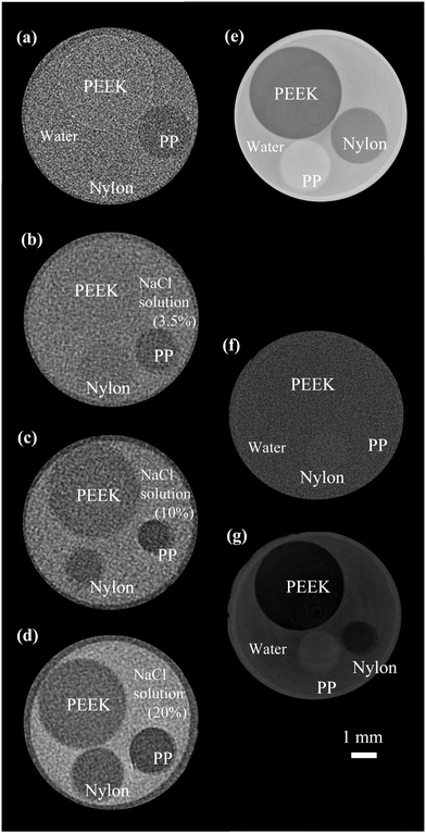
On page 3451, the notations “PP” and “Nylon” in Fig. 5(a)–(d), and (f) are in the wrong place.
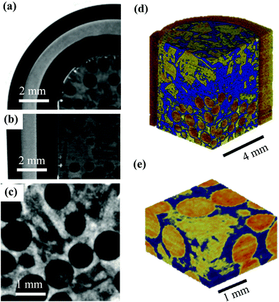
On page, 3452, Fig. 7(d) and (e) mistakenly reproduce a portion of Fig. 7 from a paper by Kerkar et al.1 The relevant parts of the figure were correct in the original submission but were replaced in error upon submission of the revised manuscript when updating the figure in response to reviewer comments.
The correct versions of Fig. 5 and 7 are shown here.
The Royal Society of Chemistry apologises for these errors and any consequent inconvenience to authors and readers.
References
- P. B. Kerkar, K. Horvat, K. W. Jones and D. Mahajan, Geochem. Geophys. Geosyst., 2014, 15, 4759–4768 CrossRef CAS.
| This journal is © the Owner Societies 2020 |


