 Open Access Article
Functional movements of the GABA type A receptor†
Csilla
Várnai‡
 a,
B. W. J.
Irwin‡
 b,
M. C.
Payne
 b,
Gábor
Csányi
 c and
P.-L.
Chau
 *d
*d
aCentre for Computational Biology, University of Birmingham, Birmingham, B15 2TT, UK
bTheory of Condensed Matter Group, Cavendish Laboratory, Department of Physics, University of Cambridge, Cambridge CB3 0HE, UK
cDepartment of Engineering, University of Cambridge, Cambridge CB2 1PZ, UK
dBioinformatique Structurale, Institut Pasteur CNRS URA 3528, CB3I CNRS USR 3756, 75724 Paris, France. E-mail: pc104@pasteur.fr
First published on 7th July 2020
Abstract
We have performed a parallel tempering crankshaft motion Monte Carlo simulation on a model of the GABA type A receptor with the aim of exploring a wide variety of local conformational space. We develop a novel method to analyse the protein movements in terms of a correlation tensor and use this to explore the gating process, that is, how agonist binding could cause ion channel opening. We find that simulated binding impulses to varying clusters of GABA binding site residues produce channel opening, and that equivalent impulses to single GABA sites produce partial opening.
1 Introduction
The family of GABAA receptors is responsible for the majority of fast neuronal inhibition in the mammalian central nervous system, and is a target of general anaesthetics, benzodiazepines, barbiturates and neurosteroids. These pentameric proteins belong to the cys-loop family of ligand-gated ion channels that includes the nicotinic acetylcholine, glycine and 5HT3 receptors. The GABAA receptors are composed of five subunits arranged pseudosymmetrically around the integral anion channel.1 The subunits, of which nineteen have thus far been identified, are separated into classes based on their sequence similarity: there are six α-subunits, three β, three γ, three ρ and single representatives of δ, ε, θ and π.2 The precise subunit isoform composition of the pentamer defines the recognition and biophysical characteristics of the particular receptor subtype.The most ubiquitous subtype, which accounts for approximately 30% of GABAA receptors in the mammalian brain,3 contains two α1-, two β2- and a single γ2-subunit.4 The GABAA receptors can be divided into three structural domains, the extracellular (EC) domain, the transmembrane (TM) domain and the intracellular (IC) domain.
Several structures of the GABAA receptor have been published. Miller and Aricescu5 determined the structure of a human β3-pentamer using X-ray crystallography. Laverty et al.6 created a chimaeric homopentameric GABAA receptor with the extracellular domain from the GLIC protein of the bacterium Gloeobacter violaceus and the transmembrane domain from the α1-subunit of the human GABAA receptor, and determined its structure by X-ray crystallography. Miller et al.7 created a chimaeric homopentameric GABAA receptor with the extracellular domain from the human β3-subunit and the transmembrane domain from the human α5-subunit and determined its structure crystallographically. Zhu et al.8 used cryo-electron microscopy to determine the structure of a truncated heteropentameric human GABAA receptor of subtype (α1)2(β2)2γ2. Phulera et al.9 performed a similar study using a truncated heteropentameric rat GABAA receptor of subtype (α1)2(β1)2γ2S. In all these unnatural receptors, the intracellular domain is absent; previous experiments show that the intracellular domain of this receptor is important in determining the conductance, the gating properties and the speed of desensitisation of the ion channel.10
Laverty et al.11 determined the structure of the GABAA receptor of subtype (α1)2(β3)2γ2, and Masiulis et al.12 determined the structures of the same GABAA receptor bound to different ligands. However, the GABAA receptor was also bound to a megabody Mb38 in all cases. Masiulis et al.12 reported that Mb38 activated the GABAA receptor, but they did not report the time elapsed between receptor activation and structure determination. Thus it was not possible to determine whether these structures were of the resting, open or desensitised receptor, though most probably they are in the desensitised state. Moreover, the intracellular domain was undetermined in all these structures.
Previous experiments have shown that, in the absence of GABA, the GABAA receptor is in the resting state. Administration of GABA will cause the receptor ion channel to open in tens of milliseconds and the receptor then spontaneously desensitises in a few hundred milliseconds.13 There have not been structural studies of the GABAA receptor to study its resting, open and desensitised states and to define how GABA binding to the receptor induces the receptor to open its central ion channel.
The GABAA receptor of subtype (α1)2(β2)2γ2 accounts for 30% of all GABAA receptors in the brain and is the target of a large number of therapeutic drugs. Ernst et al.14 and Mokrab et al.15 have made models of its structure based on similar proteins as the template, and simulations have been performed to study GABA binding and how this process leads to channel opening.
Payghan et al.16 created a model of the GABAA receptor of subtype (α1)2(β2)2γ2 using bacterial ligand-gated ion channel structures as templates. Closed-channel and open-channel states were generated, and GABA was docked to either one of, or both, binding sites of the closed-channel state. United-atom molecular dynamics simulations of 100 ns were carried out on these five structures. These authors observed changes in the pore radius when the channel opens, but they did not identify the gating mechanism. The gating mechanism describes the process whereby agonist binding leads to the opening of its central ion channel.
Carpenter et al.17 created a model of the GABAA receptor of subtype (α6)2(β3)2δ using the nAChR as template; they chose this subtype because it has the highest affinity for GABA. They then performed 100 independent molecular dynamics simulations on this protein, with a GABA molecule placed randomly 25 Å away from the centre of its binding site. Binding occurred in 19 of the 100 simulations; a consistent pathway was found to be taken by GABA, and electrostatic steering was found to be important in guiding the ligand to its binding site. Significantly, these authors noted that the C- and F-loops on either side of the binding site need not open and close to let GABA in, as there is a pathway from the cytoplasmic side of these two loops.
Ayan and Essiz18 made a homology model of the GABAA receptor of subtype (α1)2(β2)2γ2 using the Caenorhabditis elegans GluCl protein as the template. They performed a 100 ns molecular dynamics simulation on the structure embedded in a hydrated palmitoyl-oleoyl phosphatidylcholine (POPC) membrane; on removing ivermectin, the ion channel closes. Correlation analysis shows the extracellular domain β1–β2 and transmembrane domain M2–M3 loops to be important in ion channel closure.
These studies are very valuable, but none of them have systematically approached the problem of how ligand binding causes the ion channel to open. In this work, we simulate the GABAA receptor using a semi-coarse-grained protein model and an efficient Monte Carlo method19,20 which allows fast exploration of protein conformations. Despite some short-comings of our model, we shall show that the main findings of our work are not affected: using the correlated motions of the amino acids from the simulation, we present a linear response theory based method, which identifies possible gating mechanisms upon GABA binding.
2 Methods
2.1 Structure
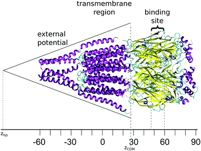
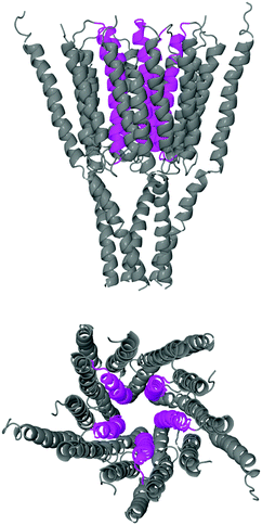
The starting structure used for the simulation is a model of the human GABAA receptor.15Fig. 1 shows a diagram of this structure with its long axis aligned to the z-axis. The z-value at each level of the protein is labelled on the axis below. The GABA binding site is surrounded by two tyrosines, two phenylalanines, a threonine and an arginine. These amino acids are situated at a z-distance of 50 Å ≤ z ≤ 60 Å. The ion channel in the transmembrane region is lined by the M2 helix; Fig. 2 shows the transmembrane and intracellular domains, with the M2 helices highlighted in magenta. The narrow part of the ion channel is situated in the transmembrane domain, at a z-distance of −25 Å ≤ z ≤ 25 Å. There are three annuli of positively-charged amino acids lining the ion channel: a lysine annulus at about z = 20 Å, and two arginine annuli at, respectively, about z = 11.5 Å and approximately z = −15.5 Å (Fig. 3, top panel). There are four annuli of amino acids inside the transmembrane part of the ion channel which contain the –OH group; they are situated at z-distances of about z = −6 Å, z = 0 Å, z = 3 Å and z = 15.5 Å (Fig. 3, bottom panel). These annuli are probably important in controlling the passage of ions through the ion channel.2.2 Simulations
For the molecular simulations, we used CRANKITE,19 a Monte Carlo simulation programme that employs a Gō-like coarse-grained force field. We chose CRANKITE because its Monte Carlo move set consists of local moves of crankshaft rotations of the protein backbone and changes of the side chain dihedral angles, the former of which allows a fast conformational sampling of proteins. CRANKITE uses a full atom representation of the protein backbone, together with explicit side chain β atoms and γ atoms, to include entropic contributions arising from the torsional flexibility of side chains. Each amino acid is treated as hydrophobic or amphipathic. The γ atoms represent the rest of the side chain, with an elongated distance between β and γ atoms to place them near the centre-of-mass of the side chain. In the energy function, volume exclusion of beads, hydrogen bonds between backbone amide H atoms and carbonyl O atoms, and a hydrophobic interaction between γ atoms are modelled explicitly. The secondary structure of the protein is held together by an additional energy term, which keeps the backbone of α-helices and β-strands at the correct backbone twist, and a Gō-like contact potential term which keeps β-sheets well-formed. The secondary structure of proteins is predetermined and does not change within a simulation.We used the force field parameters optimised previously,20 with the addition of a conical external potential on the transmembrane region to mimic the membrane, as the force field had been optimised using a set of non-transmembrane proteins. The energy contribution of this external potential on an amino acid in this region is:
 | (1) |
To explore the possible conformations of the protein while keeping its secondary structure intact, we used parallel tempering simulations with 512 temperature levels ranging from 310 K to 1023 K, sampling conformations at the lowest temperature level, every 1000 steps. In our simulation, 108 Monte Carlo steps were carried out at each temperature level, and temperature swapping took place every 1000 steps, using the Metropolis–Hastings acceptance criterion. Our simulation took 23 hours on 512 cores of the Cambridge University supercomputer CSD3.
2.3 Analysis
The parallel tempering simulation generated 105 configurations in various states at 310 K. Equilibration was determined by examining the log of the absolute value of the auto-correlation function of the energy of the system against the lag between the energies sampled, and finding where it crossed a given y-value that is no higher than the background. Fig. S1 of the ESI† shows this graph. The first 25![[thin space (1/6-em)]](https://www.rsc.org/images/entities/char_2009.gif) 000 configurations were treated as equilibration data and only the remaining 75
000 configurations were treated as equilibration data and only the remaining 75![[thin space (1/6-em)]](https://www.rsc.org/images/entities/char_2009.gif) 000 were used for data analysis. This loose cutoff was determined by monitoring the energy reported by the CRANKITE software (Fig. S1, ESI†), and the observation of spatial correlations between different parts of the protein over 1000 configuration windows (Fig. S2–S7, ESI†).
000 were used for data analysis. This loose cutoff was determined by monitoring the energy reported by the CRANKITE software (Fig. S1, ESI†), and the observation of spatial correlations between different parts of the protein over 1000 configuration windows (Fig. S2–S7, ESI†).
 | (2) |
We assume that during a CRANKITE simulation each residue has a set of principal directions which can be defined by a roughly ellipsoidal cloud of coordinates xat = (xat1, xat2, xat3), with a center of mass μa = (μa1, μa2, μa3), for a residue a, at snapshot t of the simulation which has Nt snapshots. We construct the vector correlation matrix R(1)a of all vector positions  of the residue a across frames in the simulation as
  for a single residue vector covariance matrix C(1)a defined by
 | (3) |
Consider the situation where residue a receives a vector impulse fa when it is at its equilibrium position xa. The component of the impulse in the ui direction is simply fa·ui. Hence the impulse resolved along the principal directions of a is given by ga = fa·Ua.
We then construct a rank three tensor of projections of each residue displacement from μa into each eigenvector in Ua, as Ta = Ua−1(Xa − 1 ⊗ μa)T, along with frame averaged terms τai = 〈Tait〉t We then construct a 3 × 3 correlation matrix for each pair of residues a and b, denoted R(2)ab whose elements are given by
 | (4) |
| δx(a)b = fa·UaR(2)abUb−1 | (5) |
 | (6) |
 is the set of residues. We note that the coefficients relating impulses at a to responses at b are given by a rank four tensor Kab = UaR(2)abUb−1, and these coefficients are purely determined by the CRANKITE trajectory or analogous MC/MD simulation data. This is a relatively computationally expensive step, requiring high memory. By deriving and storing these coefficients we can measure the response of any residue or any set of residues in the system to any arbitrary set of input impulses. The equation to extract responses from inputs is then
 | (7) |
In practice we have a large linear model whose coefficients are defined by spatial correlations across a simulation. The input to the model is a list of 3 dimensional impulse vectors in (x,y,z) coordinates. In our case, for the GABAA receptor which consists of 1823 amino acids residues, Fai is an (1823 × 3) matrix. In practice, when we measure the impulse response to GABA binding, Fai is non-zero for only a handful of residues at the binding site. This is then left multiplied to the coefficient tensor of Kabij of size (1823 × 1823 × 3 × 3), to produce an output matrix ΔXbj of size (1823 × 3). The output ΔXbj gives the magnitude and direction of the response for amino acid b in direction j. From the response of all residues in the transmembrane region, using eqn (2), we can estimate changes in the pore size. The correlation matrix presented here informs us about correlated amino acid motions at physiological temperatures. Reconstructing the free energy path between closed and open states is beyond the scope of this study.
Implementation. To determine the impulses required to mimic GABA binding, we compare the coordinates of the binding site amino acids of two desensitised structures of the GABAA receptor: the first structure is of the receptor complexed with a megabody (PDB code 6I53) and the other structure is of the receptor complexed with picrotoxin, GABA and the same megabody (PDB code 6HUJ). We superimposed these two structures onto one of the equilibrated configurations from our simulation. We then examined the movements of the binding site amino acids from the uncomplexed state to the state where GABA is bound. We describe the vectors corresponding to these movements as the ‘forward vectors’ (see Table 1 for the magnitudes of these vectors); they roughly denote the movements of the binding site amino acids when GABA binds.
| Amino acid from model | Amino acid from 6I53/6HUJ | Forward vector/Å |
|---|---|---|
| α1-Subunit | ||
| Phe E 92 | Phe A 65 | 0.570 |
| Arg E 94 | Arg A 67 | 0.453 |
| Leu E 145 | Leu A 118 | 0.606 |
| Thr E 157 | Thr A 130 | 0.735 |
| Phe C 92 | Phe D 65 | 0.788 |
| Arg C 94 | Arg D 67 | 0.454 |
| Leu C 145 | Leu D 118 | 0.493 |
| Thr C 157 | Thr D 130 | 0.729 |
| β2-Subunit | ||
| Tyr A 121 | Tyr B 97 | 0.904 |
| Glu A 179 | Glu B 155 | 0.862 |
| Ser A 180 | Ser B 156 | 1.047 |
| Tyr A 181 | Tyr B 157 | 1.116 |
| Phe A 224 | Phe B 200 | 0.918 |
| Ser A 225 | Ala B 201 | 0.974 |
| Thr A 226 | Thr B 202 | 0.437 |
| Tyr A 229 | Tyr B 205 | 0.987 |
| Tyr D 121 | Tyr E 97 | 0.690 |
| Glu D 179 | Glu E 155 | 0.799 |
| Ser D 180 | Ser E 156 | 0.915 |
| Tyr D 181 | Tyr E 157 | 0.830 |
| Phe D 224 | Phe E 200 | 0.844 |
| Ser D 225 | Ala E 201 | 0.895 |
| Thr D 226 | Thr E 202 | 0.736 |
| Tyr D 229 | Tyr E 205 | 0.856 |
In order to determine the number of binding site residues required to induce gating (that is, the process whereby agonist binding causes ion channel opening) we measured the responses of forward vectors on either GABA binding site 1, 2 or both 1 and 2, and for the following sets of residues: (a) the principal four residues, Phe 92 and Arg 94 from the α1-subunit and Tyr 181 and Tyr 229 from the β2-subunit, of each binding site (denoted minimal); (b) all residues within 4 Å of where GABA would have bound (limit distance 4 Å); (c) all residues within 5 Å of where GABA would have bound (limit distance 5 Å); (d) all residues within 6 Å of where GABA would have bound (limit distance 6 Å).
3 Results
We performed a parallel tempering simulation of the GABAA receptor, using the CRANKITE coarse-grained protein model resulting in a trajectory comprising of 100![[thin space (1/6-em)]](https://www.rsc.org/images/entities/char_2009.gif) 000 structures. The first 25
000 structures. The first 25![[thin space (1/6-em)]](https://www.rsc.org/images/entities/char_2009.gif) 000 structures were assumed to be associated with thermalisation, as the correlation matrix between the amino acids varies greatly until after 25
000 structures were assumed to be associated with thermalisation, as the correlation matrix between the amino acids varies greatly until after 25![[thin space (1/6-em)]](https://www.rsc.org/images/entities/char_2009.gif) 000 structures. The remaining 75
000 structures. The remaining 75![[thin space (1/6-em)]](https://www.rsc.org/images/entities/char_2009.gif) 000 structures were used to derive the coefficient tensor Kabij. Note that the correlation matrix describes the spatial correlation of amino acid positions, rather than temporal correlations, and so equilibration in time is not a problem.
000 structures were used to derive the coefficient tensor Kabij. Note that the correlation matrix describes the spatial correlation of amino acid positions, rather than temporal correlations, and so equilibration in time is not a problem.
3.1 Impulse-response suggests gating mechanism
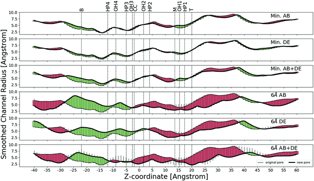
We applied ‘forward vectors’ using the correlation tensor method and noted that the movements of the binding site amino acids caused different parts of the ion channel to open.We differentiate between two binding sites: the AB site is the binding site where the β2-subunit is adjacent to the γ2-subunit, and the DE site is the binding site where the α1-subunit is adjacent to the γ2-subunit. We find that the effect of applying forward vectors to only the principal four residues of each binding site (Fig. 4) is slightly different from that of applying forward vectors to residues within a certain distance limit. The effect of increasing the limit distance from 4 Å to 6 Å is qualitatively not very different, as can be seen in Fig. 4 and Fig. S8–S19 (ESI†). We thus show results of applying the forward vectors to the principal four residues and those of applying the forward vectors to residues within 6 Å of where GABA would have bound. Results pertaining to 4 Å and 5 Å are shown in the ESI.† In these diagrams, we also indicate the following annuli that might be important in ion passage. The lysine annulus is labelled T (top), the two arginine annuli are labelled, respectively, M (middle) and B (bottom). The four annuli of amino acids bearing the OH group are labelled OH1 to OH4. Close to these four OH annuli are four hydrophobic annuli, and they are labelled HP1 to HP4.
In the following figures green corresponds to opening, and red to closing. The forward vector impulse was applied in stages with magnitude varying from 0 to 1. Some parts of the channel were seen to open or close monotonically (solid green or red bar), other parts of the channel opened at a partial application of the forward vectors and then closed again (or vice versa), this kind of motion is represented by a thin black stick above or below the solid bar.
The top profile in Fig. 4 shows the effect of applying forward vectors to the principal four amino acids of the AB site only. There is slight widening of the transmembrane domain at the cytoplasmic end and the intracellular end, and there is slight narrowing of the middle part of the transmembrane domain. The second profile from the top in Fig. 4 shows the effect of applying forward vectors to the principal four amino acids of the DE site only. There is slight widening of the transmembrane domain in most regions, but there is slight narrowing in a short section of the middle region. The third profile from the top in Fig. 4 shows the effect of applying forward vectors to the principal four amino acids of both the AB and DE sites. There is widening of the transmembrane domain in most regions, but there is narrowing in a short part of the middle region.
The fourth profile from the top in Fig. 4 shows the effect of applying forward vectors to 6 Å amino acids of the AB site only. There is slight narrowing of the extracellular half of the transmembrane part of the ion channel (0 Å < z < +30 Å), and extensive widening of the cytoplasmic half of the transmembrane part of the ion channel (−25 Å < z < 0 Å). The narrowest part of the ion channel is situated in the cytoplasmic half, so the net effect is to widen the ion channel. The fifth profile from the top in Fig. 4 shows the effect of applying forward vectors to the DE site only. The effect is similar to applying forward vectors to the AB site, except the widening of the cytoplasmic half of the transmembrane part now extends to +5 Å. The bottom profile from the top in Fig. 4 shows the effect of applying forward vectors to both binding sites. The widening effect is now restricted to −25 Å < z < −11 Å and −8 Å < z < −3 Å. There is slight constriction in the region −11 Å < z < −8 Å and also in the region −3 Å < z < +38 Å. Note that the transmembrane domain ends at about z = +25 Å, so some of the slight constriction is observed in the extracellular domain.
It can immediately be seen that the effect of moving more amino acids in the resting state is to make the ion channel movements larger. There are also changes in the movement direction as the forward vectors are applied to more amino acids. Moreover, we would expect some of the annuli to be crucial in the opening movements. The widening and narrowing effects do not seem to be related to any of the identified annuli.
In summary, we have used information from advanced Monte Carlo simulations to generate three-dimensional correlation tensor coefficients for a large linear model and subsequently analysed the response motions of the GABAA receptor under experimentally derived binding impulses. The results show how movements in the binding site region can widen or narrow the central ion channel and that impulses at a single site induce partial opening.
4 Discussion
Simulating the gating of pentameric ligand-gated ion channels is a challenge of theoretical biochemistry, but it has only been made possible by the development of simulation methods21 and the emergence of fast supercomputers. One of the more recent research has been the simulation of the GluCl channel from an open-channel state to a closed-channel state.22 In this work, the authors simulated a structure of the Caenorhabditis elegans GluCl protein that has been prised open by L-glutamate and ivermectin (PDB code: 3RIF). They performed a data collection simulation of 100 ns of this structure, and also a 140 ns of this structure with ivermectin taken away. They showed that the GluCl channel ion pore area decreased and the channel closed with changes in the tilt of the M2 helices. It is not known if the channel closed fully, as the simulation times were short and the agonist L-glutamate was still bound to the protein.Yuan et al.23 performed molecular dynamics simulations on the 5-HT3A receptor. They used the structure with PDB code 4PIR, repaired the structural defects and put it in a hydrated POPC membrane. They simulated the apo-structure twice for 700 ns, and the holo-structure (five 5-HT molecules inserted into every interface binding site) twice for 700 ns. They find that putting 5-HT into the binding site forced the 5-HT3A receptor subunits to twist and tilt in such a way that the ion channel opens but with distinct asymmetry in the receptor structure. They have also performed a correlation analysis and principal component analysis. Whilst the central ion channel is blocked by hydrophoic amino acids in the apo-structure, in the holo-structure it becomes unblocked and a continuous water structure is formed. Water and ion exit at the intracellular end via the five side portals. These portals are blocked by the M4-MA helix, but these helices move on 5-HT binding so the portals become available for water and ion passage.
To enhance the sampling of rare events such as channel opening, one can apply the ‘swarms of trajectories’ method. This was carried out on the Gloeobacter violaceus GLIC protein;24 the PDB code for its closed-channel structure is 4NPQ and that for its open-channel structure is 4HFI. The authors studied these two structures in detail and applied 35 collective variables in their simulations of gating. They were able to define the gating pathways, the free energy profiles of the conformation space and the gating kinetics. This impressive work is only possible if one knows the beginning and end structures of gating.
Guros et al.25 performed a 20 μs molecular dynamics simulation on the apo-form of the 5-HT3A receptor (PDB code: 4PIR) in a POPC/SDPC/cholesterol membrane. They performed two 15 μs simulations with the receptor immersed in 5 mM and 15 mM 5-HT, respectively, and also carried out a 15 μs simulation of the receptor embedded in a POPC-only membrane immersed in 5 mM 5-HT. They discovered that 5 mM 5-HT was adequate to activate the receptor, and noted that the receptor went through a number of pre-active states but did not observe the fully activated state (PDB code: 6HIN) probably because more simulation time was required.
Dämgen and Biggin26 created a model of the human glycine receptor using the glycine receptor of Danio rerio as the template. They placed this modelled receptor into a hydrated POPC membrane and fill the simulation box with a NaCl solution. They also placed five glycine molecules into the binding sites, and applied restraints for 150 ns to open the ion channel. Once the channel was open, they carried out a 300 ns data production simulation. They showed that the open-channel state conducted Cl− ions with a selectivity ratio over Na+ similar to that found in experiments (28![[thin space (1/6-em)]](https://www.rsc.org/images/entities/char_2009.gif) :
:![[thin space (1/6-em)]](https://www.rsc.org/images/entities/char_2009.gif) 1). Their simulations also show that hydrophobic voids between the subunits were filled by Leu residues; this was not observed in the experimental cryo-EM structure of the glycine receptor.
1). Their simulations also show that hydrophobic voids between the subunits were filled by Leu residues; this was not observed in the experimental cryo-EM structure of the glycine receptor.
These simulations are very important in giving us insight into the gating process. Nevertheless, they all require a very large amount of supercomputer time. Some of the methods used also require prior knowledge of the detailed structural changes. It would be useful to have a method that requires less computer time and can also allow us to trace the mechanistic pathway driving the gating process.
In this work, we have developed a novel method to define the effect of an impulse on the response of the protein. This is a general method which we have applied to a model of the GABAA receptor to demonstrate gating. We show that the results are consistent with existing knowledge about ion channel opening of ligand-gated ion channels.23,25,27–30 Specifically, we observe the same widening of the ion channel pore on channel gating. Significantly, we also show that a single GABA molecule can open the GABAA receptor, but that the effect of two GABA molecules binding to the receptor is greater; the magnitude of the movements is larger. This is consistent with previous experiments.31 We are currently developing a method to identify the mechanistic pathway from the binding site to the narrowest part of the ion channel. This will be the subject of another article, where we shall compare our theoretical predictions with results from previous experiments and simulations in detail.
There are, however, some issues with our study. We do not know the exact effect of GABA binding to the binding site. We have used experimental structures of receptors bound to megabodies, but the GABA-bound form is also bound to picrotoxin and both 6I53 and 6HUJ are probably in the desensitised state. To determine the forward vectors, we should ideally compare the positions of the binding site amino acids of an empty GABAA receptor in the resting state with those of a GABAA receptor bound to GABA in the open state, not in the desensitised state and with no other ligands bound. However, structures from 6I53 and 6HUJ are the closest we can get so far. In this work, applying the forward vectors calculated from 6I53 and 6HUJ suggest parts of the transmembrane ion channel close upon GABA binding, but we cannot be sure that this happens naturally as the forward vectors are determined from receptors in the desensitised state with extra ligands bound.
This work has established the feasibility of using Monte Carlo simulation results to predict protein movements. This research is a preliminary study and further work will be carried out to identify the mechanistic pathway linking the binding site to the gating amino acids. The template for our model of the GABAA receptor is the neuromuscular junction nAChR, PDB code 2BG9. The nAChR structure 2BG9 is a good template because this structure contains the complete, intact protein, and has been resolved under near-physiological conditions; the protein is embedded in its native plasma membrane in a liposome-like structure that resembles its native muscle cell. However, Mnatsakanyan and Jansen32 have shown that the identity of amino acids in the M2 and M3 helices of 2BG9 could be one turn out; the α-subunit of the nAChR has been re-modelled recently,33 and a structure of the nAChR of higher resolution confirms this problem.34
A semi-coarse-grained model such as CRANKITE is an attractive choice to model such a protein; it is less sensitive to the identity of amino acids. Moreover, coarse graining reduces the degrees of freedom, which allows faster exploration of the conformational space of the protein. This allows us to model proteins with slow motions, for which all-atom simulations are prohibitively expensive. On the other hand, one has to be careful not to oversimplify the protein model, and we need to model all relevant features of the GABAA receptor protein that are essential for the gating. In a future study, we will explore the effect of an explicit ligand, more finely tuned electrostatics and even the presence of a membrane and solvents.
Conflicts of interest
The authors declare that they have no conflicts of interest.Acknowledgements
The authors thank Nigel Unwin, Ian Martin and Katarzyna Macieszczak for useful discussions, and Stuart Rankin and Michael Rutter for help with computing. Some of the simulations in this work were carried out on the Darwin Supercomputer of the University of Cambridge High Performance Computing Service using Strategic Research Infrastructure Funding from the Higher Education Funding Council for England, and other simulations were performed using resources provided by the Cambridge Service for Data Driven Discovery (CSD3) operated by the University of Cambridge Research Computing Service, provided by Dell EMC and Intel using Tier-2 funding from the Engineering and Physical Sciences Research Council (capital grant EP/P020259/1), and DiRAC funding from the Science and Technology Facilities Council. BWJI acknowledges financial support from the UK Engineering and Physical Sciences Research Council Centre for Doctoral Training in Computational Methods for Materials Science under Grant No. EP/L015552/1. PC was partly supported by the INCEPTION project ANR-16-CONV-0005.References
- N. Nayeem, T. P. Green, I. L. Martin and E. A. Barnard, Quaternary structure of the native GABAA receptor determined by electron microscopic image analysis, J. Neurochem., 1994, 62, 815–818 CrossRef CAS PubMed.
- E. Sigel and M. E. Steinmann, Structure, function, and modulation of GABAA receptors, J. Biol. Chem., 2012, 282, 40224–40231 CrossRef PubMed.
- P. J. Whiting, GABA-A receptor subtypes in the brain: a paradigm for CNS drug discovery?, Drug Discovery Today, 2003, 8, 445–450 CrossRef CAS PubMed.
- S. J. Farrar, P. J. Whiting, T. P. Bonnert and R. M. McKernan, Stoichiometry of a ligand-gated ion channel determined by fluorescence energy transfer, J. Biol. Chem., 1999, 274, 10100–10104 CrossRef CAS PubMed.
- P. S. Miller and A. Radu Aricescu, Crystal structure of a human GABAA receptor, Nature, 2014, 512, 27–275 CrossRef PubMed.
- D. Laverty, P. Thomas, M. Field, O. J. Andersen, M. G. Gold, P. C. Biggin, M. Gielen and T. G. Smart, Crystal structures of a GABAA-receptor chimera reveal new endogenous neurosteroid-binding sites, Nat. Struct. Mol. Biol., 2017, 24, 977–985 CrossRef CAS PubMed.
- P. S. Miller, S. Scott, S. Masiulis, L. De Colibus, E. Pardon, J. Steyaert and A. Radu Aricescu, Structural basis for GABAA receptor potentiation by neurosteroids, Nat. Struct. Mol. Biol., 2017, 24, 986–992 CrossRef CAS PubMed.
- S. Zhu, C. M. Noviello, J. Teng, R. M. Walsh, J. J. Kim and R. E. Hibbs, Structure of a human synaptic GABAA receptor, Nature, 2018, 559, 67–72 CrossRef CAS PubMed.
- S. Phulera, H. Zhu, J. Yu, D. P. Claxton, N. Yoder, C. Yoshioka and E. Gouaux, Cryo-EM structure of the benzodiazepine-sensitive α1β1γ2S tri-heteromeric GABAA receptor in complex with GABA, eLife, 2018, 7, 39383 CrossRef PubMed.
- K. K. O'Toole and A. Jenkins, Discrete M3-M4 intracellular loop subdomains control specific aspects of γ-aminobutyric acid type A receptor function, J. Biol. Chem., 2011, 286, 37990–37999 CrossRef PubMed.
- D. Laverty, R. Desai, T. Uchanski, S. Masiulis, W. J. Stec, T. Malinauskas, J. Zivanov, E. Pardon, J. Steyaert, K. W. Miller and A. Radu Aricescu, Cryo-EM structure of the human α1β3γ2 GABAA receptor in a lipid bilayer, Nature, 2019, 565, 516–520 CrossRef CAS PubMed.
- S. Masiulis, R. Desai, T. Uchanski, I. Serna Martin, D. Laverty, D. Karia, T. Malinauskas, J. Zivanov, E. Pardon, A. Kotecha, J. Steyaert, K. W. Miller and A. Radu Aricescu, GABAA receptor signalling mechanisms revealed by structural pharmacology, Nature, 2019, 565, 454–459 CrossRef CAS PubMed.
- D. J. Maconochie, J. M. Zempel and J. H. Steinbach, How quickly can GABAA receptors open?, Neuron, 1994, 12, 61–71 CrossRef CAS PubMed.
- M. Ernst, S. Bruckner, S. Boresch and W. Sieghart, Comparative models of GABAA receptor extracellular and transmembrane domains: important insights in pharmacology and function, Mol. Pharmacol., 2005, 68, 1291–1300 CrossRef CAS PubMed.
- Y. Mokrab, V. N. Bavro, K. Mizuguchi, N. P. Todorov, I. L. Martin, S. M. J. Dunn, S. L. Chan and P.-L. Chau, Exploring ligand recognition and ion flow in comparative models of the human GABA type A receptor, J. Mol. Graphics Modell., 2007, 26, 760–774 CrossRef CAS PubMed.
- P. V. Payghan, I. Bera, D. Bhattacharyya and N. Ghoshal, Capturing state-dependent dynamic events of GABAA receptors: a microscopic look into the structural and functional insights, J. Biomol. Struct. Dyn., 2016, 34, 1818–1837 CrossRef CAS PubMed.
- T. S. Carpenter and F. C. Lightstone, An electrostatic funnel in the GABA-binding pathway, PLoS Comput. Biol., 2016, 12, e1004831 CrossRef PubMed.
- M. Ayan and S. Essiz, The neural γ2α1β2α1β2 gamma amino butyric acid ion channel receptor: structural analysis of the effects of the ivermectin molecular and disulfide bridges, J. Mol. Model., 2018, 24, 206 CrossRef PubMed.
- A. A. Podtelezhnikov and D. L. Wild, CRANKITE: a fast polypeptide backbone conformation sampler, Source Code Biol. Med., 2008, 3, 12 CrossRef PubMed.
- C. Várnai, N. S. Burkoff and D. L. Wild, Efficient parameter estimation of generalizable coarse-grained protein force fields using constrative divergence: a maximum likelihood approach, J. Chem. Theory Comput., 2013, 9, 5718–5733 CrossRef PubMed.
- E. Flood, C. Boiteux, B. Lev, I. Vorobyov and T. W. Allen, Atomistic simulations of membrane ion channel conduction, gating and modulation, Chem. Rev., 2019, 119, 7737–7832 CrossRef CAS PubMed.
- N. Calimet, M. Simoes, J.-P. Changeux, M. Karplus, A. Taly and M. Cecchini, A gating mechanism of penatmeric ligand-gated ion channels, Proc. Natl. Acad. Sci. U. S. A., 2013, 110, E3987–E3996 CrossRef CAS PubMed.
- S. Yuan, S. Filipek and H. Vogel, A gating mechanism of the serotonin 5-HT3 receptor, Structure, 2016, 24, 816–825 CrossRef CAS PubMed.
- B. Lev and T. W. Allen, Simulating ion channel activation mechanisms using swarms of trajectories, J. Comput. Chem., 2020, 41, 387–401 CrossRef CAS PubMed.
- N. B. Guros, A. Balijepalli and J. B. Klauda, Microsecond-timescale simulations suggest 5-HT-mediate preactivation of the 5-HT3A serotonin receptor, Proc. Natl. Acad. Sci. U. S. A., 2020, 117, 405–414 CrossRef CAS PubMed.
- M. A. Dämgen and P. C. Biggin, A refined open state of the glycine receptor obtained via molecular dynamics simulations, Structure, 2020, 28, 130–139 CrossRef PubMed.
- G. G. Wilson and A. Karlin, Acetylcholine receptor channel structure in the resting, open and desensitized states probed with the substituted-cysteine-accessibility method, Proc. Natl. Acad. Sci. U. S. A., 2001, 98, 1241–1248 CrossRef CAS.
- I. H. Yamodo, D. C. Chiara, J. B. Cohen and K. W. Miller, Conformational changes in the nicotinic acetylcholine receptor during gating and desensitization, Biochemistry, 2010, 49, 156–165 CrossRef CAS PubMed.
- N. Unwin and Y. Fujiyoshi, Gating movement of acetylcholine receptor caught by plunge-freezing, J. Mol. Biol., 2012, 422, 617–634 CrossRef CAS PubMed.
- G. Gonzalez-Gutierrez and C. Grosman, The atypical cation-conduction and gating properties of ELIC underscore the marked functional versatility of the pentameric ligand-gated ion-channel fold, J. Gen. Physiol., 2015, 146, 15–36 CrossRef CAS PubMed.
- D. A. Mathers and Y. Wang, Effect of agonist concentration on the lifetime of GABA-activated membrane channels in spinal cord neurons, Synapse, 1988, 2, 627–632 CrossRef CAS PubMed.
- N. Mnatsakanyan and M. Jansen, Experimental determination of the vertical alignment between the second and third transmembrane segments of muscle nicotinic acetylcholine receptors, J. Neurochem., 2013, 125, 843–854 CrossRef CAS PubMed.
- J. Newcombe, A. Chatzidaki, T. D. Sheppard, M. Topf and N. S. Millar, Diversity of nicotinic acetylcholine receptor positive allosteric modulators revealed by mutagenesis and a revised structural model, Mol. Pharmacol., 2018, 93, 128–140 CrossRef CAS PubMed.
- M. M. Rahman, J. Teng, B. T. Worrell, C. M. Noviello, M. Lee and A. Karlin, et al., Structure of the native muscle-type nicotinic receptor and inhibition by snake venom toxins, Neuron, 2020, 106, 1–11 CrossRef PubMed.
Footnotes |
| † Electronic supplementary information (ESI) available. See DOI: 10.1039/d0cp01128b |
| ‡ These two authors contributed equally. |
| This journal is © the Owner Societies 2020 |


