
 Open Access Article
 This Open Access Article is licensed under a
 Creative Commons Attribution 3.0 Unported Licence
A self-assembling amphiphilic dendrimer nanotracer for SPECT imaging†
Ling
Ding
 ab,
Zhenbin
Lyu
a,
Aura
Tintaru
c,
Erik
Laurini
d,
Domenico
Marson
d,
Beatrice
Louis
ef,
Ahlem
Bouhlel
ef,
Laure
Balasse
ef,
Samantha
Fernandez
ef,
Philippe
Garrigue
ef,
Eric
Mas
g,
Suzanne
Giorgio
a,
Sabrina
Pricl
dh,
Benjamin
Guillet
ef and
Ling
Peng
*a
ab,
Zhenbin
Lyu
a,
Aura
Tintaru
c,
Erik
Laurini
d,
Domenico
Marson
d,
Beatrice
Louis
ef,
Ahlem
Bouhlel
ef,
Laure
Balasse
ef,
Samantha
Fernandez
ef,
Philippe
Garrigue
ef,
Eric
Mas
g,
Suzanne
Giorgio
a,
Sabrina
Pricl
dh,
Benjamin
Guillet
ef and
Ling
Peng
*a
aAix Marseille Univ, CNRS, Centre Interdisciplinaire de Nanoscience de Marseille (UMR 7325), Equipe Labellisée Ligue Contre le Cancer, Marseille, France. E-mail: ling.peng@univ-amu.fr
bAix Marseille Univ, CNRS, CRMBM, Marseille, France
cAix Marseille Univ, CNRS, Institut de Chimie Radicalaire (UMR7273), Marseille, France
dMolecular Biology and Nanotechnology Laboratory (MolBNL@UniTS), DEA, University of Trieste, Trieste, Italy
eAix Marseille Univ, INSERM, INRA, C2VN, Marseille, France
fAix Marseille Univ, CNRS, Centre Européen de Recherche en Imagerie Médicale (CERIMED), Marseille, France
gAix Marseille Univ, CNRS, INSERM, Institut Paoli-Calmettes, Centre de Recherche en Cancérologie de Marseille (CRCM), Marseille, France
hDepartment of General Biophysics, Faculty of Biology and Environmental Protection, University of Lodz, Lodz, Poland
First published on 22nd November 2019
Abstract
Bioimaging has revolutionized modern medicine, and nanotechnology can offer further specific and sensitive imaging. We report here an amphiphilic dendrimer able to self-assemble into supramolecular nanomicelles for effective tumor detection using SPECT radioimaging. This highlights the promising potential of supramolecular dendrimer platforms for biomedical imaging.
Medical imaging plays an important role in modern medicine by providing accurate information relating to diagnosing, grading and staging diseases as well as monitoring treatment response and efficacy.1 Among the commonly used non-invasive imaging modalities such as magnetic resonance imaging (MRI), computed tomography (CT), positron emission tomography (PET), single photon emission computed tomography (SPECT), and ultrasonography (US), PET and SPECT have the highest sensitivity, and are able to visualize functional information quantitatively, which is very important for disease assessment and diagnosis and personalized medicine.1–5 Nanotechnology can further enhance the sensitivity and specificity of molecular imaging via the “Enhanced Permeation and Retention (EPR)” effect (also named passive tumor targeting).6–8 EPR results in nanoparticle specific tumor accumulation thanks to the leaky vasculature and dysfunctional lymphatic system characterizing the tumor microenvironment.9 In addition, nanosystems carrying and incorporating abundant imaging reporters can significantly amplify the contrast signal for better imaging and diagnosis. Consequently, a myriad of nanosystems have been explored for PET and SPECT imaging of tumors.2,6,10
We have recently used the self-assembling amphiphilic dendrimer Ga-1 to establish an innovative nanotracer for PET imaging (Fig. 1).11 This dendrimer is composed of a long hydrophobic alkyl chain and a hydrophilic poly(amidoamine) (PAMAM) dendron bearing the PET radionuclide 68Ga(III) complexed within the macrocyclic chelator NOTA (1,4,7-triazacyclononane-1,4,7-triacetic acid) at the peripheral units (Ga-1 in Fig. 1). It self-assembles into small and stable nanomicelles, which can effectively accumulate in tumors. These nanomicelles deliver excellent results in PET imaging of different tumors, including some which could not be detected with the standard clinical PET agent [18F]FDG (2-fluorodeoxyglucose).11 The performance of this dendrimer radiotracer is largely ascribed to the beneficial combination of its unique multivalent dendrimeric structure and the relevant EPR effect.
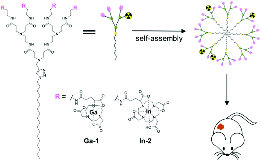 | ||
| Fig. 1 Self-assembling dendrimer nanosystems based on the amphiphilic dendrimers 1 and 2 bearing radionuclide, for positron emission tomography (PET) and single photon emission computed tomography (SPECT) imaging of tumors, respectively. | ||
Motivated by the promising PET imaging results,11 we aimed at further exploiting self-assembling nanotechnology for constructing effective dendrimer-based radiotracers for SPECT imaging. Different from PET, SPECT has the capacity to image multiple processes simultaneously by using the corresponding detection windows of different radionuclides with distinct gamma ray energies.1–3 In addition, SPECT is more readily accessible and less expensive than PET in the clinics, even if PET has higher sensitivity and resolution. Recent progress in advanced detection technology and the combination with computed tomography (CT) has considerably improved the resolution and sensitivity of SPECT, placing SPECT as a quantitative imaging modality similar to PET.3,12 Also, SPECT radiotracers generally have longer half-lives, which allows the characterization of slow kinetic processes and long biological events that take hours or days. Common radionuclides used in SPECT imaging include technetium-99m ([99mTc]Tc), indium-111 ([111In]In) and iodine-123 ([123I]I). Although [99mTc]Tc is the most widely used, [111In]In has a relatively longer half-life (2.8 days). Accordingly, in this study, we chose [111In]In as the radionuclide to develop a dendrimer radiotracer for SPECT imaging with the aim of monitoring and measuring long and slow biological processes such as tumor development and treatment.
Chelation of 111In3+ to a thermodynamically stable and kinetically inert complex is a fundamental requirement for SPECT imaging in order to prevent the release of free radionuclide.4,5 The cyclic chelator DOTA (1,4,7,10-tetraaza-cyclododecane-1,4,7,10-tetraacetic acid) and the acyclic chelator DTPA (diethylenetriaminepentaacetic acid) are the most frequently used in nuclear medicine.13 In general, DOTA forms more stable complexes with radionuclide ions than the acyclic chelator DTPA because of the entropically favorable pre-organized and rigid binding sites within the DOTA ring. Nevertheless, the process of metal-complex formation is often slow for DOTA, and requires high temperature and long reaction times. Yet, as DOTA is the “gold standard” chelator for 111In3+,13 and on the basis of our previous experience in developing the NOTA-conjugated dendrimer to complex with the radionuclide Ga3+ (Ga-1) for PET imaging (Fig. 1),11 we selected DOTA as the chelator to construct the amphiphilic dendrimer 2 which, in turn, was complexed with 111In3+ to generate In-2 for SPECT imaging (Fig. 1 and 2). Knowing that the DOTA ring is considerably larger than that of NOTA, we initially worried about the eventual synthetic difficulty stemming from steric hindrance of the DOTA terminals in 2. Gratifyingly, we could reliably synthesize the DOTA-conjugated dendrimer 2 with high yield (Fig. 2A). Also of note, we successfully prepared the stable dendrimer complex In-2 at 37 °C within 2 hours. Importantly, In-2 self-assembled into small and uniform nanomicelles for effective SPECT imaging of tumors. We present below the synthesis and characterization of the amphiphilic dendrimer 2 and its complex with In3+ as well as the nanomicelles formed with the obtained dendrimer In-2 for SPECT imaging.
 | ||
| Fig. 2 Synthesis of the amphiphilic dendrimer 2 bearing DOTA units and its chelation with the nonradioactive isotope [115In]In3+ at the terminals to deliver the dendrimer In-2. (A) Synthesis scheme: (i) (a) DOTA-GA(tBu)4, PyBOP, NMM, DMF, 30 °C, 72 h; (b) TFA, CH2Cl2, 30 °C, 24 h. (ii) [115In]InCl3, 1.0 M HCl, 37 °C, 2 h. (B) High-resolution mass spectrum (HRMS) showing the isotopic pattern of the observed triply charged species [[115In]In-2 + 3H]3+. The inset shows the calculated isotopic pattern. (C) Isothermal titration calorimetry (ITC) curve (right) for chelation of In3+ with the dendrimer 2. The left panel shows measured heat power versus time elapsed during titration. | ||
Similar to our previous synthesis of the NOTA-dendrimer 1,11 we conjugated the amine-terminated amphiphilic dendrimer with the reagent DOTA-GA(tBu)4, followed by deprotection for preparing 2 (Fig. 2A). Compared to the synthesis of 1, we halved the quantity of the reagent DOTA-GA(tBu)4 from 4 to 2 equivalents, which considerably simplified the purification procedure while maintaining the high synthesis yield of 88% for 2. The chemical structure and integrity of 2 was analyzed and confirmed using 1H, 13C and 2D NMR and high-resolution mass spectrometry (HRMS), which exhibited the signals characteristic of the chemically conjugated DOTA groups (Fig. S1 and S2, ESI†). Chelation of the stable isotope 115In3+ by 2 was performed using 115InCl3 at 37 °C, pH 5.0 for 2 hours (Fig. 2A), and the final dendrimer In-2 was obtained in pure form as a white solid after dialysis to remove the free 115In3+. The successful complexation of four 115In3+ by each molecule of the dendrimer 2 was confirmed using HRMS, which showed the isotopic pattern characteristic of the triply charged species [115In-2 + 3H]3+ in addition to the expected molecular weight peak (Fig. 2B and Fig. S3B, ESI†).14 It is important to mention that the formation of DOTA complexes with metal ions usually requires high temperature at 95 °C and long reaction time of several hours because of the slow binding kinetics of DOTA. Remarkably, we successfully chelated In3+ with the DOTA-conjugated dendrimer 2 at relatively low temperature (37 °C) within 2 h. This may be ascribed to the steric congestion created at the dendrimer terminals,15 making the DOTA entities in 2 more reactive and hence favorably promoting their complexation with In3+ rapidly and at lower temperature to form the stable complex In-2.
To corroborate the reliable synthesis of In-2, we studied the formation of the complex between dendrimer 2 and In3+ using isothermal titration calorimetry (ITC). Specifically, a solution of 2 at 100 μM was titrated with the solution of InCl3 at pH = 5.0 and 37 °C (see the ESI† for details). The left panel in Fig. 2C shows that the interaction between the DOTA cages of 2 and the In3+ cations is characterized by a robust exothermic behavior, reflecting an enthalpy-driven binding process (ΔH = −5.25 kcal mol−1) led by strong coordination interactions between the In3+ and the DOTA cages of 2. Notably, the entropic component (−TΔS = −2.61 kcal mol−1) also favors the stability of In-2. This is probably due to a synergistic effect of the hydrophobic interactions between the apolar dendrimer tails, which aggregate together with the concomitant release of water and ions from the charged surfaces when they form complexes with the cations. Accordingly, the spontaneous formation of the In-2 complex is highly thermodynamically favorable, with a Gibbs free energy (ΔG) value of −7.86 kcal mol−1. Interestingly, ITC measurements show that the number of 115In3+ in In-2 is 4.02, which confirms the ideal stoichiometry of 4![[thin space (1/6-em)]](https://www.rsc.org/images/entities/char_2009.gif) :1. Taken together, the ITC results provide evidence that the synthesis of In-2 was successful, and that In-2 is a stable complex.
:1. Taken together, the ITC results provide evidence that the synthesis of In-2 was successful, and that In-2 is a stable complex.
We next studied the self-assembly of In-2 in solution. In-2 spontaneously self-assembled into small and spherical nanoparticles with average dimensions around 18 nm, as revealed by transmission electron microscopy (TEM) (Fig. 3A). Also, dynamic light scattering (DLS) analysis confirmed the formation of small nanoparticles with sizes around 19 nm, which is typical for nanomicelles (Fig. 3B). The formed nanoparticles were stable, with the critical micelle concentration (CMC) being 60 ± 10 μM (Fig. S4, ESI†). Further molecular dynamics (MD) simulations confirmed the spontaneous aggregation of In-2 into spherical micelles. Fig. 3C illustrates a representative configuration of the stable In-2 micelles obtained at the end of the computational process starting from a random distribution of In-2 in solution. The calculated average micelle diameter was around 15 nm, in close agreement with the values obtained using experimental techniques (TEM and DLS). Along the entire MD trajectories, all the In3+/DOTA terminal groups were nicely located at the micellar periphery without any back-folding observed, as evident from the relevant radial distribution function of the terminal groups shown in Fig. 3D.
 | ||
| Fig. 3 Self-assembling of the amphiphilic dendrimer In-2 into small and uniform nanomicelles. (A) Transmission electron microscopy (TEM) image, (B) dynamic light scattering (DLS) measurement, and (C and D) computer modeling of the self-assembled nanoparticles formed by In-2. (C) Final image of the In-2 self-assembly process into spherical micelles as obtained from atomistic molecular dynamics (MD) simulations. The different parts of the In-2 molecules are represented as spheres (atom color: grey, hydrocarbon chain; yellow, DOTA cage; black, In3+), while water molecules are shown as aqua transparent spheres. The first water shell surrounding each molecule/micelle is highlighted as a light cyan transparent contour. (D) Radial distribution function of the In(III)-bearing terminals as a function of the distance from the center of mass of the In-2 micelles. | ||
Encouraged by the favorable self-assembly properties of In-2, we prepared the corresponding radioactive dendrimer complex [111In]In-2 for SPECT imaging. We obtained the [111In]In-2 complex with a satisfying radiochemical purity over 91 ± 2% along with a high molar activity of 1.09 ± 0.15 GBq μmol−1. In addition, this radiochemical purity and integrity was maintained for up to 30 hours at 37 °C in human serum (Fig. 4A). On the basis of the high radiochemical purity and stability, we performed SPECT imaging using [111In]In-2 in orthotopically xenografted mice bearing tumors derived from a human pancreatic adenocarcinoma tumor cell line, SOJ-6 (Fig. 4B). Co-registration with CT enabled precise, anatomical localization of SPECT signals for further quantification. The biodistribution of [111In]In-2 mapped by SPECT has obvious similarities with that obtained with [68Ga]Ga-1 using PET (Fig. 4B and Fig. S4, ESI†).11 [111In]In-2 showed hepatic retention and elimination through the urinary tract (Fig. 4C and Fig. S4B, ESI†), similar to what was observed for [68Ga]Ga-1 using PET,11 and many other nanoparticles.2,6–8 Notably, the hepatic uptake of [111In]In-2 was 2-fold higher than we previously observed for [68Ga]Ga-1, along with a higher kidney retention (Fig. S4B, ESI†). The tumor uptake of [111In]In-2 was almost 2-fold higher than that of [68Ga]Ga-1 (Fig. 4D and Fig. S4, ESI†), and the duration of the signal was also more stable and longer. This difference may stem from the different chelators, and the resulting negatively charged [111In]In-2 and neutral [68Ga]Ga-1, as well as the slightly different size of [111In]In-2 and neutral [68Ga]Ga-1. In addition, [111In]In has a longer half-life than [68Ga]Ga. Altogether, these results confirm the high flexibility and modularity of our dendrimer nanosystems for bioimaging.
 | ||
| Fig. 4 Radiolabeled dendrimer [111In]In-2 for SPECT imaging in a mouse orthotopic xenograft model of pancreatic adenocarcinoma (SOJ-6 cell line). (A) [111In]In-2 radiochemical purity and stability in human serum at 37 °C for at least 30 h was assessed by radio-thin layer chromatography. (B) Representative μSPECT/CT image of [111In]In-2 180 minutes after intravenous injection. Orthotopic SOJ6 tumor is highlighted by the red circle (n = 3 mice). (C) Biodistribution of [111In]In-2 quantified in each organ by μSPECT/CT 180 min after injection. Results are expressed as the mean percentage of injected dose per gram of tissue (n = 3 mice). (D) Tumor uptake comparison between [68Ga]Ga-1 and [111In]In-2 in the same mice (n = 3). | ||
In conclusion, we have developed a supramolecular dendrimer nanosystem based on self-assembly of the amphiphilic dendrimer In-2 for SPECT imaging in an orthotopic tumor-xenograft mouse model. The work present here alongside our previous studies on PET imaging11 and drug delivery,16–18 highlights that nanosystems formed from self-assembling dendrimers have great potential as novel and robust platforms for various biomedical applications. The supramolecular dendrimer nanosystem developed in this work can be further extended to radiotherapy, and to applications which combine radiotherapy and imaging,4,5 for example, those using the radionuclide [177Lu]Lu. We are working actively to realize these exciting possibilities.
We thank Michel Skandalovski (CERIMED, Aix-Marseille University) and Sandrine Pons (Faculty of Pharmacy, Aix-Marseille University) for technical support. This work was supported by the Ligue Nationale Contre le Cancer (LP, ZL), the ERA-Net EURONANOMED projects “Target4Cancer”, “NANOGLIO” and “TARBRAINFECT” (LP), H2020 NMBP “SAFE-N-MEDTECH” (814607) (LP), China Scholarship Council (LD) and Italian Association for Cancer Research (IG17413) (SP). This article is based on work from COST Action CA 17140 “Cancer Nanomedicine from the Bench to the Bedside” supported by COST (European Cooperation in Science and Technology).
Conflicts of interest
There are no conflicts to declare.Notes and references
- M. L. James and S. S. Gambhir, Physiol. Rev., 2012, 92, 897–965 CrossRef CAS PubMed.
- D. Ni, E. B. Ehlerding and W. Cai, Angew. Chem., Int. Ed., 2019, 58, 2570–2579 CrossRef CAS PubMed.
- O. Israel, O. Pellet, L. Biassoni, D. De Palma, E. Estrada-Lobato, G. Gnanasegaran, T. Kuwert, C. la Fougère, G. Mariani, S. Massalha, D. Paez and F. Giammarile, Eur. J. Nucl. Med. Mol. Imaging, 2019, 46, 1990–2012 CrossRef PubMed.
- T. J. Wadas, E. H. Wong, G. R. Weisman and C. J. Anderson, Chem. Rev., 2010, 110, 2858–2902 CrossRef CAS PubMed.
- T. I. Kostelnik and C. Orvig, Chem. Rev., 2019, 119, 902–956 CrossRef CAS PubMed.
- H. Chen, W. Zhang, G. Zhu, J. Xie and X. Chen, Nat. Rev. Mater., 2017, 2, 17024 CrossRef CAS PubMed.
- C. Li, Nat. Mater., 2014, 13, 110 CrossRef CAS PubMed.
- E. K.-H. Chow and D. Ho, Sci. Transl. Med., 2013, 5, 216rv214 Search PubMed.
- H. Maeda, J. Wu, T. Sawa, Y. Matsumura and K. Hori, J. Controlled Release, 2000, 65, 271–284 CrossRef CAS PubMed.
- E.-K. Lim, T. Kim, S. Paik, S. Haam, Y.-M. Huh and K. Lee, Chem. Rev., 2015, 115, 327–394 CrossRef CAS PubMed.
- P. Garrigue, J. Tang, L. Ding, A. Bouhlel, A. Tintaru, E. Laurini, Y. Huang, Z. Lyu, M. Zhang, S. Fernandez, L. Balasse, W. Lan, E. Mas, D. Marson, Y. Weng, X. Liu, S. Giorgio, J. Iovanna, S. Pricl, B. Guillet and L. Peng, Proc. Natl. Acad. Sci. U. S. A., 2018, 115, 11454–11459 CrossRef CAS PubMed.
- D. L. Bailey and K. P. Willowson, Eur. J. Nucl. Med. Mol. Imaging, 2014, 41, 17–25 CrossRef PubMed.
- E. W. Price and C. Orvig, Chem. Soc. Rev., 2014, 43, 260–290 RSC.
- We were unable to obtain well-resolved NMR spectra for In-2 because of the highly quadrupolar effect of the 115In nucleus. For details, please see, Handbook of High Resolution Multinuclear NMR, ed. C. Brevard and P. Granger, John Wiley and Sons, Inc., 1981 Search PubMed.
- Z. Zhou, M. Cong, M. Li, A. Tintaru, J. Li, J. Yao, Y. Xia and L. Peng, Chem. Commun., 2018, 54, 5956–5959 RSC.
- T. Wei, C. Chen, J. Liu, C. Liu, P. Posocco, X. Liu, Q. Cheng, S. Huo, Z. Liang, M. Fermeglia, S. Pricl, X.-J. Liang, P. Rocchi and L. Peng, Proc. Natl. Acad. Sci. U. S. A., 2015, 112, 2978–2983 CrossRef CAS PubMed.
- C. Chen, P. Posocco, X. Liu, Q. Cheng, E. Laurini, J. Zhou, C. Liu, Y. Wang, J. Tang, V. D. Col, T. Yu, S. Giorgio, M. Fermeglia, F. Qu, Z. Liang, J. J. Rossi, M. Liu, P. Rocchi, S. Pricl and L. Peng, Small, 2016, 12, 3667–3676 CrossRef CAS PubMed.
- Y. Dong, T. Yu, L. Ding, E. Laurini, Y. Huang, M. Zhang, Y. Weng, S. Lin, P. Chen, D. Marson, Y. Jiang, S. Giorgio, S. Pricl, X. Liu, P. Rocchi and L. Peng, J. Am. Chem. Soc., 2018, 140, 16264–16274 CrossRef CAS PubMed.
Footnote |
| † Electronic supplementary information (ESI) available. See DOI: 10.1039/c9cc07750b |
| This journal is © The Royal Society of Chemistry 2020 |