 Open Access Article
Creative Commons Attribution 3.0 Unported Licence
Correction: Bifunctional scaffolds for the photothermal therapy of breast tumor cells and adipose tissue regeneration
Xiuhui
Wang
ab,
Jing
Zhang
ab,
Jingchao
Li
ab,
Ying
Chen
ab,
Yazhou
Chen
ab,
Naoki
Kawazoe
a and
Guoping
Chen
*ab
aTissue Regeneration Materials Group, Research Center for Functional Materials, National Institute for Materials Science, 1-1 Namiki, Tsukuba, Ibaraki 305-0044, Japan. E-mail: Guoping.Chen@nims.go.jp; Fax: +81-29-860-4714; Tel: +81-29-860-4496
bDepartment of Materials Science and Engineering, Graduate School of Pure and Applied Sciences, University of Tsukuba, 1-1-1 Tennodai, Tsukuba, Ibaraki 305-8571, Japan
First published on 9th May 2019
Abstract
Correction for ‘Bifunctional scaffolds for the photothermal therapy of breast tumor cells and adipose tissue regeneration’ by Guoping Chen et al., J. Mater. Chem. B, 2018, 6, 7728–7736.
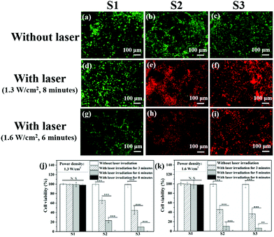
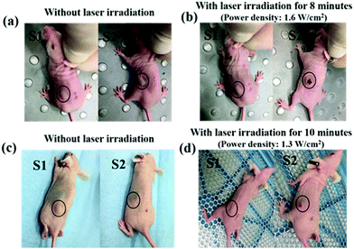
The authors regret that six incorrect images were used in Fig. 4a, d, f and i and Fig. 6a and c of the original manuscript, and the labelling of samples S1 and S2 was missing in the original Fig. 6. The correct versions of Fig. 4 and 6 are shown below. The captions for these figures remain unchanged.
The Royal Society of Chemistry apologises for these errors and any consequent inconvenience to authors and readers.
| This journal is © The Royal Society of Chemistry 2019 |


![[thin space (1/6-em)]](https://www.rsc.org/images/entities/char_2009.gif)
