
 Open Access Article
 This Open Access Article is licensed under a
 Creative Commons Attribution 3.0 Unported Licence
Correction: iTRAQ-based quantitative proteomic analysis for identification of biomarkers associated with emodin against severe acute pancreatitis in rats
Hong Xiangab,
Qingkai Zhangc,
Danqi Wangd,
Shilin Xiad,
Guijun Wange,
Guixin Zhangc,
Hailong Chenc,
Yingjie Wuab and
Dong Shang*ac
aCollege (Institute) of Integrative Medicine, Dalian Medical University, Dalian 116011, China. E-mail: shangdong@dmu.edu.cn; Fax: +86-411-83622844; Tel: +86-411-83635963
bInstitute of Gene Engineered Animal Models for Human Diseases, Dalian Medical University, Dalian 116044, China
cDepartment of General Surgery, Pancreatico-Biliary Center, The First Affiliated Hospital of Dalian Medical University, Dalian 116011, China
dClinical Laboratory of Integrative Medicine, The First Affiliated Hospital of Dalian Medical University, Dalian 116011, China
eDepartment of General Surgery, The First Affiliated Hospital of Jinzhou Medical University, Jinzhou 121000, China
First published on 23rd August 2019
Abstract
Correction for ‘iTRAQ-based quantitative proteomic analysis for identification of biomarkers associated with emodin against severe acute pancreatitis in rats’ by Hong Xiang et al., RSC Adv., 2016, 6, 72447–72457.
The authors regret that Fig. 2–4 were shown incorrectly in the original article. An incorrect section of the SAP group in the MPO-immunohistochemical staining (Fig. 2A) and HE staining (Fig. 3) experiments was used in error. In addition, Fig. 4 has been revised to show the zymogen granule, in order to better represent the ultrastructure of the pancreas. The correct versions of Fig. 2–4 are shown below.
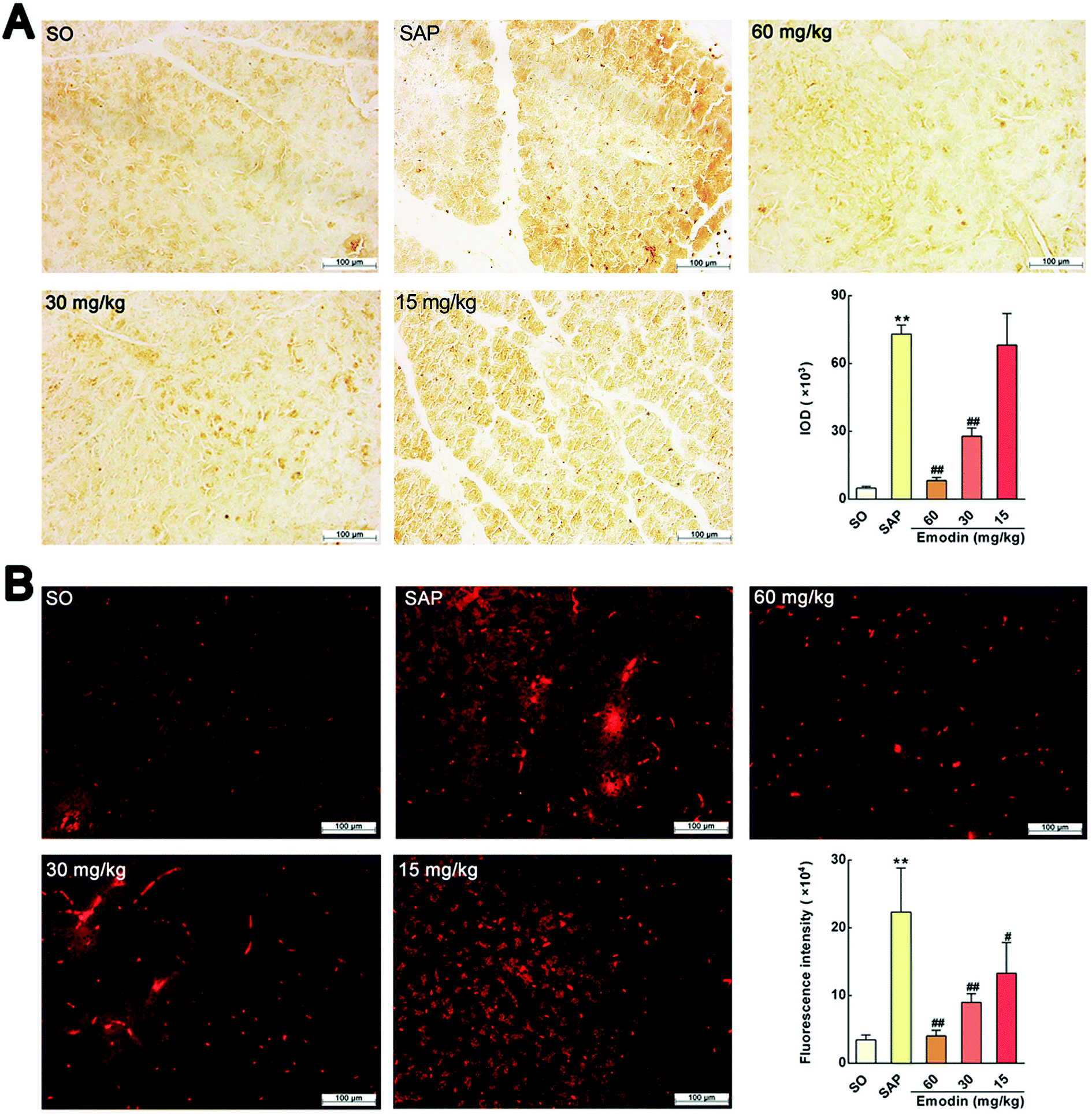 | ||
| Fig. 2 Emodin down-regulated the MPO protein expression in pancreas of SAP rats. (A) Effect of emodin on MPO-immunopositive area (brown) staining of pancreatic tissue in SAP rats by immunohistochemical detection. (B) Effect of emodin on MPO-immunopositive area (red) staining of pancreatic tissue in SAP rats by immunofluorescence detection. Images are presented at 200× magnification. The data are presented as the mean ± SD, n = 6. **P < 0.01 versus SO; #P < 0.05 versus SAP, ##P < 0.01 versus SAP. | ||
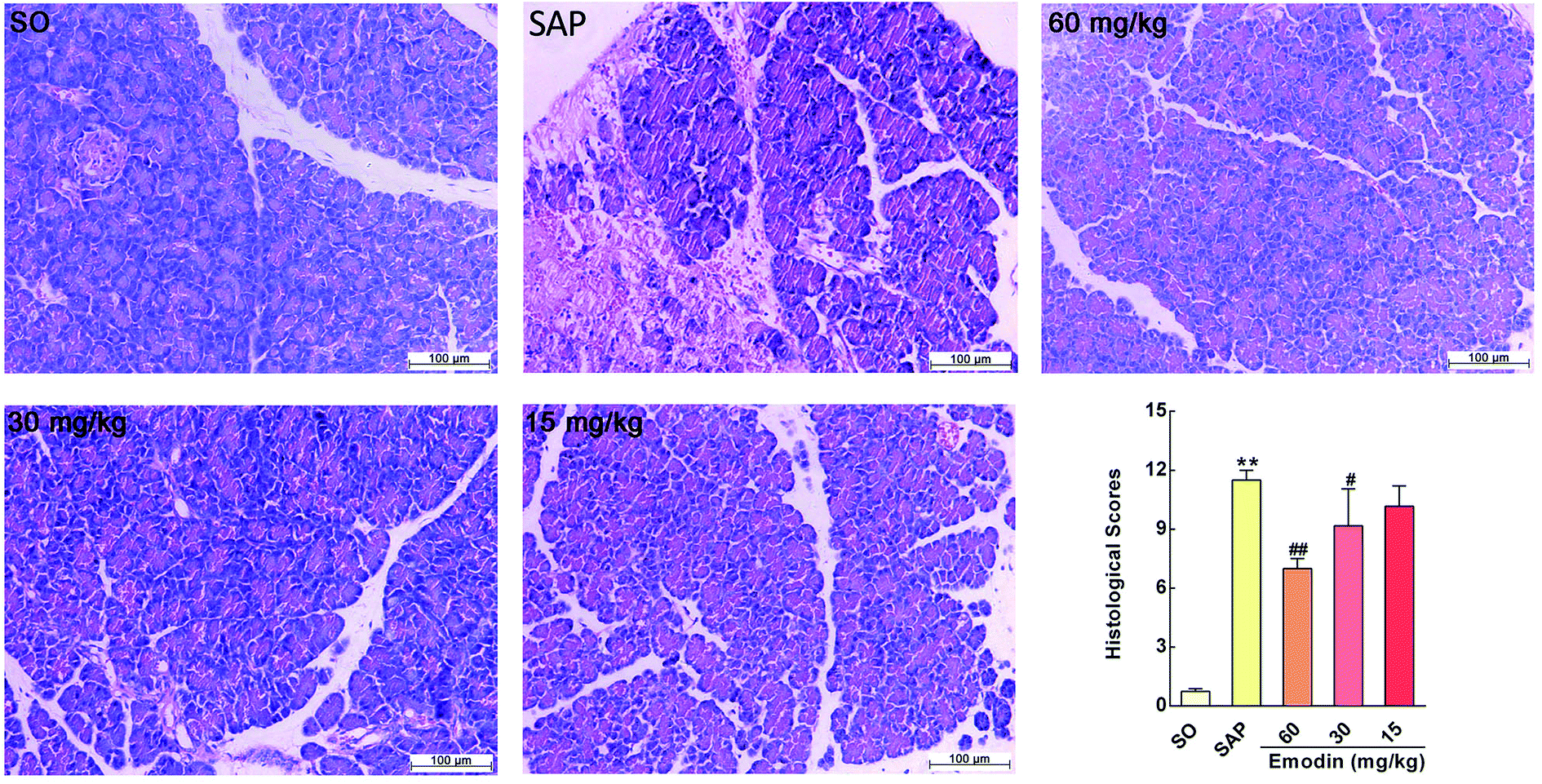 | ||
| Fig. 3 Emodin improved pancreatic histopathology of SAP rats. Effect of emodin on H&E staining of pancreatic tissue in SAP rats. Images are presented at 200× magnification. The data are presented as the mean ± SD, n = 6. **P < 0.01 versus SO; #P < 0.05 versus SAP, ##P < 0.01 versus SAP. | ||
 | ||
Fig. 4 Emodin attenuated cellular structure changes in pancreas of SAP rats. Representative images of the cells’ ultrastructure in the SO (A), SAP (B), 60 mg kg−1 emodin (C), 30 mg kg−1 emodin (D) and 15 mg kg−1 emodin (E) groups. Images are presented at 25![[thin space (1/6-em)]](https://www.rsc.org/images/entities/char_2009.gif) 000× magnification. 000× magnification. | ||
The Royal Society of Chemistry apologises for these errors and any consequent inconvenience to authors and readers.
| This journal is © The Royal Society of Chemistry 2019 |