Open Access Article
This Open Access Article is licensed under a
Creative Commons Attribution 3.0 Unported Licence

Correction: A siRNA-induced peptide co-assembly system as a peptide-based siRNA nanocarrier for cancer therapy

Wenjun Li a, Dongyuan Wang a, Xiaodong Shi a, Jingxu Li a, Yue Ma a, Yanding Wang a, Tingting Li b, Jianing Zhang a, Rongtong Zhao a, Zhiqiang Yu c, Feng Yin *a and Zigang Li *a
aState Key Laboratory of Chemical Oncogenomics, School of Chemical Biology and Biotechnology, Peking University Shenzhen Graduate School, Shenzhen, 518055, China. E-mail: lizg@pkusz.edu.cn; Fax: +86-755-2603-3174; Tel: +86-755-2603-3616
bSchool of Advanced Material, Peking University Shenzhen Graduate School, Shenzhen, 518055, China
cSchool of Pharmaceutical Sciences, Guangdong Provincial Key Laboratory of New Drug Screening, Southern Medical University, Guangzhou 510515, China

Received 29th May 2019 , Accepted 29th May 2019

First published on 6th June 2019


Abstract

Correction for ‘A siRNA-induced peptide co-assembly system as a peptide-based siRNA nanocarrier for cancer therapy’ by Wenjun Li et al., Mater. Horiz., 2018, 5, 745–752.


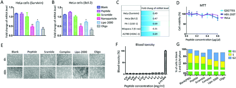
The authors regret an error in Fig. 3E in the originally published manuscript. The correct version of Fig. 3 is shown below.
image file: c9mh90031d-f3.tif
Fig. 3 Cell assay evaluation of the peptide–siRNA co-assembled nanoparticles. Scramble: peptide–siRNA (Scramble) complex. (A) RT-PCR analysis of HeLa cells transfected with peptide–siRNA (Survivin) co-assembled nanoparticles. (B) RT-PCR analysis of HeLa cells transfected with peptide–siRNA (Bcl-2) co-assembled nanoparticles. Both the delivery of Survivin and Bcl-2 siRNA showed a significant knockdown in HeLa cells. *[thin space (1/6-em)]P < 0.05, **[thin space (1/6-em)]P < 0.01 vs. PBS (blank). (C) The summary of the fold change of the mRNA level in different cell lines by RT-PCR analysis (HeLa cells, PA-1 cells, Miapaca-2 cells and A2780 cells), transfected with different peptide–siRNA nanoparticles. The detailed RT-PCR results are provided in Fig. S12 (ESI†). (D) MTT assay of peptide Wpc in Immortalized cells (HEK-293T cells), normal liver cells (QSG7701 cells) and tumor cells (HeLa cells). (E) Inverted microscope image of HeLa cells treated with peptide siRNA co-assembled nanoparticles after 4 h and 48 h. (F) Blood cytotoxicity assay of peptide Wpc in fresh mouse blood with 0.1% SDS as a positive control. (G) Percentage of HeLa cells in each mitotic phase after being treated with different samples. HeLa cells were transfected with (2.0 mg siRNA with 50 mg peptide) nanoparticles for 48 h. Then they were stained and analyzed by flow cytomerty. A clearly higher proportion of HeLa cells were in the G2 phase after being treated with peptide–siRNA nanoparticles or positive controls than any other treatments. All the experiments were conducted twice with consistent results. Error bars represent SD of the data. The statistical differences between the control group and experimental groups were analyzed by one-way ANOVA.

The Royal Society of Chemistry apologises for these errors and any consequent inconvenience to authors and readers.


This journal is © The Royal Society of Chemistry 2019