
 Open Access Article
 This Open Access Article is licensed under a
 Creative Commons Attribution 3.0 Unported Licence
Photoactive platinum(IV) complex conjugated to a cancer-cell-targeting cyclic peptide†
Huayun
Shi
 a,
Qian
Wang
b,
V.
Venkatesh
a,
Guokai
Feng
b,
Lawrence S.
Young
c,
Isolda
Romero-Canelón
ad,
Musheng
Zeng
b and
Peter J.
Sadler
*a
a,
Qian
Wang
b,
V.
Venkatesh
a,
Guokai
Feng
b,
Lawrence S.
Young
c,
Isolda
Romero-Canelón
ad,
Musheng
Zeng
b and
Peter J.
Sadler
*a
aDepartment of Chemistry, University of Warwick, Coventry CV4 7AL, UK. E-mail: p.j.sadler@warwick.ac.uk
bState Key Laboratory of Oncology in South China, Collaborative Innovation Center for Cancer Medicine, Sun Yat-Sen University Cancer Center, Guangzhou 510060, China
cMedical School, University of Warwick, Coventry CV4 7AL, UK
dSchool of Pharmacy, Institute of Clinical Sciences, University of Birmingham, Birmingham B15 2TT, UK
First published on 31st May 2019
Abstract
A conjugate of cancer-cell targeting cyclic disulphide nona-peptide c(CRWYDENAC) consisting of nine L-amino acids with the photoactive succinate platinum(IV) complex trans,trans-[Pt(N3)2(py)2(OH)(succinate)] (Pt-cP) has been synthesised and characterised. The conjugate was stable in dark, but released succinate–peptide and Pt(II) species upon irradiation with visible light, and formed photoproducts with guanine. Conjugate Pt-cP exhibited higher photocytotoxicity than parent complex trans,trans,trans-[Pt(N3)2(OH)2(py)2] (FM-190) towards cancer cells, including ovarian A2780, lung A549 and prostate PC3 human cancer cells upon irradiation with blue light (465 nm, 17.28 J cm−2) with IC50 values of 2.8–22.4 μM and the highest potency for A549 cells. Even though the dark cellular accumulation of Pt-cP in A2780 cells was lower than that of parent FM-190, Pt from Pt-cP accumulated in cancer cells upon irradiation to a level >3× higher than that from FM-190. In addition, the cellular accumulation of Pt from Pt-cP was enhanced ca. 47× after irradiation.
Photoactive diazido platinum(IV) prodrugs offer potential for improved treatment of cancer due to their high stability and low toxicity in the dark, potent photocytotoxicity, and novel mechanism of action which has the possibility to overcome cisplatin resistance in cancer cells.1–6 Among them, trans,trans,trans-[Pt(N3)2(OH)2(py)2] (FM-190) is a promising prodrug candidate,7 which can be activated by visible light with a high photocytotoxicity index. Derivatisation of the detachable axial ligands in platinum(IV) prodrugs is now a common strategy to enhance their pharmacological properties.8–12 For example, conjugation of FM-190 to αvβ3 and αvβ5 integrin-selective RGD-containing peptides introduces a preference towards SK-MEL-28 melanoma cancer cells that overexpress the αvβ3 integrin,13 and incorporation of a TEMPO radical can enhance the photocytotoxicity.14FM-190 has also been conjugated to drug delivery upconversion-luminescent nanoparticles,15 hydrogels,16 and block copolymer micelles17 for activation with longer wavelength and improved selectivity.
Integrins are not only transmembrane receptors that facilitate cell–cell and cell–matrix adhesion,18–20 but also regulators of cancer progression signalling pathways.21 Thus, integrins play an important role in cancer progression and metastasis. The overexpression of integrins on cancer cells provides a useful diagnostic and therapeutic avenue to cancer therapy.22–25 The integrin α6 receptor is reported to be overexpressed in various cancer cell lines,26–28 including ovarian, lung, prostate and other cancers, and promotes the migration, invasion and survival of cancer cells.
The tumour-specific homing cyclic peptide c(CRWYDENAC) targets the integrin α6 receptor, using the sequence RWY (Arg–Trp–Tyr) as binding site.29 RWY-grafted polymeric nanoparticles encapsulating a cisplatin prodrug display a 100-fold increase in cytotoxicity towards integrin α6-overexpressing nasopharyngeal carcinoma compared to free cisplatin.29
Here we have synthesised and characterised Pt-cP, a conjugate of a photoactive trans-diazido platinum(IV) complex with the cyclic peptide c(CRWYDENAC), via amide bond formation between the free carboxyl group of the Pt-bound axial succinate and the N-terminal amino group of the peptide. All of amino acids in the peptide have the L-configuration. Photodecomposition and photoreactions with the nucleotide guanosine 5′-monophosphate (5′-GMP) were investigated, since guanine bases are potential DNA targets for Pt(II) photoproducts. The photocytotoxicity and cellular accumulation in integrin α6-overexpressing cancer cell lines were studied in comparison with the parent complex FM-190. We show that the incorporation of the cyclic peptide as an axial ligand enhanced the photocytotoxicity and cancer cellular accumulation of this photoactive diazido platinum(IV) prodrug.
The synthetic route for photoactive platinum(IV) complex Pt-cP is summarised in Scheme 1. FM-NHS was prepared according to a procedure similar to that reported previously.15 The N-hydroxysuccinimide (NHS) active ester of trans,trans,trans-[Pt(N3)2(py)2(OH)(succinate)] with one axial carboxyl group was prepared by reaction with EDC, NHS and DMAP to generate FM-NHS and purified by column chromatography on silica gel. The coupling was carried out by stirring freshly prepared FM-NHS with cyclic peptide c(CRWYDENAC) in DMF with DIPEA under nitrogen for 36 h. The resulting yellow solid possessed good HPLC purity (94%, Fig. S1, ESI†), and was characterised by ESI-HRMS, 1H NMR and UV-vis spectroscopy. The ions [M + 2H]2+ (855.7493) and [M + H]+ (1710.4919) were detected by HR-MS (Fig. 1a and Fig. S2, ESI†). The m/z values and the isotopic mass distribution pattern of Pt are in good agreement with the calculated spectra. A full assignment of the 1H NMR peaks for the cyclic peptide peptide in DMSO-d6 was not attempted, but the doublet at 8.81 ppm (J = 5.4 Hz) and the triplets at 8.25 ppm (J = 7.6 Hz) and 7.81 ppm (J = 6.6 Hz) can be assigned to the α, γ and β protons, respectively, of the pyridine ligands (Fig. S3, ESI†). The singlet at 10.7 ppm is ascribed to the NH of the Trp indole ring and the NH amide proton signals appear between 6.5 and 9.0 ppm. In addition, in a mixture of 80% DMSO-d6 and 20% D2O (v/v), aromatic protons assigned to pyridine, and Trp and Tyr amino acid residues confirm the conjugation of the Pt(IV) fragment to the cyclic peptide (Fig. S4, ESI†). The absorption band at 290 nm (30![[thin space (1/6-em)]](https://www.rsc.org/images/entities/char_2009.gif) 435 M−1 cm−1) for Pt-cP in phenol red-free RPMI-1640 cell culture medium with 5% DMSO (v/v) present to aid solubility, is mainly assignable to a LMCT (N3 → PtIV) transition (Fig. 1b). The cyclic peptide displayed an absorption band at 278 nm due to Trp and Tyr side chains, and also contributed to the absorption band for Pt-cP at 290 nm.
435 M−1 cm−1) for Pt-cP in phenol red-free RPMI-1640 cell culture medium with 5% DMSO (v/v) present to aid solubility, is mainly assignable to a LMCT (N3 → PtIV) transition (Fig. 1b). The cyclic peptide displayed an absorption band at 278 nm due to Trp and Tyr side chains, and also contributed to the absorption band for Pt-cP at 290 nm.
 | ||
| Scheme 1 The synthetic route for photoactive conjugate Pt-cP. (1) Succinic anhydride, DMF, 348 K, overnight; (2) EDC, NHS, DMAP, DMF, N2, 298 K, overnight; (3) c(CRWYDENAC), DIPEA, DMF, N2, 298 K, 36 h. | ||
 | ||
| Fig. 1 (a) Observed and calculated HR-ESI mass spectra for the molecular ion [M + 2H]2+ of Pt-cP; (b) UV-vis spectra of the conjugate Pt-cP (red) and the free cyclic peptide (black) in phenol red-free RPMI-1640 cell culture medium at 298 K. | ||
The dark stability and photodecomposition of Pt-cP in phenol red-free RPMI-1640 were monitored by UV-vis spectroscopy. The absorption spectra of Pt-cP exhibited little change in the dark over 2 h, indicating its dark stability (Fig. 2a). However, a gradual decrease in intensity of absorption at 290 nm was observed upon irradiation with blue light (420 nm), which suggested the photo-reduction of Pt(IV) to Pt(II) and release of azide ligands (Fig. 2b).
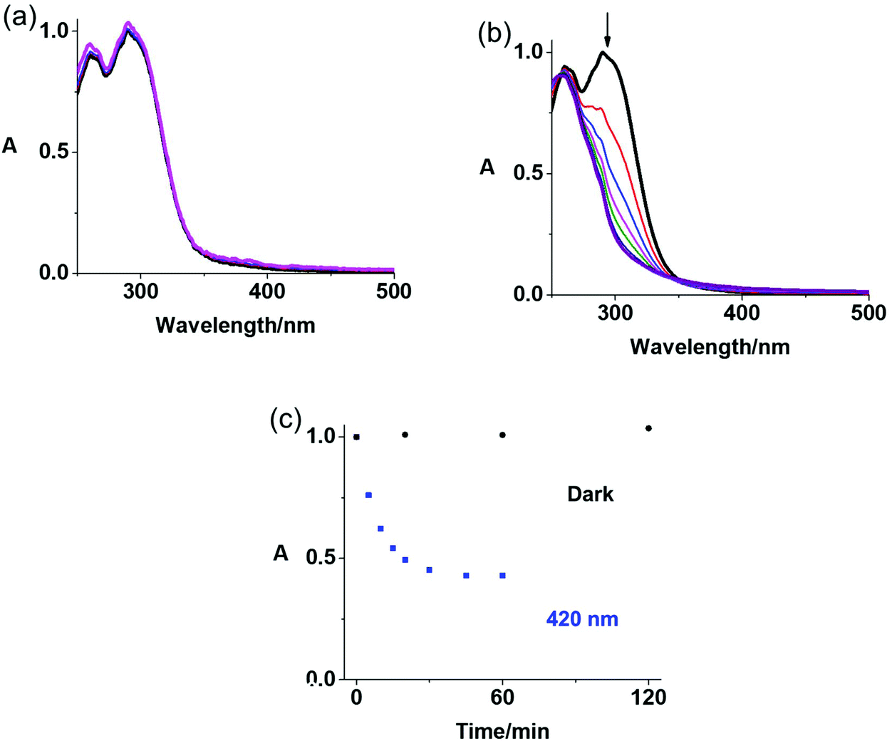 | ||
| Fig. 2 (a) Dark stability over 2 h and (b) photodecomposition with blue light (420 nm, over 1 h) of conjugate Pt-cP in phenol red-free RPMI-1640 determined by UV-vis spectroscopy; (c) time dependence of the absorbance at 290 nm. | ||
The photoproducts from reactions of Pt-cP and 5′-GMP (guanosine 5′-monophosphate) were investigated by LC-MS (Fig. S5 and Table S1, ESI†). An aqueous solution of Pt-cP (30 μM) and 2 mol equiv. of 5′-GMP was irradiated with blue light (420 nm) for 1 h at 298 K. Then the products were analysed by reverse-phase LC-MS. Upon 1 h irradiation, the peak assigned as Pt-cP (retention time = 12.7 min) disappeared and the intact succinate–c(CRWYDENAC) moiety (m/z = 1257.28) was released (Fig. S5†). The Pt-GMP adducts {PtII(N3)(py)2(GMP)}+ (758.22) and {PtII(OC(O)H)(py)2(GMP)}+ (762.16) were detected (GMP in MS formula is considered neutral unless otherwise stated, and the formic acid arises from the mobile phase). No apparent differences between the Pt-GMP adducts formed by the photoreaction between Pt-cP and 5′-GMP compared with similar reactions of the parent complex FM-190 were observed, which suggested that conjugation of cyclic peptide in an axial position did not affect the photochemical reactions of the platinum(IV) centre and binding to the DNA/RNA base guanine after irradiation.7
Conjugate Pt-cP exhibited promising dark stability and photocytotoxicity towards several human cancer cell lines, including A2780 ovarian, A549 lung and PC3 prostate human cancer cell lines. The dose-dependent inhibition of cell viability determined by the sulforhodamine B (SRB) colorimetric assay for conjugate Pt-cP in comparison with the parent complex FM-190 both in the dark and after irradiation, is summarised in Table 1. Both complexes were relatively non-toxic towards all cancer cell lines in the dark with IC50 values >100 μM. However, the cytotoxicity of both complexes was significantly enhanced after 1 h irradiation with blue light (465 nm, 17.28 J cm−2). Importantly, the photocytotoxicity of conjugate Pt-cP (IC50 = 6.6 μM for A2780, 2.8 μM for A549, and 22.4 μM for PC3) was greater than that of the parent complex FM-190 (IC50 = 7.1 μM for A2780, 51.9 μM for A549, and 55.6 μM for PC3) with photocytotoxicity indices (PI) of >15.2, 35.7 and 4.5 towards A2780 ovarian, A549 lung and PC3 prostate cancer cells, respectively. These results indicate that the conjugation with the cyclic peptide c(CRWYDENAC) enhances the photocytotoxicity of this platinum(IV) prodrug without reducing its dark stability. Notably, under the conditions used (short treatment times), the clinical drug cisplatin was inactive (Table 1). Low dark cytotoxicity (IC50 > 100 μM) of Pt-cP in healthy MRC5 lung cells was observed, which might allow selectivity towards cancer cells to be achieved by spatially-directed irradiation (Table 1).
| Cell line | IC50a (μM) |
|||
|---|---|---|---|---|
| Pt-cP | FM-190 | CDDP | ||
| a Data are from three independent experiments. b Data are adapted from ref. 14 and 30. | ||||
| A2780 | Dark | >100 | >100b | >100b |
| Irrad. | 6.6 ± 0.2 | 7.1 ± 0.4b | >100b | |
| PI | >15.2 | >14.1 | — | |
| A549 | Dark | >100 | >100 | >100 |
| Irrad. | 2.8 ± 0.2 | 51.9 ± 2.5 | >100 | |
| PI | >35.7 | >1.9 | — | |
| PC3 | Dark | >100 | >100 | >100 |
| Irrad. | 22.4 ± 2.2 | 55.6 ± 0.9 | >100 | |
| PI | >4.5 | >1.8 | — | |
| MRC5 | Dark | >100 | >100b | >100b |
Cellular accumulation of metallodrugs often plays an important role in their antiproliferative potency. Pt accumulation by A2780 ovarian and A549 lung cancer cells in the dark was investigated when they were exposed to photoactive platinum(IV) complexes at the same Pt concentration (10 μM) for 1 h (Table 2). The cellular accumulation of Pt from conjugate Pt-cP (2.5 ng per 106 cells) in A549 cells was 2.5× higher than that from the parent complex, which is consistent with the high photocytotoxicity of the conjugate in A549 cells. In contrast, a very low Pt accumulation was detected in A2780 cells after treatment with Pt-cP (0.17 ng per 106 cells). For comparison, parent complex FM-190 exhibited similar accumulation in both cell lines, which indicates the selectivity of the conjugate. However, similar photocytotoxicity was observed for Pt-cP and FM-190 in A2780 cells.
To investigate the effect of light exposure on accumulation, duplicate plates of A2780 ovarian cancer cells were exposed to conjugate Pt-cP for 1 h in the dark at the IC50 concentration (6.6 μM). Then some plates were irradiated with blue light (465 nm) for 1 h, while other plates treated with the same drug were left in the dark for comparison (Table 3). It is notable that the cellular accumulation of Pt was ca. 47× enhanced after irradiation (9.4 ng Pt per 106 cells after irradiation, 0.2 ng Pt per 106 cells in the dark), probably because the Pt(II) photoproducts are more reactive towards intracellular biomolecules than the Pt(IV) prodrug and were less readily effluxed from the cells.31 In addition, the cellular accumulation of parent complex FM-190 (as Pt) in the absence of light (0.9 ng Pt per 106 cells) was ca. 3× higher than in the presence of light (2.7 ng Pt per 106 cells), on treatment of cells at IC50 concentration (7.1 μM). Platinum accumulation of the conjugate was lower than that of the parent complex before irradiation. However, the amount of Pt from conjugate Pt-cP accumulated in A2780 cells is >3× higher than that from parent complex FM-190 (Table 3) after irradiation. This increased accumulation resulted in the similar photocytotoxicity of conjugate Pt-cP with FM-190, and suggested that the cyclic peptide might deliver Pt to different parts of the cell compared to FM-190 alone, as anticipated.
| Platinum accumulation (ng per 106 cells)a | ||
|---|---|---|
| Dark | Irradiated | |
| a All data were determined from triplicate samples and their statistical significance evaluated by a two-tail t-test with unequal variances. *p < 0.05, **p < 0.01, ***p < 0.001. | ||
| Pt-cP | 0.20 ± 0.03* | 9.41 ± 0.03*** |
| FM-190 | 0.9 ± 0.2* | 2.7 ± 0.1*** |
In summary, we have prepared and characterised a photoactive conjugate between a trans-diazido platinum(IV) prodrug and a receptor-targeting cyclic RWY (Arg–Trp–Tyr) nona-peptide Pt-cP. The conjugate exhibited high dark stability, but was potently photocytotoxic towards several human cancer cell lines with IC50 values of 2.8–22.4 μM. The highest photocytotoxicity and accumulation of the conjugate was observed for A549 cells. Light irradiation promoted the cellular accumulation of Pt from Pt-cP significantly (ca. 3× that of parent prodrug FM-190). This work suggests that cancer cell-targeting cyclic peptides can improve the photo-cytotoxicity and photo-accumulation of photoactive platinum(IV) complexes, and their photo-selectivity towards cancer cells.
Conflicts of interest
Peptide cP is the subject of US patent US9809622B2.Acknowledgements
This research was supported by the EPSRC (grants EP/G006792, EP/F034210/1 to PJS), ERC (grant 247450 to PJS), a Chancellor's International PhD Scholarship from the University of Warwick (for HS), and the Royal Society (grant no. IE131109 for LSY, Newton International Fellowship and follow-on funding AL170006\1 for VV). We also thank Dr Lijiang Song for his help with mass spectrometry.References
- M. Imran, W. Ayub, I. S. Butler and Z. ur-Rehman, Coord. Chem. Rev., 2018, 376, 405–429 CrossRef CAS.
- K. Mitra, Dalton Trans., 2016, 45, 19157–19171 RSC.
- P. Müller, B. Schröder, J. A. Parkinson, N. A. Kratochwil, R. A. Coxall, A. Parkin, S. Parsons and P. J. Sadler, Angew. Chem., Int. Ed., 2003, 42, 335–339 CrossRef PubMed.
- F. S. Mackay, J. A. Woods, P. Heringová, J. Kašpárková, A. M. Pizarro, S. A. Moggach, S. Parsons, V. Brabec and P. J. Sadler, Proc. Natl. Acad. Sci. U. S. A., 2007, 104, 20743–20748 CrossRef CAS PubMed.
- Y. Zhao, N. J. Farrer, H. Li, J. S. Butler, R. J. McQuitty, A. Habtemariam, F. Wang and P. J. Sadler, Angew. Chem., Int. Ed., 2013, 52, 13633–13637 CrossRef CAS PubMed.
- J. Kasparkova, H. Kostrhunova, O. Novakova, R. Křikavová, J. Vančo, Z. Trávníček and V. Brabec, Angew. Chem., Int. Ed., 2015, 54, 14478–14482 CrossRef CAS PubMed.
- N. J. Farrer, J. A. Woods, L. Salassa, Y. Zhao, K. S. Robinson, G. Clarkson, F. S. Mackay and P. J. Sadler, Angew. Chem., Int. Ed., 2010, 49, 8905–8908 CrossRef CAS PubMed.
- T. C. Johnstone, K. Suntharalingam and S. J. Lippard, Chem. Rev., 2016, 116, 3436–3486 CrossRef CAS PubMed.
- Y. Yuan, R. T. K. Kwok, B. Tang and B. Liu, J. Am. Chem. Soc., 2014, 136, 2546–2554 CrossRef CAS PubMed.
- Y. Zheng, K. Suntharalingam, T. C. Johnstone, H. Yoo, W. Lin, J. G. Brooks and S. J. Lippard, J. Am. Chem. Soc., 2014, 136, 8790–8798 CrossRef CAS PubMed.
- S. G. Awuah, Y. Zheng, P. M. Bruno, M. T. Hemann and S. J. Lippard, J. Am. Chem. Soc., 2015, 137, 14854–14857 CrossRef CAS PubMed.
- E. Petruzzella, J. P. Braude, J. R. Aldrich-Wright, V. Gandin and D. Gibson, Angew. Chem., Int. Ed., 2017, 56, 11539–11544 CrossRef CAS PubMed.
- A. Gandioso, E. Shaili, A. Massaguer, G. Artigas, A. González-Cantó, J. A. Woods, P. J. Sadler and V. Marchán, Chem. Commun., 2015, 51, 9169–9172 RSC.
- V. Venkatesh, C. J. Wedge, I. Romero-Canelón, A. Habtemariama and P. J. Sadler, Dalton Trans., 2016, 45, 13034–13037 RSC.
- Y. Min, J. Li, F. Liu, E. K. L. Yeow and B. Xing, Angew. Chem., Int. Ed., 2014, 53, 1012–1016 CrossRef CAS PubMed.
- V. Venkatesh, N. K. Mishra, I. Romero-Canelón, R. R. Vernooij, H. Shi, J. P. C. Coverdale, A. Habtemariam, S. Verma and P. J. Sadler, J. Am. Chem. Soc., 2017, 139, 5656–5659 CrossRef CAS PubMed.
- D. Zhou, J. Guo, G. B. Kim, J. Li, X. Chen, J. Yang and Y. Huang, Adv. Healthcare Mater., 2016, 5, 2493–2499 CrossRef CAS PubMed.
- R. O. Hynes, Cell, 1992, 69, 11–25 CrossRef CAS PubMed.
- J. S. Desgrosellier and D. A. Cheresh, Nat. Rev. Cancer, 2010, 10, 9–22 CrossRef CAS PubMed.
- Y. H. Soung, H. J. Gil, J. L. Clifford and J. Chung, Curr. Protein Pept. Sci., 2011, 12, 23–29 CrossRef CAS PubMed.
- A. E. Aplin, A. K. Howe and R. L. Juliano, Curr. Opin. Cell Biol., 1999, 11, 737–744 CrossRef CAS PubMed.
- J. R. Marthick and J. L. Dickinson, Prostate Cancer, 2012, 2012, 298732 CrossRef PubMed.
- C. W. Huang, Z. Li, H. Cai, T. Shahinian and P. S. Conti, Mol. Imaging, 2011, 10, 284–294 CAS.
- A. Massaguer, A. González-Cantó, E. Escribano, S. Barrabés, G. Artigas, V. Moreno and V. Marchán, Dalton Trans., 2015, 44, 202–212 RSC.
- L. Wei, F. Yin, W. Zhang and L. Li, Medicine, 2017, 96(12), e6345 CrossRef CAS PubMed.
- Z. T. Colburn and J. C. R. Jones, Am. J. Respir. Cell Mol. Biol., 2017, 56(4), 443–452 CrossRef CAS PubMed.
- I. C. Sroka, H. Chopra, L. Das, J. M. C. Gard, R. B. Nagle and A. E. Cress, J. Cell. Biochem., 2016, 117(2), 491–499 CrossRef CAS PubMed.
- L. Das, T. A. Anderson, J. M. C. Gard, I. C. Sroka, S. R. Strautman, R. B. Nagle, C. Morrissey, B. S. Knudsen and A. E. Cress, J. Cell. Biochem., 2017, 118(5), 1038–1049 CrossRef CAS PubMed.
- G. Feng, M. Zhang, H. Wang, J. Cai, S. Chen, Q. Wang, J. Gong, K. W. Leong, J. Wang, X. Zhang and M. Zeng, Adv. Therap., 2019, 1900018 CrossRef.
- H. Shi, I. Romero-Canelón, M. Hreusova, O. Novakova, V. Venkatesh, A. Habtemariam, G. J. Clarkson, J. Song, V. Brabec and P. J. Sadler, Inorg. Chem., 2018, 57, 14409–14420 CrossRef CAS PubMed.
- D. Guo, S. Xu, Y. Huang, H. Jiang, W. Yasen, N. Wang, Y. Su, J. Qian, J. Li, C. Zhang and X. Zhu, Biomaterials, 2018, 177, 67–77 CrossRef CAS PubMed.
Footnote |
| † Electronic supplementary information (ESI) available. See DOI: 10.1039/c9dt00909d |
| This journal is © The Royal Society of Chemistry 2019 |