
 Open Access Article
 This Open Access Article is licensed under a
 Creative Commons Attribution 3.0 Unported Licence
Correction: Enzyme-mediated dual-targeted-assembly realizes a synergistic anticancer effect
Dingze
Mang‡
a,
Shijin
Zhang‡
a,
Xia
Wu
a,
Xunwu
Hu
a,
Toshiaki
Mochizuki
b,
Guanying
Li
a and
Ye
Zhang
*a
aBioinspired Soft Matter Unit, Okinawa Institute of Science and Technology Graduate University, 1919-1 Tancha, Onna-son, Okinawa, 904-0495, Japan. E-mail: ye.zhang@oist.jp
bImaging Section, Okinawa Institute of Science and Technology Graduate University, 1919-1 Tancha, Onna-son, Okinawa, 904-0495, Japan
First published on 13th June 2019
Abstract
Correction for ‘Enzyme-mediated dual-targeted-assembly realizes a synergistic anticancer effect’ by Dingze Mang et al., Chem. Commun., 2019, 55, 6126–6129.
The authors regret that the supporting information figure citations in the text on page 6129 are incorrect as the ESI available online has been updated. The correct citations are as follows. On page 6129 the sentence beginning, “Compared to the 90% closure rate…” should read, “Compared to the 90% closure rate of the control wound healing experiment, single-administration of 1 at 50 μM and 100 μM concentrations resulted in 48% and 30% closure rates (Fig. S9 and S10, ESI†), while single-administration of 2 resulted in 40% and 20% closure rates, respectively (Fig. S9 and S11, ESI†). Co-administration at the same total concentrations led to 38% and 16% closure rates, respectively (Fig. S9 and S12, ESI†). Apparently, co-administration shows slightly higher inhibition efficacy than both single-administrations in a 2D cell culture. In a 3D cell culture, co-administration exhibited strong inhibition efficacy in the migration of HeLa spheroids (Fig. 5B), superior to single-administrations. HeLa spheroids under the treatment of 1 at concentrations of 50 μM and 100 μM exhibit 87% and 78% of the total area of the control experiment after 72 hours, respectively (Fig. 5C and Fig. S13, ESI†).” Additionally, the sentence beginning, “In the 3D cell culture…” should read “In the 3D cell culture, PLAP expression of HeLa cells is twice as high as in the 2D cell culture (Fig. S14, ESI†).”
The authors regret that Fig. 2 is incorrect. The images in Fig. 2A do not indicate a clear fluorescence intensity difference, and the labels for Fig. 2B and C are incorrect. The correct version of Fig. 2 is presented here.
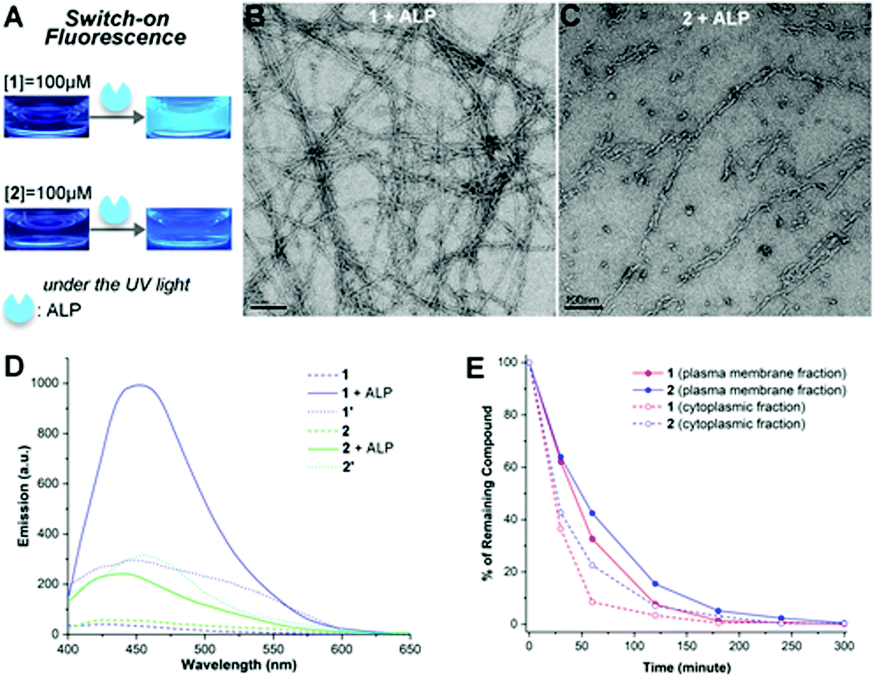 | ||
| Fig. 1 (A) Optical images of molecules 1 and 2 before and after the ALP catalysed hydrolysis in PBS buffer at a concentration of 100 μM under UV light. TEM images of 1 (B) and 2 (C) in PBS buffer at a concentration of 100 μM towards the end of their ALP catalysed hydrolysis. Scale bar is 100 nm. (D) Emission spectra of 1 and 2 before (labelled as 1 and 2) and after (labelled as 1 + ALP and 2 + ALP) the ALP catalysed hydrolysis, and 1′ and 2′ in PBS buffer at a concentration of 100 μM excited at 350 nm. (E) Kinetic profiles of enzyme catalysed hydrolysis of 1 and 2 in the plasma membrane fraction and cytosolic fraction of HeLa cells at 37 °C. | ||
The authors apologise that parts of the data presented in Fig. 3 may be incorrect. They believe contamination of the cells used for the experiment in Fig. 3D may have occurred. To verify the results, the experiments were repeated using HeLa cells from a confirmed cell source. The results obtained for lysosome-targeted assembly were the same, however the authors would like to replace Fig. 3. The correct version of Fig. 3 is presented below.
 | ||
Fig. 2 (A) The fluorescence image of HeLa cells incubated with 1 (100 μM, 12 h) and co-stained with CellMask Red. (B) The SEM image of HeLa cells incubated with 1 (100 μM, 12 h). (C) The fluorescence image of HeLa cells incubated with 2 (100 μM, 12 h) and co-stained with LysoTracker Red. (D) The TEM image of lysosomes isolated from HeLa cells incubated with 2 (100 μM, 12 h). The fluorescence image of HeLa cells incubated with a mixture of 1 and 2 (1![[thin space (1/6-em)]](https://www.rsc.org/images/entities/char_2009.gif) :1, total concentration 100 μM, 12 h) without (E) and with (F) PLAP inhibition, and co-stained with CellMask Red. :1, total concentration 100 μM, 12 h) without (E) and with (F) PLAP inhibition, and co-stained with CellMask Red. | ||
Additionally, the second author should have been shown as “Shijin Zhang‡a” to indicate equal contribution.
The Royal Society of Chemistry apologises for these errors and any consequent inconvenience to authors and readers.
| This journal is © The Royal Society of Chemistry 2019 |