
 Open Access Article
 This Open Access Article is licensed under a Creative Commons Attribution-Non Commercial 3.0 Unported Licence
Drug orientations within statin-loaded lipoprotein nanoparticles by 19F solid-state NMR†
Sophie
Lau
,
Naomi
Stanhope
,
John
Griffin
 ,
Eleri
Hughes
and
David A.
Middleton
*
,
Eleri
Hughes
and
David A.
Middleton
*
Department of Chemistry, Lancaster University, Lancaster LA1 4YB, UK. E-mail: d.middleton@lancaster.ac.uk
First published on 14th October 2019
Abstract
NMR measurements of 19F chemical shift anisotropy and 1H–19F dipolar couplings provide unprecedented information on the molecular orientations of two fluorine-containing statin drugs within the heterogeneous environment of reconstituted high-density lipoprotein (rHDL) nanoparticles, a drug delivery system under clinical investigation.
The high cost of bringing a new pharmaceutical to market – an estimated $2.6 billion – reflects the fact that 85–90% of drug candidates do not progress through clinical trials.1 One reason for this high attrition rate is the failure of drugs to engage effectively with their molecular target(s) in human patients,2 and this has motivated efforts to develop smart, nanoscale formulations to lengthen drug circulation times in vivo, access impervious tissue and increase the probability of drug-target interactions. Nanoparticle-mediated drug delivery offers the opportunity to rejuvenate clinical failures and repurpose existing drugs, and the US Food and Drug Administration has approved around 60 nanoparticle formulations to date.3 Correspondingly, as increasingly sophisticated formulations enter development there is a growing need for analytical research tools and quality-control methods to characterise nanoparticles and their cargo at the atomic and molecular levels and to explore the mechanisms of drug delivery.
Here we use 19F NMR as a sensitive probe of the molecular orientations of fluorine-containing drugs as they undergo anisotropic motion within a reconstituted high-density lipoprotein (rHDL) nanoparticle formulation. rHDL nanoparticles are attractive drug delivery vehicles that are well-tolerated in vivo, do not invoke an immune response and are capable of delivering lipophilic drugs to tissues via specific receptors.4,5 The clinically-tolerated rHDL preparations CER-001 and CSL112 are nanodiscs comprising a phosphatidylcholine or sphingomyelin/phosphatidylglycerol lipid bilayer surrounded by a helical belt of recombinant apolipoprotein A-I (apoA-I), the 28 kDa major protein component of natural HDL.6,7 The position of the native cholesteryl ester (CE) cargo within the core of natural HDL is important for cellular transfer via the SR-B1 receptor,8 and packaging by the nanodiscs may similarly influence the cellular uptake of drugs. Transfer of CE, vitamins and drugs from HDL into cells is in part mediated by scavenger receptors (e.g., SR-B1), which have channel-forming extracellular domains that selectively bind certain lipophilic molecules and transfer them to the cytoplasm.9 A drug's orientation within the rHDL lipid matrix will influence how the drug is presented to a receptor and so may affect the rate and selectivity of transfer.10
We exploit 19F chemical shifts and 1H–19F dipolar couplings11 to compare the orientations of two lipophilic fluoroaromatic drugs within the lipid bilayer component of rHDL particles. We chose as our drugs two fluorine-bearing statins, fluvastatin (log![[thin space (1/6-em)]](https://www.rsc.org/images/entities/char_2009.gif) P = 4.17) and rosuvastatin (logP = 1.47) (Scheme 1); statin-loaded rHDL nanoparticles have been shown to reduce atherosclerotic lesions in an atherosclerosis mouse model.12 We prepared nanoparticles of rHDL comprising palmitoyl oleoyl phosphatidylcholine (POPC), fluvastatin or rosuvastatin and full-length apoA-I in a 100:10:1 molar ratio using a detergent-mediated dialysis method. Native gel electrophoresis indicates that the particle size ranges from 8–10 nm and transmission electron microscopy (TEM) confirms the characteristic discoidal morphology of the particles (Fig. 1A, left and middle). A circular dichroism (CD) spectrum (Fig. 1A, right) indicates that apoA-I adopts the expected predominantly (84%) α-helical conformation. It should be noted that similar-sized truncated apoA-I/lipid nanodiscs are widely used in biological NMR to analyse the structures of membrane-embedded proteins, as their rapid tumbling times conveniently provide high-resolution spectra.13 Here the isotropic molecular tumbling of nanodiscs in solution would obscure the effects of local anisotropic motions of the drug that convey information about drug orientation, so we precipitated the nanoparticles from aqueous solution to eliminate particle tumbling. The fluid hydrated lipid bilayer is preserved in the precipitate, as indicated by a 31P NMR spectrum of the POPC phosphate groups with the characteristic 45 ppm width and axially symmetric line shape (Fig. 1B, left).14,15 A 15N magic angle spinning NMR spectrum of precipitated rHDL particles constructed from 15N-labelled apoA-I confirms that protein is also present in the precipitate and the backbone chemical shifts are consistent with the expected predominantly α-helical structure (Fig. 1B, right).
P = 4.17) and rosuvastatin (logP = 1.47) (Scheme 1); statin-loaded rHDL nanoparticles have been shown to reduce atherosclerotic lesions in an atherosclerosis mouse model.12 We prepared nanoparticles of rHDL comprising palmitoyl oleoyl phosphatidylcholine (POPC), fluvastatin or rosuvastatin and full-length apoA-I in a 100:10:1 molar ratio using a detergent-mediated dialysis method. Native gel electrophoresis indicates that the particle size ranges from 8–10 nm and transmission electron microscopy (TEM) confirms the characteristic discoidal morphology of the particles (Fig. 1A, left and middle). A circular dichroism (CD) spectrum (Fig. 1A, right) indicates that apoA-I adopts the expected predominantly (84%) α-helical conformation. It should be noted that similar-sized truncated apoA-I/lipid nanodiscs are widely used in biological NMR to analyse the structures of membrane-embedded proteins, as their rapid tumbling times conveniently provide high-resolution spectra.13 Here the isotropic molecular tumbling of nanodiscs in solution would obscure the effects of local anisotropic motions of the drug that convey information about drug orientation, so we precipitated the nanoparticles from aqueous solution to eliminate particle tumbling. The fluid hydrated lipid bilayer is preserved in the precipitate, as indicated by a 31P NMR spectrum of the POPC phosphate groups with the characteristic 45 ppm width and axially symmetric line shape (Fig. 1B, left).14,15 A 15N magic angle spinning NMR spectrum of precipitated rHDL particles constructed from 15N-labelled apoA-I confirms that protein is also present in the precipitate and the backbone chemical shifts are consistent with the expected predominantly α-helical structure (Fig. 1B, right).
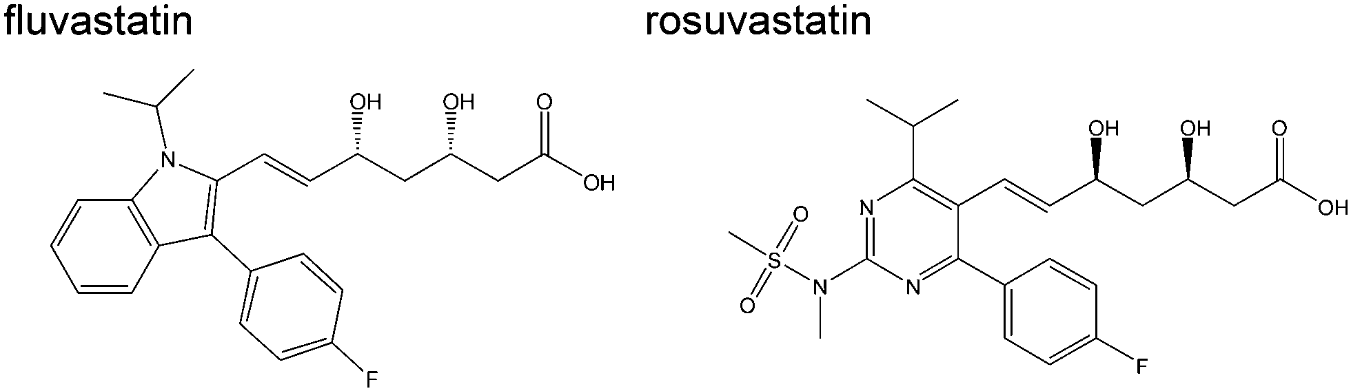 | ||
| Scheme 1 Chemical structures of fluorine-containing statins. | ||
 | ||
| Fig. 1 Encapsulation of statins within rHDL particles. A: native gel electrophoresis (left) and TEM (middle) confirm the presence of rHDL particles approximately 8–10 nm in diameter in the absence or presence of fluvastatin. CD spectroscopy (right) indicates the presence of predominantly helical protein. B: 31P NMR (left) confirms the presence of lamellar POPC lipids in PEG precipitated rHDL and 15N CP-MAS NMR (right) indicates that the helical structure of apoA-I is retained in the PEG precipitate. The red line is a simulated line shape comprising the predicted backbone 15N chemical shifts of all residues of apoA-I in a fully helical conformation. C: rapid dynamics of a statin embedded in a lipid bilayer of rHDL that may scale the 19F chemical shift anisotropy. The principal axes of the 19F chemical shift tensor are shown with the origin at the centre of mass of the molecule. Angles α and β define the 19F principal axes relative to the principal rotation axis, which is parallel with the bilayer normal, and the axis of the aromatic ring rotation is parallel with the direction of 19F chemical shift tensor component δ22. Off-axis excursions are represented by an order parameter Smol = cos(θ). | ||
Statins embedded in the rHDL lipid bilayer will undergo anisotropic motions that scale the 19F chemical shift anisotropy in a predictable way that depends on the drug's orientation in the planar matrix.11 Molecules rotate about a principal axis that runs parallel with the bilayer normal and the NMR line shape is sensitive to the orientations of the 19F chemical shift tensor elements δ11, δ22 and δ33 relative to this axis, as defined by angles α and β (Fig. 1C). If the chemical shift tensor orientation relative to the molecular geometry is known, one can relate the measured chemical shift anisotropy to the average orientation of the drug in the lipid bilayer. DFT calculations predict that the 19F chemical shift tensor is oriented with the least-shielded component δ11 exactly in plane with the aromatic ring and δ22 directed along the fluorine–carbon bond (Fig. S1, ESI†), as seen for other fluorophenyl compounds.11 Additional random motional fluctuations of the molecule about the two axes perpendicular to the main axis must also be considered in the analysis, as must internal rotations of the fluoroaromatic ring (Fig. 1C). Random fluctuations are described by an order parameter, Smol, which describes the maximum angular excursion θ from the principal axis through Smol = cos(θ)
Proton-decoupled 19F NMR spectra confirm that the two statins are present in the rHDL formulations (fluvastatin in Fig. 2A and rosuvastatin Fig. S2A, ESI†). Whereas the 19F chemical shift anisotropy Δδ of the two statins in the solid state is ∼60 ppm, with an asymmetry parameter η of ∼0.7 (Fig. S1 and Table S1, ESI†), the spectra of the rHDL formulations indicate an approximate 10-fold reduction in the observed anisotropy and an apparent axially symmetric chemical shift tensor (η = 0) (Table S2, ESI†). These features confirm that the molecules undergo anisotropic rotation and give a scaled chemical shift anisotropy, Δδav, of 6.2 ppm for fluvastatin and 8.3 ppm for rosuvastatin. The absence of narrow peaks at the isotropic chemical shifts of the statins indicates that the drugs are not distributed between the nanoparticles and the residual aqueous phase but are confined within the lipid matrix. Calculations of the expected value of Δδav for different values of α and β (varying each from 0° to 180°) reveal that the maximum possible value of Δδav is 4.6 ppm for fluvastatin and 4.9 ppm for rosuvastatin (Fig. S2B, ESI†) when rotation of the fluorophenyl ring is considered in addition to the overall molecular rotation. These Δδav values are smaller than the observed values and hence free rotation of the fluorophenyl ring must be slow on the NMR (μs) time scale, possibly because of a high energy barrier resulting from steric clashes between the aromatic and heptenoic acid protons.
 | ||
| Fig. 2 The average orientations of statins with the lipid bilayers of rHDL particles from 19F NMR lineshape analysis. A: spectrum of fluvastatin in rHDL (black). The value of Δδav (6.1 ppm) was obtained by least-squares fitting of a simulated lineshape (red). B: the measured Δδav is consistent with the pairs of α and β values shown by the continuous lines. Discrete values of α (in 5° increments) and corresponding β values (denoted by the filled and open circles, in 10° increments for clarity) were used to simulate proton-coupled spectra as described below. C: each of the discrete [α, β] pairs define a specific statin orientation relative to a principal axis of rotation. From each orientation the rotationally-averaged dipolar couplings dav between 19F and the 4 protons (H1–H4) in the fluorophenyl ring were calculated. D: proton-coupled spectra were simulated for each [α, β] pair and the corresponding dipolar coupling values. Chi-squared values represent the agreement between each simulated spectrum and the experimental dipolar-coupled spectrum. E: the line of closest agreement with the proton-coupled spectrum (chi-squared minimum) is consistent with 4 [α, β] values given in Table S3 (ESI†). F and G: average orientations of fluvastatin and rosuvastatin in rHDL bilayers. Vertical arrows denote the principal axis of rotation, which is parallel with the bilayer normal. | ||
Fig. 2B and Fig. S3B (ESI†) show that the Δδav values measured from the proton-decoupled spectra are consistent with a continuum of α and β values, with α ranging from 0–180° and β ranging from 40–65°. To restrict further the range of possible molecular orientations defined by these angles, we obtained proton-coupled 19F NMR spectra of the rHDL formulations to obtain line shapes that are modulated by 1H–19F dipolar interactions within the statin fluoroaromatic ring. The magnitude of the dipolar couplings is, like Δδav, sensitive to the orientation of the statins relative to their principal rotation axis. We calculated the dynamically-averaged dipolar coupling constants, dav, for 36 discrete pairs of α and β along the continuum (Fig. 2C and Fig. S3C, ESI†). We then simulated a series of proton-coupled 19F NMR line shapes based on the calculated couplings and the measured Δδav values for each statin to find the values of α and β achieving the closest agreement with the experimental line shapes according to chi-square analysis (Fig. 2D and Fig. S3–S5, ESI†). For fluvastatin, the closest agreement is obtained with four [α, β] combinations and for rosuvastatin with two [α, β] combinations (Fig. 2E and Fig. S2E, Table S3, ESI†). Discrepancies between the simulated and experimental spectra may reflect minor contributions from longer-range inter- and intramolecular couplings, which could not be included in the simulations. For each pair of angles, errors of ±5° in α and ±1.5° in β reflect the uncertainty in Smol, the true value of which we assume lies between the extremes of 0.5 (θ = 60°) and 1.0 (θ = 0°). The value of Smol reported for sterol molecules is ∼0.8.16,17
After taking into consideration the out-of-plane fluorophenyl ring conformation of the two statins, as determined from crystal structures and by energy minimisation (Fig. S6, ESI†) we translated the angles into two possible mean orientations of fluvastatin about its principal axis of rotation in the lipid bilayer (Fig. 2F, top) and one orientation of rosuvastatin (Fig. 2F, bottom), as well as their mirror images. The two molecules adopt similar orientations in the lipid matrix. Solution-state 1H–1H NOESY NMR spectra of fluvastatin in dodecylphosphocholine micelles revealed cross peaks between the aromatic protons of the drug and headgroup and hydrocarbon chain protons of the lipid, suggesting that the aromatic ring serves as a hydrophobic anchor and the dihydroxyheptenoic acid is associated with the lipid headgroups.18,19 Here, a NOESY NMR spectrum of fluvastatin in POPC multilamellar vesicles (MLVs), obtained with magic-angle spinning, revealed cross peaks between protons in the fluoroaromatic ring and hydrocarbon chain protons (Fig. S7, ESI†), confirming that the ring serves as a hydrophobic anchor. This may explain the restricted rotation of the ring.
Lastly, we asked whether the statins adopt similar orientations in spherical, protein-free POPC MLVs. In contrast to the rHDL spectra, the 19F proton-decoupled and coupled spectra of the MLVs (Fig. 3) each comprise two components in approximately equal proportion. The narrow component in the spectra of rosuvastatin (Fig. 3B) does not arise from an isotropic population of the drug (i.e., that is rapidly tumbling in solution or smaller vesicles) because the component is offset from the isotropic chemical shift. All spectra could be simulated by two anisotropic components, one with Δδav > 0 and the other with Δδav < 0 (Table S2, ESI†), indicating that each statin adopts two different average orientations in the MLV bilayers. The spectra illustrate how highly sensitive this method is to minor alterations in molecular orientation, because analysis of the two spectral components for each statin reveals very close values of α and β for the molecules in each population (Table S3, ESI†). The sensitivity of the α and β values to orientation also reveal that fluvastatin and rosuvastatin adopt very subtly different orientations in POPC MLVs and in rHDL.
 | ||
| Fig. 3 Proton-decoupled and proton-coupled 19F NMR spectra of fluvastatin (A) and rosuvastatin (B) embedded in POPC MLVs. Experimental spectra (black) are overlaid with best-fitting simulated-two component lineshapes (red). The two values of Δδav obtained from the two components of each spectrum are given in Table S2 (ESI†) and the values of α and β calculated from the spectra are given in Table S3 (ESI†). Dotted lines indicate the isotropic chemical shifts. | ||
There is still much to learn about the molecular packaging of drugs by nanoparticles, yet these details may influence how drugs transfer to the recipient tissue. Here we have shown an NMR method exploiting chemical shifts and dipolar couplings to determine the mean orientations of drugs within the lipid matrix of rHDL nanoparticles. About 30% of small molecule drugs contain fluorine20 and so the 19F NMR method could be applicable to up to 750 approved and 1600 experimental drugs, depending on their suitability for HDL encapsulation. The high sensitivity of the 19F NMR line shape to just minor adjustments of molecular orientation makes it a useful method for structure–activity studies relating the molecular orientations of drugs to the rates of transfer to tissue, and for analysing the reproducibility of formulations.
Conflicts of interest
There are no conflicts to declare.Notes and references
- C. H. Wong, K. W. Siah and A. W. Lo, Biostatistics, 2019, 20, 273–286 CrossRef PubMed.
- T. J. Hwang, D. Carpenter, J. C. Lauffenburger, B. Wang, J. M. Franklin and A. S. Kesselheim, JAMA Intern. Med., 2016, 176, 1826–1833 CrossRef PubMed.
- D. Bobo, K. J. Robinson, J. Islam, K. J. Thurecht and S. R. Corrie, Pharm. Res., 2016, 33, 2373–2387 CrossRef CAS PubMed.
- R. Kuai, D. Li, Y. E. Chen, J. J. Moon and A. Schwendeman, ACS Nano, 2016, 10, 3015–3041 CrossRef CAS PubMed.
- A. G. Lacko, N. A. Sabnis, B. Nagarajan and W. J. McConathy, Front. Pharmacol., 2015, 6, 247 Search PubMed.
- S. Diditchenko, A. Gille, I. Pragst, D. Stadler, M. Waelchli, R. Hamilton, A. Leis and S. D. Wright, Arterioscler., Thromb., Vasc. Biol., 2013, 33, 2202–2211 CrossRef CAS PubMed.
- C. H. Keyserling, R. Barbaras, R. Benghozi and J. L. Dasseux, Clin. Drug Invest., 2017, 37, 483–491 CrossRef CAS PubMed.
- S. T. Thuahnai, S. Lund-Katz, D. L. Williams and M. C. Phillips, J. Biol. Chem., 2001, 276, 43801–43808 CrossRef CAS PubMed.
- D. Neculai, M. Schwake, M. Ravichandran, F. Zunke and R. F. Collins, Nature, 2013, 504, 172–176 CrossRef CAS PubMed.
- W. V. Rodrigueza, S. T. Thuahnai, R. E. Temel, S. Lund-Katz, M. C. Phillips and D. L. Williams, J. Biol. Chem., 1999, 274, 20344–20359 CrossRef CAS PubMed.
- E. Hughes, J. M. Griffin, M. P. Coogan and D. A. Middleton, Phys. Chem. Chem. Phys., 2018, 20, 18207–18215 RSC.
- R. Duivenvoorden, J. Tang, D. P. Cormode, A. J. Mieszawska, D. Izquierdo-Garcia, C. Ozcan, M. J. Otten, N. Zaidi, M. E. Lobatto, S. M. van Rijs, B. Priem, E. L. Kuan, C. Martel, B. Hewing, H. Sager, M. Nahrendorf, G. J. Randolph, E. S. G. Stroes, V. Fuster, E. A. Fisher, Z. A. Fayad and W. J. M. Mulder, Nat. Commun., 2014, 5, 3065 CrossRef PubMed.
- A. Viegas, T. Viennet and M. Etzkorn, Biol. Chem., 2016, 397, 1335–1354 CAS.
- E. E. Burnell, P. R. Cullis and B. Dekruijff, Biochim. Biophys. Acta, 1980, 603, 63–69 CrossRef CAS.
- P. M. Macdonald, Q. Saleem, A. Lai and H. H. Morales, Chem. Phys. Lipids, 2013, 166, 31–44 CrossRef CAS PubMed.
- N. Matsumori, Y. Kasai, T. Oishi, M. Murata and K. Nomura, J. Appl. Chem. Sci., 2008, 130, 4757–4766 CAS.
- O. Soubias, F. Jolibois, S. Massou, A. Milon and V. Reat, Biophys. J., 2005, 89, 1120–1131 CrossRef CAS PubMed.
- L. F. Galiullina, O. V. Aganova, I. A. Latfullin, G. S. Musabirova, A. V. Aganov and V. V. Klochkov, Bionanoscience, 2016, 6, 352–354 CrossRef.
- L. F. Galiullina, O. V. Aganova, I. A. Latfullin, G. S. Musabirova, A. V. Aganov and V. V. Klochkov, Biochem. Biophys. Acta, 2017, 1859, 295–300 CAS.
- Y. Zhou, J. Wang, Z. N. Gu, S. N. Wang, W. Zhu, J. L. Acena, V. A. Soloshonok, K. Izawa and H. Liu, Chem. Rev., 2016, 116, 422–518 CrossRef CAS PubMed.
Footnote |
| † Electronic supplementary information (ESI) available: Methods and materials, additional spectra and calculations. See DOI: 10.1039/c9cc05344a |
| This journal is © The Royal Society of Chemistry 2019 |