Neha
Bhardwaj
ab,
Sanjeev K.
Bhardwaj
ab,
Deepanshu
Bhatt
ab,
Satish K.
Tuteja
c,
Ki-Hyun
Kim
*d and
Akash
Deep
*ab
aCentral Scientific Instruments Organisation (CSIR-CSIO), Sector 30 C, Chandigarh, 160030, India. E-mail: dr.akashdeep@csio.res.in
bAcademy of Scientific and Innovative Research, CSIR-CSIO, Sector 30 C, Chandigarh, India
cIWE1-Institut für Werkstoffe der Elektrotechnik RWTH Aachen University, Germany
dDepartment of Civil and Environmental Engineering, Hanyang University, 222 Wangsimni-Ro, Seoul 04763, Korea. E-mail: kkim61@hanyang.ac.kr
First published on 7th January 2019
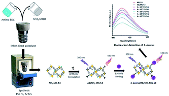
In this research, a new luminescent bioprobe was developed for the detection of S. aureus based on bio-conjugation of an amine functionalized metal–organic framework (NH2-MIL-53(Fe)) with an anti-S. aureus antibody (Ab). The formation of the desired bioprobe (Ab/NH2-MIL-53), in its liquid phase, has been verified with several spectroscopic and structural characterization techniques. The bioprobe was incubated with varying concentrations of S. aureus bacteria. The resulting antibody conjugated bioprobe (Ab/NH2-MIL-53) maintained a strong inverse correlation in which decreases in the fluorescence intensities were accompanied by an increase in the bacterial count. Thus, the potential of the herein developed probe has been successfully demonstrated for the detection of S. aureus with a low limit of detection (85 CFU mL−1) over a wide concentration range (4 × 10²–4 × 10⁸ CFU mL−1). It was further found to be reliable with regard to inter-/intra- precision assays and long-term stability. The feasibility of the method was further supported through the detection of S. aureus spiked in environmental samples (e.g., river water and cream pastry).
Generally, the food industries rely upon chemical and microbiological techniques to assess their product quality. Traditional culture-based methods, including microbiological and biochemical analyses, are still the most practiced options.6 The polymerase chain reaction (PCR) is another popular method used for the quantitative determination of bacteria. Despite their proven utility, the above mentioned conventional methods are time-consuming and labour intensive. To practice these methods, a single bacterial cell must be grown into a visible colony to amplify the signal.3 The applications of such techniques are rather inconvenient in situations demanding more frequent, rapid, and on-site screening of the pathogen contamination.7 Hence, a significant focus of current research is shifted to develop rapid and sensitive methods for the detection of foodborne pathogens. The efforts are directed to devise methods and systems that can be operated even by an untrained person. This is important to ensure more routine monitoring of the food quality and to prevent the infections and losses caused by contaminated food.
Biosensors are one of the most viable options as a point-of-care and portable solution for fast detection of bacteria with relative ease.8–10 During the last decade, nanomaterials have emerged as promising functional materials in biosensor fabrication.10–12 The use of nanomaterials in biosensor development enabled higher loading of biomolecules due to several meritful properties (e.g., high surface area).8,11,13 This ultimately facilitated the enhancement of sensitivity toward the bio-analytical assay. The nanomaterials also possess many other useful properties including small size, quantum confinement effects, high surface reactivity, and enhanced magnetic/electrical/optical properties, which can be tailored to optimize the desired specifications of the sensors. For achieving the desired specificity of biosensing applications, nanomaterials are generally conjugated with biomolecules, e.g. antibodies, enzymes, bacteriophages, nucleic acids, etc.14–17
Fluorescence based sensing is an excellent tool for detection of bacteria in food samples with advantages of high sensitivity, facile fabrication, and multiplexed detection.2,6,18,19 Several researchers have reported the development of fluorescent biosensors for S. aureus based on the application of nanomaterial–antibody/aptamer interfaces.20–22 For instance, an aptamer functionalized graphene oxide (GO) based biosensor was used in combination with a polydimethylsiloxane (PDMS)/paper hybrid to develop a microfluidic system for single-step detection of S. aureus.20 The fluorescence quenching property of GO was exploited for the assay development. In the presence of target bacteria (S. aureus), the fluorescently tagged aptamer molecules became detached from GO, resulting in the restoration of the fluorescence signal. The above developed assay demonstrated a limit of detection (LOD) of 8 × 10² CFU mL−1.20 He et al. (2014) proposed a dual color flow cytometric sensing platform using a combination of fluorescent silica nanoparticles (FSiNPs) and fluorescent dye (SYBR Green I).21 The FSiNP labelled aptamer was employed as a recognition probe while SYBER Green I was used to stain the nucleic acids. The above detection system for S. aureus offered LODs of 150 and 76 CFU mL−1 in buffer and milk samples, respectively.21 A similar sensing platform composed of aptamer conjugated fluorescent silica nanoparticles (Apts/FNPs) was developed for the detection of S. aureus in spiked water samples.22 The assay was based on positive dielectrophoresis (pDEP) driven on-line determination of S. aureus. The bacteria sample was incubated with Apts/FNPs prior to the injection into the microfluidic channel. An applied AC voltage resulted in the accumulation of Apts/FNP labelled S. aureus over the electrode gap owing to the pDEP force. The accumulated S. aureus were later detected using fluorescence microscopy imaging with detection limits of 9.3 × 101 and 2.7 × 10² CFU mL−1 in deionized and spiked water samples, respectively.22
Due to their diverse structural configurations and exciting optical properties, the metal–organic frameworks (MOFs) have attracted immense attention for biosensing applications.14,23,24 MOFs are hybrid materials consisting of metal ions linked to the organic ligands through coordination bonds to form 1-, 2-, or 3-D network structures. They are supramolecular assemblies with tuneable properties and functionality depending on the type of metal or ligand used during synthesis. Recently, luminescent MOFs have been proposed for several biological applications, including drug delivery, imaging, and molecular sensing.23–28 The luminescent MOFs show a number of peculiar advantages over other fluorescent molecules, e.g. crystallinity, nano-to-micron sized structures, stable fluorescence (with respect to time and temperature), and readily available functional groups for the conjugation of biorecognition species.29–31
For the first time, the present work reports the use of an optical (luminescent) biosensor, employing a non-toxic, biocompatible, and water-stable MOF, i.e., NH2-MIL-53 (Fe) as a fluorescent marker. To this end, this MOF has been interfaced with an anti-S. aureus antibody and the resulting biosensor has been investigated for the detection of S. aureus in different samples, including some real (spiked) samples. The specific binding of the bacteria with the bioprobe (i.e., antibody conjugated MOF) caused a reduction in the fluorescence intensity in proportion to the amount of bacteria added. This is attributable to the competitive absorption of the excitation energy by both the bioprobe and the attached bacteria (Graphical abstract). Hence, upon capturing the bacteria, the fluorescent bioprobe experienced a reduction in excitation energy which subsequently resulted in a decreased emission intensity. As our results have highlighted, the application of this new Ab/NH2-MIL-53 biosensor has allowed the sensitive detection of S. aureus over a wide concentration range at a considerably low limit of detection of 85 CFU mL−1. The results of this sensing technique, when applied for the detection of S. aureus in spiked water and cream pastry samples, agreed well with those of the standard colony counting method.
The validation of the data was carried out against the standard colony counting method. The samples with spiked bacteria were diluted in different fractions (e.g. 1×, 102×, 103×, and 104×) and then cultured to form colonies. A plate with easily countable number of colonies was analyzed to determine the bacterial concentration in all the prepared samples.
PXRD patterns of both NH2-MIL-53 and Ab/NH2-MIL-53 are shown in Fig. 3. PXRD of NH2-MIL-53 was characterized with all the typical peaks matching well with the established structural data.32 As it is highlighted, the intrinsic crystallinity of the MOF was preserved even after the bio-conjugation with antibodies. Further, the FE-SEM investigations (Fig. 4) also verified that the Ab/NH2-MIL-53 complex inherited the primary structural properties of the parent fluorescent MOF.
![]() | ||
Fig. 5 (a) PL spectra of the Ab/NH2-MIL-53 complex against 40–4 × 10⁸ CFU mL−1 of S. aureus; (b) corresponding calibration curve. |
The Ab/NH2-MIL-53 bioprobe has allowed the detection of S. aureus by showing a concentration dependent quenching in the PL intensity (Fig. 5(a)). This observed phenomenon in quenching can be attributed to the attenuation in the effective excitation energy available to the Ab/NH2-MIL-53 probe after the bacteria–antibody binding. In other words, it may also reflect the results of the competitive absorption of light between the Ab/NH2-MIL-53 bioprobe and the captured bacteria. The calibration curve for the above sensor response is plotted and is shown in Fig. 5(b). Clearly, the Ab/NH2-MIL-53 bioprobe was capable of providing a logarithmically linear sensor response over a wide range (4 × 10² to 4 × 10⁸ CFU mL−1) of analyte concentrations. The limit of detection (LOD) was calculated with the formula of 3σ/S (where σ and S corresponded to the standard deviation estimated for a blank sample and the slope of a calibration curve, respectively). Based on σ and S values of 3.5 and 0.1235, respectively, the LOD of the present Ab/NH2-MIL-53 based luminescent biosensing method was calculated to be 85 CFU mL−1. It is worth noting that the above value of LOD obtained with the Ab/NH2-MIL-53 system is better than or comparable with that of most of the earlier reported optical biosensors for S. aureus (Table 1).
Order | Sensing material | Important sensor characteristics | Ref. | |
---|---|---|---|---|
Range (CFU mL−1) | LOD (CFU mL−1) | |||
1 | Ab/Quantum dot | 103–105 | 103 | 18 |
2 | (GO/PDMS)/paper | 104–106 | 8 × 10² | 20 |
3 | FSiNPs/SYBR Green I | 3.4 × 10²–3.4 × 10⁴ (buffer) | 1.5 × 10² (buffer) | 21 |
6 × 10²–6 × 10⁵ (milk) | 7.6 × 10² (milk) | |||
4 | Aptamers/FNPs | 5 × 101–1 × 10⁶ (deionized water) | 9.3 × 101 (deionized water) | 22 |
7 × 101–7 × 10⁶ (spiked water) | 2.7 × 10² (spiked water) | |||
5 | Ab/NH 2 -MIL-53 | 40–4 × 10 8 | 85 | Present work |
The specificity of the present sensor toward the target analyte (S. aureus, 4 × 10² CFU mL−1) in the presence of other possible interfering bacteria (E. coli and S. arlettae, 4 × 10⁶ CFU mL−1) has also been tested. The results of this study are shown in Fig. 6(a). Due to the specific nature of the antibodies used, the Ab/NH2-MIL-53 bioprobe could very well tolerate the potential interferences of other bacteria as tested by above-mentioned ones.
The spiked samples of river water and cream pastry have also been analyzed with the Ab/NH2-MIL-53 bioprobe. The results of the PL based analysis along with those obtained with the standard colony counting method are given in Table 2. The performance and accuracy of the Ab/NH2-MIL-53 bioprobe matched nicely with the data of the standard colony counting method.
Known concentration of S. aureus taken in the aqueous sample (CFU mL−1) | Concentration of S. aureus as detected by different methods | |
---|---|---|
Standard colony counting method (CFU mL−1) | Ab/NH2-MIL-53 (CFU mL−1) | |
a Calculations for the colony counting method: CFU/mL = (number of colonies appeared on the plate × 10 × reciprocal of counted dilution)/(vol. of bacterial suspension). | ||
4 × 10² | 3.9 × 10² | 3.9 (±0.03) × 102 |
4 × 10⁴ | 3.9 × 10⁴ | 3.9 (±0.02) × 104 |
4 × 10⁶ | 3.9 × 10⁶ | 4.0 (±0.05) × 106 |
4 × 10⁸ | 3.9 × 10⁸ | 4.0 (±0.06) × 108 |
The reproducibility of the Ab/NH2-MIL-53 bioprobe was determined by carrying out intra- and inter- assay precision measurements (Table 3). For intra-assay precision, the response of the bioprobe was checked against a fixed concentration of S. aureus (4 × 10⁴ CFU mL−1) for five replicate determinations. The value of the relative standard deviation (RSD) was computed to be 4.3%. The inter-assay precision was determined by measuring a fixed concentration of S. aureus (4 × 10⁴ CFU mL−1) with four different batches of the bioprobe prepared under identical conditions. The analysis of S. aureus with the above prepared different sets of the bioprobe exhibited an RSD of 6.47%. Both the above intra-assay and inter-assay studies gave a fair idea about the reproducibility of the Ab/NH2-MIL-53 bioprobe.
Order | Sample name | PL quenching (%) | RSD (%) |
---|---|---|---|
Intra-assay precision measurements | |||
1 | Replicate -1 | 12.51 | 4.3 |
2 | Replicate -2 | 13.15 | |
3 | Replicate -3 | 11.84 | |
4 | Replicate -4 | 12.93 | |
5 | Replicate -5 | 12.16 | |
![]() |
|||
Inter-assay precision measurements | |||
1 | Batch-1 | 12.51 | 6.3 |
2 | Batch-2 | 11.47 | |
3 | Batch-3 | 13.36 | |
4 | Batch-4 | 12.72 |
The long-term stability of the sensing material was investigated by recording its PL response as a function of the storage period. The Ab/NH2-MIL-53 complex was stored at 4 °C and used for the analysis of S. aureus (4 × 10⁴ CFU mL−1) at periodic intervals. As the data shown in Fig. 7 revealed, the Ab/NH2-MIL-53 bioprobe was able to provide a stable sensing response even when it was kept over 60 days.
![]() | ||
Fig. 7 Results showing the stability of the response attained from the stored Ab/NH2-MIL-53 complex (60 days) toward S. aureus (4 × 10⁴ CFU mL−1). |
This journal is © The Royal Society of Chemistry 2019 |