
 Open Access Article
 This Open Access Article is licensed under a
 Creative Commons Attribution 3.0 Unported Licence
Correction: Mitochondrial dysfunction-induced apoptosis in breast carcinoma cells through a pH-dependent intracellular quercetin NDDS of PVPylated-TiO2NPs
Thondhi
Ponraj
*a,
Raju
Vivek
bc,
Manickam
Paulpandi
a,
Chandrababu
Rejeeth
c,
Varukattu Nipun
Babu
a,
Karuppaiya
Vimala
d,
Krishnan
Anand
e,
Subramani
Sivaselvam
f,
Alagarsamy
Vasanthakumar
g,
Nagamony
Ponpandian
f and
Soundarapandian
Kannan
*d
aProteomics and Molecular Cell Physiology Lab, Department of Zoology, School of Life Sciences, Bharathiar University, Coimbatore 641 046, India. E-mail: ponsprotein17@yahoo.com
bChemical Biology, Nano Drug Delivery Systems, Bio-Innovation Center, Rajiv Gandhi Centre for Biotechnology, Thiruvananthapuram, India
cSchool of Biomedical Engineering, Shanghai Jiao Tong University, Med-X Research Institute, 1954 Huashan Road, Xuhui District, China
dDepartment of Zoology, Periyar University, Salem 636 011, India. E-mail: skperiyaruniv@gmail.com
eDiscipline of Medical Biochemistry, School of Laboratory Medicine and Medical Sciences, University of KwaZulu-Natal, Durban 4001, South Africa
fDepartment of Nanoscience and Technology, Bharathiar University, Coimbatore – 641 046, India
gDivision of Bio-materials and Nanomedicine, Department of Human Genetics and Molecular Biology, School of Life Sciences, Bharathiar University, Coimbatore – 641 046, India
First published on 28th June 2018
Abstract
Correction for ‘Mitochondrial dysfunction-induced apoptosis in breast carcinoma cells through a pH-dependent intracellular quercetin NDDS of PVPylated-TiO2NPs’ by Thondhi Ponraj et al., J. Mater. Chem. B, 2018, 6, 3555–3570.
The authors regret that incorrect microscopy images were used in the middle row (50 μg/mL) of Fig. 11 of the original manuscript. The corrected version of Fig. 11 is shown below. Please note that the caption for Fig. 11 remains unchanged.
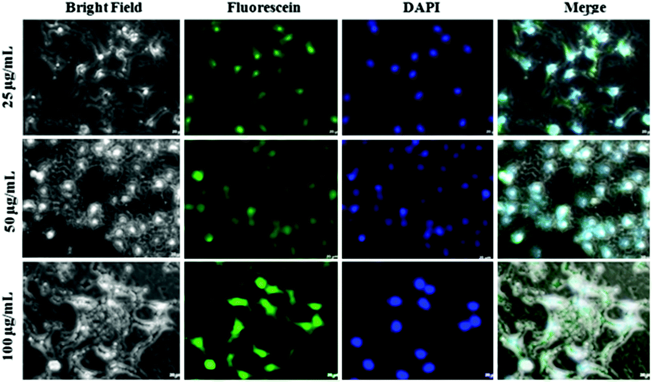 | ||
| Fig. 11 Fluorescence microscope images of MCF-7 cells incubated with the Qtn-PVPylated-TiO2NPs nanocombinations labeled with fluorescein for 3 h at different concentrations (25 mg mL−1 (top panel); 50 mg mL−1 (middle panel), and 100 mg mL−1 (bottom panel) in MCF-7 cells). Bright field; green fluorescence from fluorescein; blue fluorescence from DAPI in the nuclei (cell nuclei were stained with DAPI); and the merged images. | ||
In addition, the authors wish to point out that ref. 18 in the original manuscript is incorrect and should appear as listed below.
18. L. Wang, R. Vivek, W. Wu, G. Wang and J. Ye Wang, ACS Biomater. Sci. Eng., 2018, 4(5), 1880–1890.
The Royal Society of Chemistry apologises for these errors and any consequent inconvenience to authors and readers.
| This journal is © The Royal Society of Chemistry 2018 |