
 Open Access Article
 This Open Access Article is licensed under a
 Creative Commons Attribution 3.0 Unported Licence
Correction: An injectable hyaluronic acid/PEG hydrogel for cartilage tissue engineering formed by integrating enzymatic crosslinking and Diels–Alder “click chemistry”
Feng
Yu
ab,
Xiaodong
Cao
*abc,
Yuli
Li
abc,
Lei
Zeng
ac,
Bo
Yuan
ab and
Xiaofeng
Chen
*abc
aSchool of Materials Science and Engendering, South China University of Technology, Guangzhou, 510641, PR China. E-mail: caoxd@scut.edu.cn; Fax: +86-20-87111752; Tel: +86-20-22236066
bNational Engineering Research Centre for Tissue Restoration and Reconstruction, Guangzhou, 510006, PR China. E-mail: chenxf@scut.edu.cn; Fax: +86-20-22236083; Tel: +86-20-22236283
cGuangdong Province Key Laboratory of Biomedical Engineering, South China University of Technology, Guangzhou 510640, PR China
First published on 6th July 2018
Abstract
Correction for ‘An injectable hyaluronic acid/PEG hydrogel for cartilage tissue engineering formed by integrating enzymatic crosslinking and Diels–Alder “click chemistry”’ by Feng Yu et al., Polym. Chem., 2014, 5, 1082–1090.
The authors regret that the incorrect images were used in Fig. 8A, B and C to represent three different culture time points. The correct version of Fig. 8 is shown below.
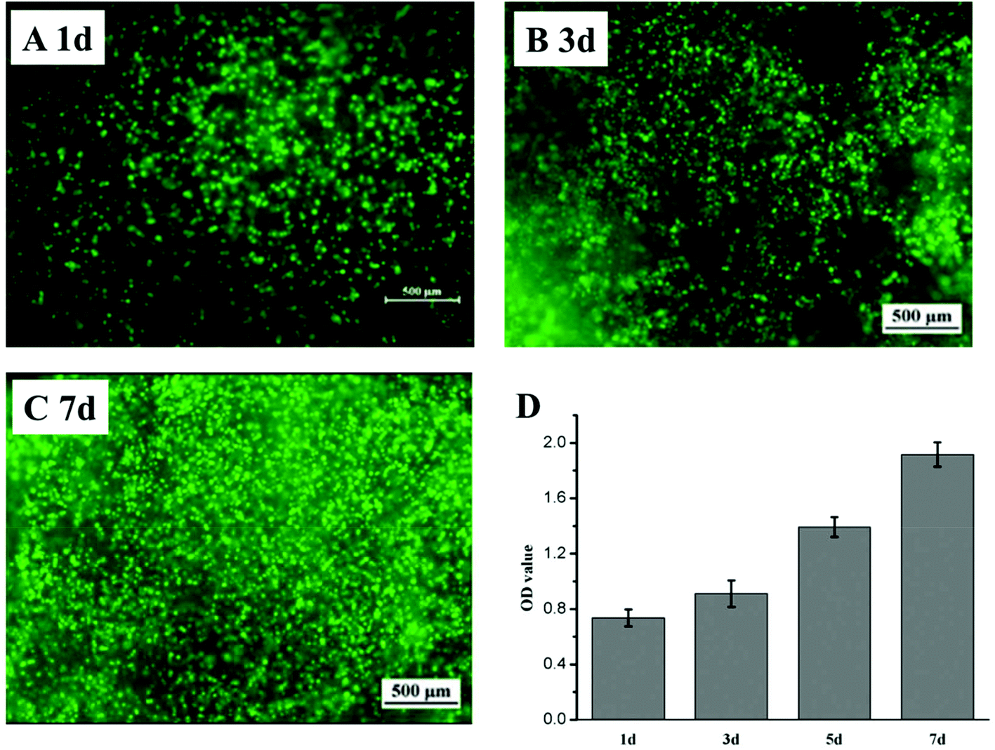 | ||
| Fig. 8 Live–dead and CCK-8 assay of ATDC-5 cells encapsulated in HA/PEG DS1 hydrogel (living cells appearing green, 4 magnifications). (A–C) The live–dead assay of ATDC-5 cells encapsulated in hydrogels after 1, 3, 7 days culture with 40% image brightness increasing. (D) A CCK-8 cytotoxicity assay was performed to evaluate the cells proliferation after 1, 3, 5 and 7 days culture. | ||
In order to further prove the authenticity of the experimental data, additional supplementary information is also provided here.
Fig. S1(A–C) are the original images generated using fluorescence microscopy. Because of the high cell culture density encapsulated in the hydrogel (3 × 10⁶ cells per ml), the phenomenon of cell proliferation after 1 d, 3 d and 7 d culture cannot be distinguished and observed especially in low brightness. Therefore, the image brightness was increased to 20% and 40% to make sure that more cells (marked with green fluorescence) could appear in the image and so that the cell proliferation after different culture days could be observed by the naked eye (Fig. S1(D–I)).
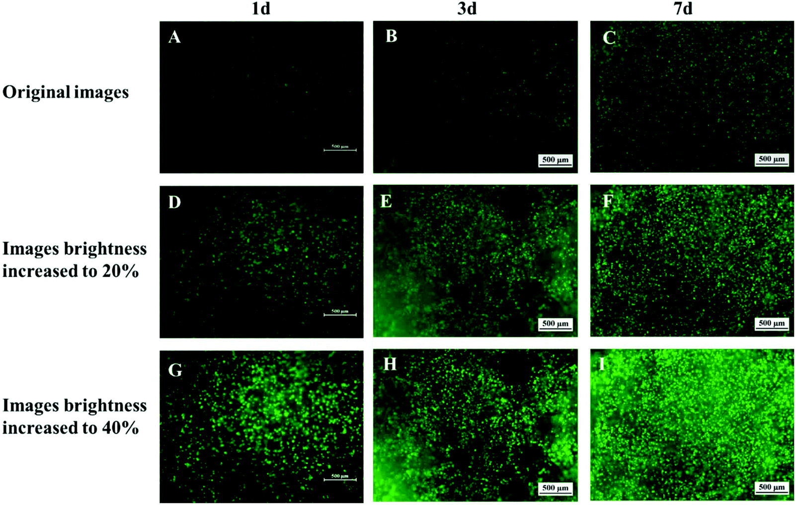 | ||
| Fig. S1 Live–dead assay of encapsulated ATDC-5 cells in HA/PEG DS1 hydrogel (living cells appearing green, 4 magnifications). (A–I) The live–dead assay of ATDC-5 cells encapsulated in hydrogels after 1, 3, 7 days culture with different brightness. A–C are the original images. D–F are the images with 20% brightness increasing. G–I are the images with 40% brightness increasing. | ||
In addition, a CCK-8 assay is an efficient and quantitative way to prove the cell proliferation. Here, the detailed data are also provided in Table S1 to further supplement the data shown in Fig. 8D.
| OD value | 1 day | 3 days | 5 days | 7 days |
|---|---|---|---|---|
| Parallel group 1 | 0.68284 | 0.981505 | 1.39048 | 1.848 |
| Parallel group 2 | 0.723215 | 1.02393 | 1.431505 | 1.94505 |
| Parallel group 3 | 0.731945 | 0.921341 | 1.429301 | 1.79301 |
| Parallel group 4 | 0.703852 | 0.843448 | 1.321341 | 1.96406 |
| Average value | 0.710463 | 0.942556 | 1.393157 | 1.88753 |
The Royal Society of Chemistry apologises for these errors and any consequent inconvenience to authors and readers.
| This journal is © The Royal Society of Chemistry 2018 |