 Open Access Article
Creative Commons Attribution 3.0 Unported Licence
QM/MM simulations identify the determinants of catalytic activity differences between type II dehydroquinase enzymes†
Emilio
Lence‡
 ab,
Marc W.
van der Kamp‡
 *ac,
Concepción
González-Bello
 *b and
Adrian J.
Mulholland
*a
aCentre for Computational Chemistry, School of Chemistry, University of Bristol, Cantock's Close, BS8 1TS Bristol, UK. E-mail: adrian.mulholland@bristol.ac.uk; Tel: +44 (0)117 9289097
bCentro Singular de Investigación en Química Biolóxica e Materiais Moleculares (CIQUS), Departamento de Química Orgánica, Universidade de Santiago de Compostela, Jenaro de la Fuente s/n, 15782 Santiago de Compostela, Spain. E-mail: concepcion.gonzalez.bello@usc.es; Tel: +34 881 815726
cSchool of Biochemistry, University of Bristol, University Walk, BS8 1TD Bristol, UK. E-mail: marc.vanderkamp@bristol.ac.uk; Tel: +44 (0)117 3312147
First published on 16th May 2018
Abstract
Type II dehydroquinase enzymes (DHQ2), recognized targets for antibiotic drug discovery, show significantly different activities dependent on the species: DHQ2 from Mycobacterium tuberculosis (MtDHQ2) and Helicobacter pylori (HpDHQ2) show a 50-fold difference in catalytic efficiency. Revealing the determinants of this activity difference is important for our understanding of biological catalysis and further offers the potential to contribute to tailoring specificity in drug design. Molecular dynamics simulations using a quantum mechanics/molecular mechanics potential, with correlated ab initio single point corrections, identify and quantify the subtle determinants of the experimentally observed difference in efficiency. The rate-determining step involves the formation of an enolate intermediate: more efficient stabilization of the enolate and transition state of the key step in MtDHQ2, mainly by the essential residues Tyr24 and Arg19, makes it more efficient than HpDHQ2. Further, a water molecule, which is absent in MtDHQ2 but involved in generation of the catalytic Tyr22 tyrosinate in HpDHQ2, was found to destabilize both the transition state and the enolate intermediate. The quantification of the contribution of key residues and water molecules in the rate-determining step of the mechanism also leads to improved understanding of higher potencies and specificity of known inhibitors, which should aid ongoing inhibitor design.
Introduction
It is well known that the catalytic efficiency of homologous enzymes can vary significantly depending on the species. In most cases the active site of homologous enzymes is highly conserved, and it is generally not obvious how other differences in amino acid sequence may affect activity. Understanding such differences can be used for designing specific inhibitors against enzymes, for example for developing new drug candidates to combat pathogenic bacteria that have developed resistance against existing antibiotics. The emergence and worldwide spread of multidrug-resistant microbial strains is one of the most important challenges for public health in the 21st century.1–3 The lack of effective antibiotics is currently undermining our ability to deal with infectious diseases, and to manage complications in vulnerable patients undergoing general surgery, organ transplantation, dialysis and chemotherapy for cancer, for which the ability to treat secondary infections is crucial. It has been estimated that if antibiotic resistance continues rising at this rate we will pass from the current 700![[thin space (1/6-em)]](https://www.rsc.org/images/entities/char_2009.gif) 000 deaths annually to about 10 million (one every 3 seconds) in 2050.4,5 Most of the antibiotics in clinical use target the same type of bacterial functions and resistance to them is now widespread and well known. It is therefore not surprising that much effort is currently being devoted to search not only for more effective antibiotics, but also to develop novel chemical entities disabling unexploited essential processes in bacteria. In this context, it is relevant to study the biochemical behavior of those unexplored targets in detail.1–3,6–8
000 deaths annually to about 10 million (one every 3 seconds) in 2050.4,5 Most of the antibiotics in clinical use target the same type of bacterial functions and resistance to them is now widespread and well known. It is therefore not surprising that much effort is currently being devoted to search not only for more effective antibiotics, but also to develop novel chemical entities disabling unexploited essential processes in bacteria. In this context, it is relevant to study the biochemical behavior of those unexplored targets in detail.1–3,6–8
A particular goal is to develop more specific antimicrobials, i.e. drugs that target particular types of organism, to treat specific infections and diseases. This will require understanding of the essential differences between the same target from different organisms. For enzyme targets, it will be important to understand the differences in catalysis and mechanism. Understanding and analysis of differences in activity and specificity should assist in structure-based design of more specific antimicrobials, targeting specific enzymes. The origins of such differences are generally not obvious from structural data alone: for example, differences in (e.g. carbapenemase) activity between various Class A beta-lactamases cannot be discerned from their structures.9 However, molecular simulations can identify differences, effectively acting as a ‘computational assay’ of biological activity,10 and – crucially – analyze the physical origins of these differences at the molecular level. For example, simulations of reactions with combined quantum mechanics/molecular mechanics (QM/MM) methods,11 distinguish Class A beta-lactamases that can efficiently breakdown carabapenem antibiotics from enzymes that cannot.12 Modelling of reactions in enzymes with QM/MM methods can identify mechanisms13 and provide atomically detailed knowledge about the key interactions of the substrate(s), reaction intermediate(s) and transition state(s). It can further identify specific role(s) of active site amino acids in catalysis14 and conformational changes that the enzyme undergoes in a catalytic cycle.15 Alongside explaining differences in catalytic efficiency between enzymes, this detailed knowledge potentially provides new perspectives for the structure- and mechanism-based design of enzyme inhibitors.
Here, we present an extensive QM/MM simulation study of the reaction free energy profile of two homologous enzymes of a recognized target for antibiotic drug design, the type II dehydroquinase enzyme (DHQ2). The approach used, providing detailed understanding of catalytic and mechanistic differences, could be used generally for designing specific inhibitors against enzymes present in certain pathogenic bacteria vs. non-pathogenic ones, as well as aiding in the rationalization of experimentally observed differences in inhibitor efficacy among homologous enzymes. In particular, the present study compares the DHQ2 from Mycobacterium tuberculosis (MtDHQ2) with that from Helicobacter pylori (HpDHQ2). We identify, from QM/MM simulations, the key factors that explain the 50-fold catalytic efficiency difference between the two homologous enzymes.
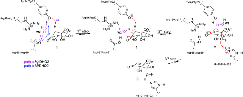
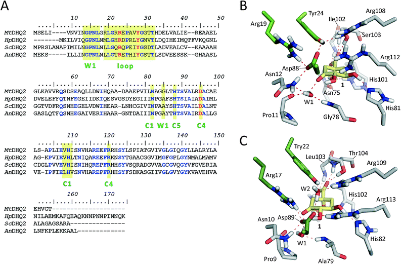
DHQ2 (3-dehydroquinate dehydratase, EC 4.2.1.10) is the third enzyme of the shikimic acid pathway through which erythrose-4-phosphate and phosphoenol pyruvate are converted into chorismic acid. The latter is the precursor of important aromatic metabolites such as the aromatic amino acids, folate cofactors, ubiquinone and vitamins E and K.16 DHQ2, which is encoded by the aroD/aroQ gene and does not have any counterpart in human cells, is an essential enzyme for Mycobacterium tuberculosis, which causes tuberculosis, as well as for Helicobacter pylori, which is a major cause of gastric and duodenal ulcers and has been classified as a class I carcinogen.17,18 The enzyme is also present in the non-pathogenic bacterium Streptomyces coelicolor,19 as well as the fungi Aspergillus nidulans,20 and Neurospora crassa.21 DHQ2 is a dodecamer (tetramer of trimers), with a trimer as the minimum catalytic unit.22 It catalyzes the reversible dehydration of 3-dehydroquinic acid (1) to form 3-dehydroshikimic acid (3) (Scheme 1). The enzymatic mechanism consists of an overall anti elimination of water involving the loss of the more acidic pro-S hydrogen from C2 in 1via the enolate intermediate 2.23–25 Three residues have been identified by chemical modification and site-directed mutagenesis studies as being essential for enzyme activity. Two of them are located in a flexible loop (henceforth the substrate-covering loop) that forms a lid that closes the active site for catalysis: an arginine26 (Arg19/Arg17 in MtDHQ2 and HpDHQ2, respectively; this order will be used throughout) and a tyrosine27 (Tyr24/Tyr22).28 The third residue is an aspartate (Asp88′/Asp89′) from the neighboring enzyme subunit (residues from this subunit will be marked with an accent mark).25 The resolution of several crystal structures of DHQ2 in complex with the product and some reversible competitive inhibitors, in combination with diverse biochemical and computational studies allows a detailed description of the active site and provides a good overall knowledge of the enzymatic mechanism. The substrate is anchored to the active site by several hydrogen bonding interactions through the carboxylate group and the hydroxyl groups (Fig. 1B and C). Specifically, the carboxylate group, which is key for recognition, establishes four strong hydrogen bonding interactions involving the main chain amide NH groups of Leu102/Ile103 and Ser103/Thr104, the side chain hydroxyl group of Ser103/Thr104, and the side chain amide of the conserved Asn75/Asn76.28 The latter also forms a second hydrogen bond between its carbonyl group and the C1 hydroxyl group, thereby positioning the hydroxyl group to accept a proton from the conserved His101/His102. The main hydrogen bonding interactions of the secondary hydroxyl groups are with His81/His82 and Arg112/Arg113 (C5 hydroxyl group), and Asp88′/Asp89′ (C4 hydroxyl group). In addition, the C3 carbonyl group is anchored to the active site through a structural water molecule (henceforth W1) that also hydrogen bonds with residues Pro11/Pro9, Asn12/Asn10 and Gly78/Ala79. Previous computational studies involving QM/MM Steered Molecular Dynamics (SMD) simulations of the reaction showed that the reaction is initiated by the essential aspartate (Asp88′/Asp89′).25 This residue acts as the general base to deprotonate the essential tyrosine to the catalytic tyrosinate form, which triggers the enzyme-catalyzed chemical reaction. For the HpDHQ2 enzyme, the deprotonation occurs with the assistance of a water molecule (henceforth W2), while for MtDHQ2, the tyrosine is directly deprotonated by the aspartate residue. The latter is supported by results from solvent isotope effects and proton inventory studies.25 The necessary reduction in pKa of Tyr24/Tyr22 has been proposed to be achieved by the proximity of two conserved arginine residues, Arg108/Arg109 and Arg112/Arg113 and a cation–π interaction with the essential Arg19/Arg17.29 MD simulation studies revealed that the latter residues are also responsible for the release of the product from the active site.25 Importantly, the essential arginine controls the correct conformation of the tyrosinate for the removal of the pro-S hydrogen from C2 in 1. The final step is the acid-catalyzed elimination of the C1 hydroxyl group that is mediated by the conserved histidine His101/His102 acting as a proton donor.
The previously reported computational studies on the mechanism of DHQ2 either included a small model of the active site (lacking the essential arginine or a detailed description of how the catalytic tyrosinate is generated)24 or used limited sampling of conformations on the reaction pathway using QM/MM SMD25 studies. However, the over 50-fold catalytic efficiency difference between the M. tuberculosis (kcat/Km = 22 × 104 s−1 M−1)30 and H. pylori (kcat/Km = 0.4 × 10⁴ s−1 M−1)30 enzymes could not be explained; the active site of the DHQ2 enzymes is highly conserved (Fig. 1A).
Here, we present an extensive semiempirical QM/MM Umbrella Sampling simulation study, with corrections up to the correlated ab initio (MP2) level, to explore the reaction free energy profile of these two homologous enzymes. Detailed analysis of the key rate-determining step, which involves the formation of the enolate intermediate 2, allowed us to identify relevant differences in this step that explain the experimentally observed difference in catalytic efficiency of the two studied enzymes, MtDHQ2 and HpDHQ2. The results shown here give a detailed picture of the mechanism and identify relevant interactions that could be used for future inhibitor design.
Results and discussion
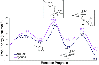
QM/MM (SCC-DFTB/ff03) umbrella sampling simulations were performed for the two DHQ2 enzymes, MtDHQ2 and HpDHQ2, and for the three reaction steps: (a) generation of the catalytic tyrosinate, (b) formation of the enolate intermediate 2, and (c) enolate dehydration (Scheme 1). In general, the resulting free energy profiles at this level of theory indicate that the three-step mechanism is feasible, with an overall energy barrier of 15.4 and 16.0 kcal mol−1 for MtDHQ2 and HpDHQ2, respectively. The free energy profiles at the SCC-DFTB/ff03 level suggest that the enolate dehydration (3rd step) is the rate-determining step (Fig. S1†), whereas experimental isotope effects indicate that this is due to the formation of the enolate intermediate 2 (2nd step).23 Small model calculations indicated that this discrepancy is likely due to inaccuracies of SCC-DFTB for the reaction (and activation) energy of the 2nd step, i.e. due to inaccurate proton affinities. Our results show that SCC-DFTB underestimates the stability of the enolate intermediate 2 with respect to the substrate (with tyrosinate) by between 6–8 kcal mol−1, when compared with more accurate methods such as MP2 and B3LYP (Fig. S2†). Thus, corrections at the MP2/6-31+G(d,p) level to the energy profile for the 2nd step, give free energy profiles for the three step mechanism with overall barriers of 14.0 and 17.0 kcal mol−1 for MtDHQ2 and HpDHQ2, respectively (Fig. 2). These energy values correlate reasonably well with values derived from the experimentally determined apparent catalytic rates (using the Arrhenius equation with a unity pre-factor): 16.5 and 17.4 kcal mol−1 for MtDHQ2 and HpDHQ2, respectively. Notably, the 2nd step is now rate-determining for both enzymes, in agreement with the experimental data. A more detailed description of the corrections carried out, as well as the main differences between the two enzymes along the reaction path identified in the herein reported QM/MM (SCC-DFTB/ff03) umbrella sampling simulations is provided below.1st step: Generation of the catalytic tyrosinate
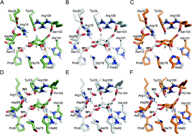
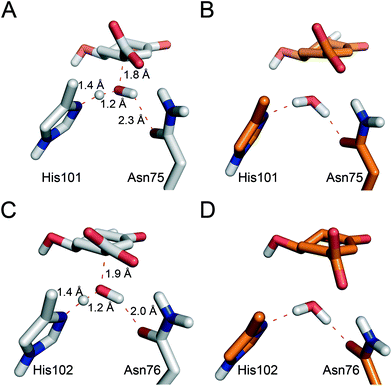
Reaction barriers of 6.8 and 8.8 kcal mol−1 for MtDHQ2 and HpDHQ2, respectively, were found for this step (Fig. 2). The ∼2 kcal mol−1 difference between the enzymes can be explained by the distinct nature of the process (Fig. 3). While for MtDHQ2 the essential Tyr24 is deprotonated directly by the essential Asp88′, for HpDHQ2, the process is mediated by a water molecule (W2), which is located between Tyr22 and Asp89′. W2 is observed in diverse crystal structures of binary HpDHQ2/inhibitor complexes such as PDB 4B6R (Fig. 4).31 It seems that W2 supplements the inability of the essential Tyr22 to be located close enough to the catalytic Asp89′ for direct proton transfer. This is mainly due to the salt bridge present during the whole reaction between Asp18 and Arg20, both located in the substrate-covering loop, which controls and limits active site flexibility. This strong interaction, which is not present in MtDHQ2 (Fig. 1A, Glu20 and Ala22 are present in equivalent positions), has been proposed to control and reduce the plasticity of this loop (Fig. S2†).31 In addition, the presence of Glu20 in the loop of MtDHQ2, replacing the shorter Asp18 in HpDHQ2, favours the interaction between Glu20 and the neutral Asp88′. In fact, we observed that after performing the Umbrella Sampling simulation for the 2nd step (forming the substrate 1), this interaction leads to a large distance between Tyr24 and the neutral Asp88′ (average distance of 5.1 Å between the Tyr24 hydroxyl O and the Asp88′ carboxylic acid H). To avoid this issue, we introduced an additional reaction coordinate (and performed QM/MM umbrella sampling along this coordinate) to reduce the distance between Tyr24 and Asp88′ and to break the interaction between Glu20 and neutral Asp88′.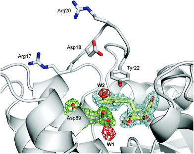 | ||
| Fig. 4 Unbiased electron density for (2S)-2-perfluorobenzyl-3-dehydroquinic acid (yellow, inhibitor), W1, W2 and Asp89′ in the binary HpDHQ2/inhibitor complex (chains A and C, PDB code 4B6R,31 1.9 Å). A maximum-likelihood weighted 2Fo − Fc map contoured at 1σ is shown up to 1.6 Å around the inhibitor molecule (blue), W1 and W2 (red) and Asp89′ (green). | ||
Significant differences in the behaviour of the structural water molecule W1 for both enzymes were identified. While for the MtDHQ2 enzyme, the four hydrogen bonding interactions that anchor W1 to the active site (also observed in the Michaelis complex and involving residues Pro11, Gly78 and Asn12 and the C3 ketone in 1) remained unchanged during the whole mechanism, this is not the case for HpDHQ2 (Fig. 3B vs. C). For HpDHQ2, only the interactions involving residues Pro9 and Ala79 remained present during the whole mechanism since Asn10 forms a hydrogen bond with Asp89′ (Fig. 3C). As a consequence, W1 rotates during the 2nd step to form a hydrogen bond with the O3 atom of the forming enolate, an equivalent interaction as observed for the MtDHQ2 enzyme.
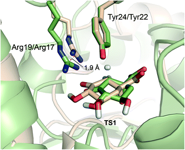
Overall, the reaction energy for the 1st step is almost identical (0.7 kcal mol−1 difference, Fig. 2). The capability of both enzymes for stabilization of the catalytic tyrosinate is therefore similar. For both enzymes, the formation of the catalytic tyrosinate occurs with a parallel arrangement between the phenol group of the essential tyrosine and the guanidinium group of the essential arginine Arg19/Arg17 (Fig. 3A and D). This arrangement helps to maximize the cation–π interaction between both residues, which has been proposed to (a) control the appropriate position of the tyrosinate for abstraction of the pro-S hydrogen atom from C2 in 1; and (b) to reduce the pKa of the tyrosine, along with the conserved Arg108/Arg109 residue.29 The latter residue is likely to set up the catalytic tyrosine or W2 (in MtDHQ2 and HpDHQ2, respectively), for deprotonation. Once the tyrosinate was formed, this parallel arrangement between Tyr24/Tyr22 and Arg19/Arg17 was partially lost, although both residues remain close (Fig. 3A and D).
2nd step: Enolate formation
As aforementioned, simulations of the full reaction with SCC-DFTB fail to predict the 2nd step as the partial rate determining step of the reaction path, due to the overestimation of the energy for hydrogen abstraction, as well as the underestimation of the reaction barrier by SCC-DFTB (Fig. S3†); such limitations are typical of this efficient but approximate density-functional theory method (with the recent third-order parametrization showing promising improvement).32 To overcome this issue, several high-level QM methods (ab initio MP2, and DFT: B3LYP and MPW1K) were employed to correct the free energy profile of this 2nd step, using energy calculations on SCC-DFTB/MM potential energy profiles. All showed the same trends, so only the ab initio MP2 results are discussed here (Fig. S4†). Moreover, these QM methods also provide an energy profile for this step with the typical proton-transfer curve, which was not observed using SCC-DFTB. These corrections not only allow the correct identification of the partially rate-determining 2nd step, but also serve to assess the nature of the process and the difference between the two homologous enzymes. Once corrected, the formation of 2 is thermodynamically favorable for both enzymes, by −3.8 and −0.6 kcal mol−1 (for MtDHQ2 and HpDHQ2, respectively). As a result, MtDHQ2 stabilizes the enolate intermediate 2 more effectively than HpDHQ2, with a relative difference compared to the Michaelis complex of 3.9 kcal mol−1. Further, the energy barrier for the transition state (henceforth TS1) in MtDHQ2 is lower than for HpDHQ2 (11.7 vs. 14.0 kcal mol−1).We evaluate the electrostatic contributions to stabilisation of enolate intermediate 2 and TS1 of key residues (Arg19/Arg17 and Asp88′/Asp89′) and the structural and catalytic water molecules W1 and W2 (Tables 1, S1 and S3†). Significant differences between the two enzymes are identified for each (Fig. 5).
| Residue/water molecule | TS1 | Enolate intermediate 2 | ||
|---|---|---|---|---|
| MtDHQ2 | HpDHQ2 | MtDHQ2 | HpDHQ2 | |
| a Energy values were obtained from single point MP2/6-31+G(d,p) calculations and as a difference of the relative energy (with respect to the reactants) of the QM region without the residue minus the energy of the full QM region. Positive values indicate stabilization with respect to the reactants (substrate and tyrosinate state). | ||||
| Arg19/Arg17 | −2.74 | 4.30 | 6.20 | 15.21 |
| Asp88′/Asp89′ | 1.51 | −2.07 | 1.67 | −4.56 |
| W1 | 1.13 | 0.89 | 4.03 | 2.89 |
| W2 | n/a | −8.60 | n/a | −16.89 |
3rd step: Enolate dehydration
No significant differences between the two enzymes were identified for this step either in energy or geometry (Fig. 7). Similar free energy barriers (8.4 and 7.9 kcal mol−1) and reaction energies (−7.8 and −6.7 kcal mol−1) were obtained for MtDHQ2 and HpDHQ2, respectively. In contrast, Blomberg et al.24 reported a great variability for the energies barriers calculated for this step and for the energy of the whole process for the Streptomyces coelicolor enzyme. In particular, the energy barrier for the two step mechanism (enolate formation and enolate dehydration) found previously with DFT ranged between 14.4 and 22.5 kcal mol−1 and showing a maximum energy for the transition state of the enolate dehydration (TS2), and the energy of the transformation ranged between −8.2 to 9.2 kcal mol−1. Our model is more consistent with the experimental data, with a barrier located in the TS1 for the formation of the enolate 2, and an overall energy of −9.3 and −4.3 kcal mol−1 (for MtDHQ2 and HpDQH2, respectively) for the three step mechanism.Taken together, the calculations reported herein allow the quantification of small differences in the energy barriers of catalytic processes carried by homologous enzymes, which is mainly related to kcat. The differences in recognition (Km) between both enzymes, which in this case are also experimentally significant and have an important impact on the overall process catalyzed by the enzyme (kcat/Km), cannot be quantified by this type of computational study. However, the knowledge in atomic detail of the geometry of the Michaelis complex, transition states and intermediates of the catalytic process in both enzymes and more importantly, the relevant differences found between the mechanisms of both enzymes in terms of interactions between the enzyme and reacting species provides a good understanding of the experimentally observed differences in efficiency [M. tuberculosis (fast) vs. H. pylori (slow)].
Conclusions
In the present study we have shown that a combined approach using QM/MM umbrella sampling simulations at the SCC-DFTB/ff03 level of theory with MP2/6-31+G(d,p) corrections on the key reaction step of the reaction catalyzed by the DHQ2 enzyme provides free energy profiles in agreement with experimental data. No significant energetic differences were found for the generation of the catalytic tyrosinate and the enolate dehydration steps in the MtDHQ2 and HpDHQ2 homologues. The 2nd step is rate-determining, in line with experiment (and in contrast with previously reported computational studies), and it is this step which gives rise to observed differences in activity between the two enzymes. Quantification of the contribution of relevant residues in the rate-determining step allowed the identification of the key factors responsible for the 50-fold catalytic efficiency difference between two homologous enzymes, MtDHQ2 and HpDHQ2. The more efficient stabilization of the enolate intermediate by the M. tuberculosis enzyme results in a thermodynamically more favorable 2nd step (ΔG −3.8 kcal mol−1) than for its H. pylori homologue (−0.6 kcal mol−1). For HpDHQ2, a conserved water molecule (W2) between the catalytic residues Asp89′ and Tyr22 was found to destabilize both the transition state (TS1) and the enolate product 2 of the 2nd step. The water molecule W2 that is required for the 1st step in HpDHQ2 (probably to compensate for the inability to locate both residues close enough for the generation of the catalytic tyrosinate) leads to a less optimal arrangement of the active site for the subsequent steps of the enzymatic conversion. The presence of W2 (and the lack of the Asp–Glu interaction observed in MtDHQ2) also contributes to the destabilising influence of Asp89′ (compared to stabilization by Asp88′ in MtDHQ2). The requirement for W2 in HpDHQ2 is probably determined by the more limited flexibility of the substrate-covering loop in this enzyme, due to the formation of an internal salt bridge between two residues of this loop, Asp18 and Arg20. Comparison of the amino acid sequence of the catalytic loop in diverse DHQ2 homologous enzymes reveals that MtDHQ2 is the only one with an apolar residue in one of the corresponding positions (Ala22; Fig. 1A), which appears to be crucial for its larger flexibility.31Our quantification of the contributions of the key residues in the rate-determining step of the mechanism provides a better understanding of the higher inhibitory potencies of known mechanism-based inhibitors, i.e. analogs of the natural substrate and the enolate intermediate. Whereas the crystal structures of the binary DHQ2/ligand complexes for both enzymes show a highly similar binding mode (e.g. PDB codes 4B6O31 and 2Y7135 for MtDHQ2 and PDB codes 4B6R31 and 2WKS34 for HpDHQ2), the inhibitors present in these structures have significantly distinct inhibitory potencies. The identified more efficient stabilization of the enolate intermediate and TS1 by the M. tuberculosis enzyme explains the difference in inhibition data and will be helpful for ongoing inhibitor design. The computational studies presented here highlight the potential of QM/MM simulations as a tool for understanding differences in catalytic efficiency between homologous enzymes.
Experimental
System preparation
Initial structures were prepared as in ref. 25. For MtDHQ2, the coordinates from the crystal structure of the M. tuberculosis enzyme in complex with the product, 3-dehydroshikimic acid (3) (Protein Databank (PDB) code 3N59),38 were used. For HpDHQ2, the crystal structure PDB 2XB9,29 which has the inhibitor (2R)-2-(4-methoxybenzyl)-3-dehydroquinic acid in the active site, was employed. For MtDHQ2, the reaction mechanism was studied in the forward (formation of 3) and backward direction (formation of 1). Because the latter provided more reliable results with less motion of the key residues (in line with efficient reaction), both enzymatic conversions are studied and compared in the direction of formation of 1 here. The ligand present in the PDB 2XB9 structure was replaced by the product, 3-dehydroshikimic acid (3). Except the structural water molecule (W1), all crystallographic waters were removed. The addition of missing hydrogen atoms and protonation states of residues were assigned using the H++ web server at pH 7.0 (http://biophysics.cs.vt.edu/H++),39 except for Asp88′/Asp89′ that was treated as protonated due to mechanistic considerations. The ff0340 AMBER force field and GAFF41 were used to assign bonded and non-bonded parameters to the protein and both 3-dehydroquinic acid (1) and 3-dehydroshikimic acid (3), respectively. Each complex was immersed in a truncated octahedron of TIP3P42 water molecules and neutralised with Na+ ions43 (∼20![[thin space (1/6-em)]](https://www.rsc.org/images/entities/char_2009.gif) 000 water molecules and 15 Na+ ions for the MtDHQ2 model and ∼30
000 water molecules and 15 Na+ ions for the MtDHQ2 model and ∼30![[thin space (1/6-em)]](https://www.rsc.org/images/entities/char_2009.gif) 000 water molecules and 16 Na+ ions for the HpDHQ2 model were added).
000 water molecules and 16 Na+ ions for the HpDHQ2 model were added).
Molecular dynamics simulations
Simulations were performed using the sander and pmemd modules of AMBER 12.44 Periodic boundary conditions were applied and electrostatic interactions were treated using the smooth PME (particle mesh Ewald) method45 with a grid spacing of 1 Å. The cut-off distance for the nonbonded interactions was 9 Å, the SHAKE46 algorithm was applied to all bonds and an integration step of 2.0 fs was used throughout. After an initial energy minimization of the water molecules and counter ions, both systems were heated to 300 K in 25 ps using positional restraints of 50.0 kcal mol−1 Å−2 for the protein Cα atoms. The HpDHQ2 model was then equilibrated over 200 ps with the positional restraints, which were subsequently reduced by 5.0 kcal mol−1 Å−2 every 20 ps, followed by 20 ps without restraints. The MtDHQ2 model (with coordinates from 3 already present in the crystal structure) was equilibrated for 100 ps using restraints of 5.0 kcal mol−1 Å−2 for the protein Cα atoms (except for amino acids Asp89′, Arg19 and Glu20). Subsequently, restraints were reduced by 1.0 kcal mol−1 Å−2 every 20 ps, and another 20 ps without restraints. To maintain a catalytically relevant conformation, a restraint of 100 kcal mol−1 Å−2 was used to keep the dihedrals CB, CG, OD2, HD2 of Asp88′ at 180° and CA, CB, CG, OD2 of Asp89′ between 120° and −60°. Two further one-sided distance restraints of 15 kcal mol−1 Å−2 were used: one to keep the CGAsp89′–CDGlu20 distance to within 4 Å and another to keep the CDGlu20–CZArg108 distance to at least 6.5 Å, in order to avoid distortion of the active site. For both models, the final equilibration snapshot was used for mechanistic studies with QM/MM MD.QM/MM MD simulations
For both enzymes, the QM region included the equivalent atoms of the product, 3-dehydroshikimic acid (3), the side chains of residues Arg19/Arg17, Tyr24/Tyr22, His101/His102 and Asp88′/Asp89′, and the product and structural (W1) and product (henceforth W3) water molecules. For the HpDHQ2 model, a second water molecule, W2, that was observed located between Tyr22 and Asp89′ was also included. Hydrogen ‘link atoms’47,48 were used to model bonds across the QM/MM boundary, specifically between Cγ and Cδ of Arg19/Arg17, Cα and Cβ of Tyr24/Tyr22, His101/His102 and Asp88′/Asp89′. W3 was manually docked in the carboxylate recognition pocket, establishing one hydrogen bond with the Oδ1 oxygen atom of Asn75/Asn76 and another one with the Nδ1 nitrogen atom of His101/His102. QM/MM calculations were performed using sander from AMBER 12 (version 12.19). SCC-DFTB49 was used for the QM region.QM/MM umbrella sampling MD simulations were run for each reaction step, harmonically restraining the reaction coordinate with a force constant of 100 kcal mol−1 Å−2. Each simulation (window) consisted of 5 ps (10![[thin space (1/6-em)]](https://www.rsc.org/images/entities/char_2009.gif) 000 steps with an integration step of 0.5 fs) of sampling and the reaction coordinate was increased (or decreased) by 0.1 Å between neighboring windows, using the last geometry of the previous window as starting point. Values of the reaction coordinate were collected for all simulation steps. The free-energy profiles for each step were obtained by combining the statistics from all simulations for that reaction using the weighted histogram analysis method (WHAM).50–52 The following reaction coordinates were used: (i) third step – for both enzymes, a 2D umbrella sampling simulation involving the formation of the C1–oxygen bond in 1, and the proton transfer from W3 to His101/His102 was used: r1a = d(C1–OW3) and r1b = d(ND1His–H1W3) − d(H1W3–OW3). (ii) second step – for both enzymes, a single reaction coordinate for the hydrogen addition to C2 in 1 by Tyr24/Tyr22 was used: r2 = d(HHTyr–C2) − d(HHTyr–HOTyr). Moreover, several additional restraints were employed to avoid sampling rearrangements not related to the chemical steps of interest (e.g. ligand unbinding, see ESI† for details). (iii) first step – For the MtDHQ2 model, a single reaction coordinate for protonation of the catalytic tyrosinate Tyr24 was employed: r3a = d(OHTyr22–HD2Asp89′) − d(HD2Asp89′–OD2Asp89′), and an additional reaction coordinate was needed for the approach of the neutral Asp89′ to the tyrosinate anion (see ESI† for details). For the HpDHQ2 model, 2D umbrella sampling with two reaction coordinates was required: (1) deprotonation of W2 by tyrosinate Tyr22, and (2) protonation of W2 by neutral Asp88′, r3a = d(OHTyr22–H1W2) − d(H1W2–OW2) and r3a = d(HD2Asp88′–OW2) − d(HD2Asp88′–OD2Asp88′).
000 steps with an integration step of 0.5 fs) of sampling and the reaction coordinate was increased (or decreased) by 0.1 Å between neighboring windows, using the last geometry of the previous window as starting point. Values of the reaction coordinate were collected for all simulation steps. The free-energy profiles for each step were obtained by combining the statistics from all simulations for that reaction using the weighted histogram analysis method (WHAM).50–52 The following reaction coordinates were used: (i) third step – for both enzymes, a 2D umbrella sampling simulation involving the formation of the C1–oxygen bond in 1, and the proton transfer from W3 to His101/His102 was used: r1a = d(C1–OW3) and r1b = d(ND1His–H1W3) − d(H1W3–OW3). (ii) second step – for both enzymes, a single reaction coordinate for the hydrogen addition to C2 in 1 by Tyr24/Tyr22 was used: r2 = d(HHTyr–C2) − d(HHTyr–HOTyr). Moreover, several additional restraints were employed to avoid sampling rearrangements not related to the chemical steps of interest (e.g. ligand unbinding, see ESI† for details). (iii) first step – For the MtDHQ2 model, a single reaction coordinate for protonation of the catalytic tyrosinate Tyr24 was employed: r3a = d(OHTyr22–HD2Asp89′) − d(HD2Asp89′–OD2Asp89′), and an additional reaction coordinate was needed for the approach of the neutral Asp89′ to the tyrosinate anion (see ESI† for details). For the HpDHQ2 model, 2D umbrella sampling with two reaction coordinates was required: (1) deprotonation of W2 by tyrosinate Tyr22, and (2) protonation of W2 by neutral Asp88′, r3a = d(OHTyr22–H1W2) − d(H1W2–OW2) and r3a = d(HD2Asp88′–OW2) − d(HD2Asp88′–OD2Asp88′).
High level QM corrections and residue contributions for the 2nd step
For both enzymes, snapshots of the 0.0 Å window in the 2nd step were minimized in a three step procedure: (1) 500 steps (20 steepest descent followed by 480 conjugate gradient) with the same restraints used in umbrella sampling but with a force constant of 2500 kcal mol−1 Å−2; (2) minimization using the LBFGS method (ntmin = 3) and a convergence criterion of 0.002 kcal mol−1 (drms = 0.002) keeping only restraints to the reaction coordinate; (3) minimization with positional restraints of 50 kcal mol−1 Å−2 to any residue, water molecule or ion that was more than 5.0 Å away from the QM region and the restraint of the reaction coordinate. The reaction coordinate (between −1.1 to 1.0 Å for MtDHQ2 and −1.2 to 1.5 Å for HpDHQ2) was explored forwards and backwards until a converged and smooth (adiabatic) potential energy profile along the reaction pathway was obtained. Coordinates for the QM region (with H link atoms) along the reaction pathway were then extracted and single point energy calculations in vacuum with Gaussian0953 with SCC-DFTB and MP2 (with the 6-31+G(d,p) basis set) were performed. SCS-MP2,54 B3LYP55,56 and MPW1K57 single point energy calculations were also performed. Corrections to the potential and free energy surfaces (PES and FES) were applied by subtracting the energy calculated for the QM region using SCC-DFTB and adding the energy from the MP2 (or other) calculations. Similar approaches have been applied successfully previously to other enzyme-catalyzed reactions;58,59 it corrects for limitations of the lower-level method e.g. in the calculation of proton affinities. Individual electrostatic contributions for residues Asp88′/89′ and Arg19/Arg17 and water molecules W1 and W2 (only HpDHQ2) were then evaluated by calculating the difference between the energy profile with or without the corresponding residue present. These energies were calculated as single points with the same methods as before.Due to the more complicated reaction coordinate for the 3rd step (involving carbon–oxygen bond breaking as well as a proton transfer, which necessitates two-dimensional umbrella sampling) as well as relevant solvent rearrangements, a similar energy correction based on single-point energies from a potential energy profile is not reliable for this reaction step. Nevertheless, we performed such a correction to give an approximate indication of how the reaction is affected by more accurate QM treatment (Table S7†). The approximate corrections would lead to free-energy barriers for step 3 (relative to the reactant state) of ∼11.5 kcal mol−1 for MtDHQ2 and ∼16.1 kcal mol−1 for HpDHQ2. In cases such as this, simple potential energy profiles of enzyme reactions suffer from extreme variability due to conformation variability and solvation changes and are not an effective approach to modelling the mechanism. Single structures do not represent the ensemble of structures, and structural changes involved in reaction. Indeed, if this approach were applicable, we would have followed such an approach, as we have done in some previous work. It is not in this case, as we have established by test calculations. Instead, it is essential to sample conformational changes and generate appropriate structural ensembles for the reacting system through QM/MM molecular dynamics simulations. This necessitates the use of a low-level QM/MM method, in this case SCC-DFTB, with corrections where necessary (see below). We have discussed these factors (and approaches to modelling enzyme reactions in general) in ref. 11 and 60–62 as well as in a large number of published applications (ref. 12 and 63).
Specific errors for sampling can be obtained by repeating the reaction simulations multiple times. Where we have done this in the past using a similar approach (and known enzyme–substrate complexes), we find standard deviations of 0.1–1 kcal mol−1.12
Conflicts of interest
There are no conflicts to declare.Abbreviations
| DHQ2 | Type II dehydroquinase |
| MtDHQ2 | Type II dehydroquinase from Mycobacterium tuberculosis |
| HpDHQ2 | Type II dehydroquinase from Helicobacter pylori |
Acknowledgements
Financial support from the Spanish Ministry of Economy and Competiveness (SAF2016-75638-R), the Xunta de Galicia (Centro singular de investigación de Galicia accreditation 2016–2019, ED431G/09) and the European Union (European Regional Development Fund - ERDF) is gratefully acknowledged. EL thanks the Xunta de Galicia for his postdoctoral fellowship. MWvdK is a BBSRC David Phillips Fellow (BB/M026280/1) and he and AJM thank EPSRC for funding (grant numbers EP/G007705/1; EP/M022609/1; EP/M013219/1). We are grateful to the Advanced Computing Research Centre of the University of Bristol and Centro de Supercomputación de Galicia (CESGA) for use of the computational facilities.Notes and references
- E. D. Brown and G. D. Wright, Nature, 2016, 529, 336–343 CrossRef PubMed.
- M. A. Fischbach and C. T. Walsh, Science, 2009, 325, 1089–1093 CrossRef PubMed.
- K. Bush, P. Courvalin, G. Dantas, J. Davies, B. Eisenstein, P. Huovinen, G. A. Jacoby, R. Kishony, B. N. Kreiswirth, E. Kutter, S. A. Lerner, S. Levy, K. Lewis, O. Lomovskaya, J. H. Miller, S. Mobashery, L. J. V. Piddock, S. Projan, C. M. Thomas, A. Tomasz, P. M. Tulkens, T. R. Walsh, J. D. Watson, J. Witkowski, W. Witte, G. Wright, P. Yeh and H. I. Zgurskaya, Nat. Rev. Microbiol., 2011, 9, 894–896 CrossRef PubMed.
- D. A. Goff, R. Kullar, E. J. C. Goldstein, M. Gilchrist, D. Nathwani, A. C. Cheng, K. A. Cairns, K. Escandón-Vargas, M. V. Villegas, A. Brink, D. van den Bergh and M. Mendelson, Lancet Infect. Dis., 2017, 17, e56–e63 CrossRef PubMed.
- https://www.cdc.gov/drugresistance/threat-report-2013/ (last access July 25th, 2017).
- T. Kostyanev, M. J. M. Bonten, S. O'Brien, H. Steel, S. Ross, B. François, E. Tacconelli, M. Winterhalter, R. A. Stavenger, A. Karlén, S. Harbarth, J. Hackett, H. S. Jafri, C. Vuong, A. MacGowan, A. Witschi, G. Angyalosi, J. S. Elborn, R. deWinter and H. Goossens, J. Antimicrob. Chemother., 2016, 71, 290–295 CrossRef PubMed.
- http://www.carb-x.org/ (last access July 25th, 2017).
- https://www.imi.europa.eu/content/nd4bb (last access July 25th, 2017).
- F. Fonseca, E. I. Chudyk, M. W. van der Kamp, A. Correia, A. J. Mulholland and J. Spencer, J. Am. Chem. Soc., 2012, 134, 18275–18285 CrossRef PubMed.
- C. J. Woods, M. Malaisree, B. Long, S. McIntosh-Smith and A. J. Mulholland, Sci. Rep., 2013, 3, 3561 CrossRef PubMed.
- M. W. van der Kamp and A. J. Mulholland, Biochemistry, 2013, 52, 2708–2728 CrossRef PubMed.
- E. I. Chudyk, M. A. L. Limb, C. Jones, J. Spencer, M. W. van der Kamp and A. J. Mulholland, Chem. Commun., 2014, 50, 14736–14739 RSC.
- M. J. Byrne, N. R. Lees, L. C. Han, M. W. van der Kamp, A. J. Mulholland, J. E. M. Stach, C. L. Willis and P. R. Race, J. Am. Chem. Soc., 2016, 138, 6095–6098 CrossRef PubMed.
- K. E. Ranaghan, J. E. Hung, G. J. Bartlett, T. J. Mooibroek, J. N. Harvey, D. N. Woolfson, W. A. van der Donk and A. J. Mulholland, Chem. Sci., 2014, 5, 2191–2199 RSC.
- M. W. van der Kamp, R. Chaudret and A. J. Mulholland, FEBS J., 2013, 280, 3120–3131 CrossRef PubMed.
- C. Abell, Enzymology and Molecular Biology of the Shikimate Pathway, in Comprehensive Natural Products Chemistry, ed. U. Sankawa, Pergamon, Elsevier Science Ltd., Oxford, 1999, vol. 1, pp. 573–607 Search PubMed.
- G. Lamichhane, J. S. Freundlich, S. Ekins, N. Wickramaratne, S. T. Nolan and W. R. Bisha, mBio, 2011, 2, e00301 CrossRef PubMed.
- Data base for essential genes: http://www.essentialgene.org/.
- P. J. White, J. Young, I. S. Hunter, H. G. Nimmo and J. R. Coggins, Biochem. J., 1990, 265, 735–738 CrossRef PubMed.
- A. R. Hawkins, N. H. Giles and J. R. Kinghorn, Biochem. Genet., 1982, 20, 271–286 CrossRef PubMed.
- J. A. Hautala, J. W. Jacobson, M. E. Case and N. H. Giles, J. Biol. Chem., 1975, 250, 6008–6014 Search PubMed.
- N. C. Price, D. J. Boam, S. M. Kelly, D. Duncan, T. Krell, D. G. Gourley, J. R. Coggins, V. Virden and A. R. Hawkins, Biochem. J., 1999, 338, 195–202 CrossRef PubMed.
- J. M. Harris, C. Gonzalez-Bello, C. Kleanthous, A. R. Hawkins, J. R. Coggins and C. Abell, Biochem. J., 1996, 319, 333–336 CrossRef PubMed.
- L. Blomberg, M. Mangold, J. Mitchell and J. Blumberger, J. Chem. Theory Comput., 2009, 5, 1284–1294 CrossRef PubMed.
- C. Coderch, E. Lence, A. Peón, H. Lamb, A. R. Hawkins, F. Gago and C. González-Bello, Biochem. J., 2014, 458, 547–557 CrossRef PubMed.
- T. Krell, A. R. Pitt and J. R. Coggins, FEBS Lett., 1995, 360, 93–96 CrossRef PubMed.
- T. Krell, M. J. Horsburgh, A. Cooper, S. M. Kelly and J. R. Coggins, J. Biol. Chem., 1996, 271, 24492–24497 CrossRef PubMed.
- A. W. Roszak, D. A. Robinson, T. Krell, I. S. Hunter, M. Fredrickson, C. Abell, J. R. Coggins and A. J. Lapthorn, Structure, 2002, 10, 493–503 CrossRef PubMed.
- A. Peón, J. M. Otero, L. Tizón, V. F. V. Prazeres, A. L. Llamas-Saiz, G. C. Fox, M. J. Van Raaij, H. Lamb, A. R. Hawkins, F. Gago, L. Castedo and C. González-Bello, ChemMedChem, 2010, 5, 1726–1733 CrossRef PubMed.
- Data from: L. D. B. Evans, A. W. Roszak, L. J. Noble, D. A. Robinson, P. A. Chalk, J. L. Matthews, J. R. Coggins, N. C. Price and A. J. Lapthorn, FEBS Lett., 2002, 530, 24–30 CrossRef PubMed.
- E. Lence, L. Tizón, J. M. Otero, A. Peón, V. F. V. Prazeres, A. L. Llamas-Saiz, G. C. Fox, M. J. van Raaij, H. Lamb, A. R. Hawkins and C. González-Bello, ACS Chem. Biol., 2013, 8, 568–577 CrossRef PubMed.
- M. Gruden, L. Andjeklović, A. Kuriappan Jissy, S. Stepanović, M. Zlatar, Q. Cui and M. Elstner, J. Comput. Chem., 2017, 38, 2171–2185 CrossRef PubMed.
- V. F. V. Prazeres, L. Castedo, H. Lamb, A. R. Hawkins and C. González-Bello, ChemMedChem, 2009, 4, 1980–1984 CrossRef PubMed.
- V. F. V. Prazeres, L. Tizón, J. M. Otero, P. Guardado-Calvo, A. L. Llamas-Saiz, M. J. Van Raaij, L. Castedo, H. Lamb, A. R. Hawkins and C. González-Bello, J. Med. Chem., 2010, 53, 191–200 CrossRef PubMed.
- L. Tizón, J. M. Otero, V. F. V. Prazeres, A. L. Llamas-Saiz, G. C. Fox, M. J. Van Raaij, H. Lamb, A. R. Hawkins, J. A. Ainsa, L. Castedo and C. González-Bello, J. Med. Chem., 2011, 54, 6063–6084 CrossRef PubMed.
- B. Blanco, A. Sedes, A. Peón, H. Lamb, A. R. Hawkins, L. Castedo and C. González-Bello, Org. Biomol. Chem., 2012, 10, 3662–3676 Search PubMed.
- A. Peón, C. Coderch, F. Gago and C. González-Bello, ChemMedChem, 2013, 8, 740–747 CrossRef PubMed.
- M. V. B. Dias, W. C. Snee, K. M. Bromfield, R. J. Payne, S. K. Palaninathan, A. Ciulli, N. I. Howard, C. Abell, J. C. Sacchettini and T. L. Blundell, Biochem. J., 2011, 436, 729–739 CrossRef PubMed.
- J. C. Gordon, J. B. Myers, T. Folta, V. Shoja, L. S. Heath and A. Onufriev, Nucleic Acids Res., 2005, 33, W368–W371 CrossRef PubMed.
- Y. Duan, C. Wu, S. Chowdhury, M. C. Lee, G. M. Xiong, W. Zhang, R. Yang, P. Cieplak, R. Luo, T. Lee, J. Caldwell, J. M. Wang and P. Kollman, J. Comput. Chem., 2003, 24, 1999–2012 CrossRef PubMed.
- J. Wang, R. M. Wolf, J. W. Caldwell, P. A. Kollman and D. A. Case, J. Comput. Chem., 2004, 25, 1157–1174 CrossRef PubMed.
- W. L. Jorgensen, J. Chandrasekhar, J. D. Madura, R. W. Impey and M. L. Klein, J. Chem. Phys., 1983, 79, 926–935 CrossRef.
- J. Aqvist, J. Phys. Chem., 1990, 94, 8021–8024 CrossRef.
- D. A. Case, J. T. Berryman, R. M. Betz, D. S. Cerutti, T. E. Cheatham, III, T. A. Darden, R. E. Duke, T. J. Giese, H. Gohlke, A. W. Goetz, N. Homeyer, S. Izadi, P. Janowski, J. Kaus, A. Kovalenko, T. S. Lee, S. LeGrand, P. Li, T. Luchko, R. Luo, B. Madej, K. M. Merz, G. Monard, P. Needham, H. Nguyen, H. T. Nguyen, I. Omelyan, A. Onufriev, D. R. Roe, A. Roitberg, R. Salomon-Ferrer, C. L. Simmerling, W. Smith, J. Swails, R. C. Walker, J. Wang, R. M. Wolf, X. Wu, D. M. York and P. A. Kollman, AMBER 2012, University of California, San Francisco, 2012 Search PubMed.
- T. Darden, D. York and L. Pedersen, J. Chem. Phys., 1993, 98, 10089–10092 CrossRef.
- J.-P. Ryckaert, G. Ciccotti and H. J. C. Berendsen, J. Comput. Phys., 1977, 23, 327–341 CrossRef.
- M. J. Field, P. A. Bash and M. Karplus, J. Comput. Chem., 1990, 11, 700–733 CrossRef.
- N. Reuter, A. Dejaegere, B. Maigret and M. Karplus, J. Phys. Chem. A, 2000, 104, 1720–1735 CrossRef.
- M. Elstner, D. Porezag, G. Jungnickel, J. Elsner, M. Haugk, T. Frauenheim, S. Suhai and G. Seifert, Phys. Rev. B: Condens. Matter Mater. Phys., 1998, 58, 7260–7268 CrossRef.
- S. Kumar, D. Bouzida, R. H. Swendsen, P. A. Kollman and J. M. Rosenberg, J. Comput. Chem., 1992, 13, 1011–1021 CrossRef.
- S. Kumar, J. M. Rosenberg, D. Bouzida, R. H. Swendsen and P. A. Kollman, J. Comput. Chem., 1995, 16, 1339–1350 CrossRef.
- B. Roux, Comput. Phys. Commun., 1995, 91, 275–282 CrossRef.
- M. J. Frisch, G. W. Trucks, H. B. Schlegel, G. E. Scuseria, M. A. Robb, J. R. Cheeseman, G. Scalmani, V. Barone, B. Mennucci, G. A. Petersson, H. Nakatsuji, M. Caricato, X. Li, H. P. Hratchian, A. F. Izmaylov, J. Bloino, G. Zheng, J. L. Sonnenberg, M. Hada, M. Ehara, K. Toyota, R. Fukuda, J. Hasegawa, M. Ishida, T. Nakajima, Y. Honda, O. Kitao, H. Nakai, T. Vreven, J. A. Montgomery, Jr., J. E. Peralta, F. Ogliaro, M. Bearpark, J. J. Heyd, E. Brothers, K. N. Kudin, V. N. Staroverov, R. Kobayashi, J. Normand, K. Raghavachari, A. Rendell, J. C. Burant, S. S. Iyengar, J. Tomasi, M. Cossi, N. Rega, J. M. Millam, M. Klene, J. E. Knox, J. B. Cross, V. Bakken, C. Adamo, J. Jaramillo, R. Gomperts, R. E. Stratmann, O. Yazyev, A. J. Austin, R. Cammi, C. Pomelli, J. W. Ochterski, R. L. Martin, K. Morokuma, V. G. Zakrzewski, G. A. Voth, P. Salvador, J. J. Dannenberg, S. Dapprich, A. D. Daniels, Ö. Farkas, J. B. Foresman, J. V. Ortiz, J. Cioslowski and D. J. Fox, Gaussian 09, Revision A.2, Gaussian, Inc., Wallingford CT, 2009 Search PubMed.
- S. Grimme, J. Chem. Phys., 2003, 118, 9095–9102 CrossRef.
- A. D. Becke, J. Chem. Phys., 1993, 98, 5648–5652 CrossRef.
- P. J. Stephens, F. J. Devlin, C. F. Chabalowski and M. J. Frisch, J. Phys. Chem., 1994, 98, 11623–11627 CrossRef.
- B. J. Lynch, P. l. Fast, M. Harris and D. G. Truhlar, J. Phys. Chem. A, 2000, 104, 4811–4815 CrossRef.
- J. C. Hermann, C. Hensen, L. Ridder, A. J. Mulholland and H. D. Holtje, J. Am. Chem. Soc., 2005, 127, 4454–4465 CrossRef PubMed.
- A. L. Bowman, I. M. Grant and A. J. Mulholland, Chem. Commun., 2008, 4425–4427 RSC.
- R. Lonsdale and A. J. Mulholland, Curr. Top. Med. Chem., 2014, 14, 1339–1347 CrossRef PubMed.
- R. Lonsdale, J. N. Harvey and A. J. Mulholland, Chem. Soc. Rev., 2012, 41, 3025–3038 RSC.
- R. Lonsdale, K. E. Ranaghan and A. J. Mulholland, Chem. Commun., 2010, 46, 2354–2372 RSC.
- D. Callegari, K. E. Ranaghan, C. J. Woods, R. Minari, M. Tiseo, M. Mor, A. J. Mulholland and A. Lodola, Chem. Sci., 2018, 9, 2740–2749 RSC.
Footnotes |
| † Electronic supplementary information (ESI) available: Fig. S1–S5, Tables S1–S3 and extra details on umbrella sampling simulations. See DOI: 10.1039/c8ob00066b |
| ‡ These authors contributed equally to this work. |
| This journal is © The Royal Society of Chemistry 2018 |

