 Open Access Article
Creative Commons Attribution 3.0 Unported Licence
Correction: Metal enhanced fluorescence biosensing: from ultra-violet towards second near-infrared window
Sarah Madeline
Fothergill
,
Caoimhe
Joyce
and
Fang
Xie
*
Department of Materials and London Centre for Nanotechnology, Imperial College London, Exhibition Road, London SW7 2AZ, UK. E-mail: f.xie@imperial.ac.uk
First published on 9th November 2018
Abstract
Correction for ‘Metal enhanced fluorescence biosensing: from ultra-violet towards second near-infrared window’ by Sarah Madeline Fothergill et al., Nanoscale, 2018, DOI: 10.1039/c8nr06156d.
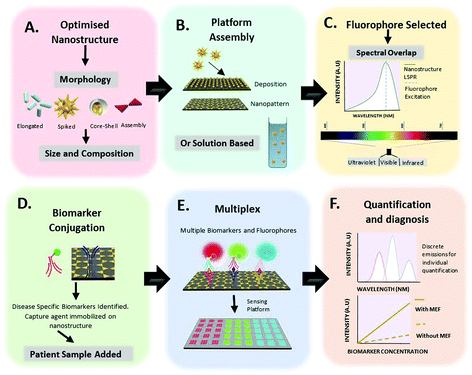
The authors have noticed that Fig. 1D in the originally published review article was missing a label for ‘Patient Sample Added’. The corrected version of Fig. 1 is therefore shown below.
The Royal Society of Chemistry apologises for these errors and any consequent inconvenience to authors and readers.
| This journal is © The Royal Society of Chemistry 2018 |

