
Nanofiber membrane supported lung-on-a-chip microdevice for anti-cancer drug testing†

Abstract
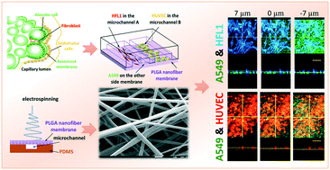
Organ-on-a-chip technology can simulate the physiological and pathological microenvironment of tissues and organs in vitro, thus offering the potential of dispensing with animal models to predict the toxicity and efficacy of therapies. In this study, taking the alveolar microenvironment as a model, we developed a lung-on-a-chip with a poly(lactic-co-glycolic acid) (PLGA) electrospinning nanofiber membrane as the chip substrate and cell scaffold. The PLGA nanofiber membrane, with a controlled thickness of ∼3 μm, is porous and permeable to molecules, has good biocompatibility, and offers a means to simulate the alveolar respiratory membrane. On the chip, we carried out cell culture and co-culture of human non-small cell lung cancer cells (A549) and human fetal lung fibroblasts (HFL1), and evaluated gefitinib, an epidermal growth factor receptor (EGFR)-targeted anti-tumor drug. We further probed the possible sources of A549 cell drug resistance in the presence of HFL1 cells. In addition, we co-cultured A549, HFL1, and human umbilical vein endothelial cells (HUVECs), and found that A549 cells could lead to endothelial cell apoptosis or death, and then the occurrence of tumor invasion. This established lung-on-a-chip is simple, effective, and easy to operate. It is expected to have important applications in personalized treatment of lung tumors and to play a potential role in other clinical treatments and tissue engineering.
- This article is part of the themed collections: In celebration of Chinese New Year 2020 and Organ-, body- and disease-on-a-chip systems
 Please wait while we load your content...

