Issue 51, 2018

Raman fingerprints of amyloid structures

Abstract

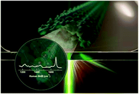
Structural differences in pathological and functional amyloid fibrils have been investigated by Raman microspectroscopy. Second-derivative analyses of amide-I and amide-III bands distinguish parallel in-register β-sheets from a β-solenoid. Further, spatially resolved Raman spectra reveal molecular heterogeneity in amyloid structures.

Graphical abstract: Raman fingerprints of amyloid structures

Supplementary files

Article information

Article type
Communication
Submitted
20 Apr 2018
Accepted
08 May 2018
First published
14 May 2018

Chem. Commun., 2018,54, 6983-6986

Raman fingerprints of amyloid structures

J. D. Flynn and J. C. Lee, Chem. Commun., 2018, 54, 6983 DOI: 10.1039/C8CC03217C

To request permission to reproduce material from this article, please go to the Copyright Clearance Center request page.

If you are an author contributing to an RSC publication, you do not need to request permission provided correct acknowledgement is given.

If you are the author of this article, you do not need to request permission to reproduce figures and diagrams provided correct acknowledgement is given. If you want to reproduce the whole article in a third-party publication (excluding your thesis/dissertation for which permission is not required) please go to the Copyright Clearance Center request page.

Read more about how to correctly acknowledge RSC content.

Social activity

Spotlight

Advertisements