 Open Access Article
Creative Commons Attribution 3.0 Unported Licence
Mass spectrometric detection of iron nitrosyls, sulfide oxidation and mycothiolation during nitrosylation of the NO sensor [4Fe–4S] NsrR†
Jason C.
Crack
a,
Chris J.
Hamilton
b and
Nick E.
Le Brun
 *a
*a
aCentre for Molecular and Structural Biochemistry, School of Chemistry, University of East Anglia, Norwich Research Park, Norwich, NR4 7TJ, UK. E-mail: n.le-brun@uea.ac.uk
bSchool of Pharmacy, University of East Anglia, Norwich Research Park, Norwich, NR4 7TJ, UK
First published on 23rd May 2018
Abstract
The bacterial nitric oxide (NO)-sensing transcriptional regulator NsrR binds a [4Fe–4S] cluster that enables DNA-binding and thus repression of the cell's NO stress response. Upon exposure to NO, the cluster undergoes a complex nitrosylation reaction resulting in a mixture of iron-nitrosyl species, which spectroscopic studies have indicated are similar to well characterized low molecular weight dinitrosyl iron complex (DNIC), Roussin's Red Ester (RRE) and Roussin's Black Salt (RBS). Here we report mass spectrometric studies that enable the unambiguous identification of NsrR-bound RRE-type species, including a persulfide bound form that results from the oxidation of cluster sulfide. In the presence of the low molecular weight thiols glutathione and mycothiol, glutathionylated and mycothiolated forms of NsrR were readily formed.
Proteins that contain iron–sulfur (Fe–S) clusters play key roles in an array of biochemical processes from respiration and photosynthesis to DNA replication. The clusters are generally fragile and are particularly reactive towards nitric oxide (NO),1 which is generated in macrophages during the initial immune response against invasion by microbes.2 Therefore, it is likely that Fe–S proteins are primary targets of NO at high (cytotoxic) concentrations, and perhaps also at lower (signalling) concentrations. Indeed, detection of paramagnetic dinitrosyl iron complexes (DNICs; [Fe(I)(NO)2(SR)2]− in which RS− are protein cysteinates) by EPR3–6 gave the first evidence of the in vivo reactivity of NO, and at least some of these species contained iron that was originally part of an Fe–S cluster.
Several bacterial NO sensor proteins, including NsrR and WhiB-like (Wbl) family proteins, contain an Fe–S cluster.1,7,8 NsrR, a [4Fe–4S] cluster-containing member of the Rrf2 family of transcriptional regulators, functions as part of the cell's response to nitrosative stress.9–12 Recently, the structure of the [4Fe–4S] cluster bound form of NsrR was solved, the first for any Fe–S cluster Rrf2 family member.13
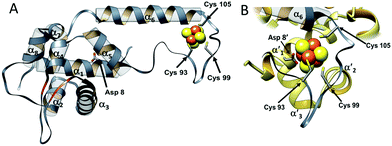
The overall structure of NsrR (Fig. 1) is similar to that of apo-IscR14,15 and CymR,16 consisting of an elongated protein fold with a DNA-binding domain comprising four α-helices and two anti-parallel β-strands (α1, α2, α3, β1, β2, α4), which includes a winged helix-turn-helix motif. This is connected to a dimerization helix (α6, α7) via a loop containing three Cys residues that are conserved in IscR and other Fe–S cluster containing Rrf2 proteins. The cluster is coordinated by the three conserved Cys residues (Cys93, 99 and 105) and Asp8 from the other subunit of the NsrR dimer. The [4Fe–4S] NsrR structure, along with that of apo-NsrR, revealed the structural basis for cluster-dependent DNA binding.13 A network of H-bonds connects the Cys-containing cluster binding loop with the DNA recognition helix of the other subunit; loss of the cluster results in a less ordered cluster loop and partial loss of the H-bond network, resulting in a relative shift of the recognition helix.
 | ||
| Fig. 1 The structure of S. coelicolor [4Fe–4S] NsrR. (A) Ribbon depiction of one of the subunits of the NsrR dimer. α-Helices are labelled (α1–α7) and strands β1 and β2 are shown in red. Fe and S atoms are shown in brown and yellow, respectively. The locations of the Cys ligands (residues 93, 99, and 105) are shown. (B) The [4Fe–4S] cluster binding loop in more detail. The fourth ligand, Asp8, originates from the α′1 helix of the opposite monomer (PDB: 5N07).13 | ||
In pathogens, resistance to host-generated nitrosative stress is essential in order to establish infection.17 Recent advances in purifying and handling sufficiently high concentrations of often extremely sensitive Fe–S proteins have facilitated the application of a range of biophysical techniques to study their reactions with NO. In some cases, the kinetics and thermodynamics of cluster nitrosylation have been investigated; S. coelicolor WhiD and NsrR,12,18 and Escherichia coli FNR19 have been shown to react rapidly with NO in a complex multi-step processes involving up to ∼8 NO molecules per cluster. These and other studies have revealed that paramagnetic DNIC species are, in general, not the major products of Fe–S cluster nitrosylation; instead, EPR silent multinuclear iron species that appear to be similar to the well known inorganic complexes Roussin's Red Ester (RRE; [Fe2(NO)4(SR)2]) and Roussin's Black Salt (RBS; [Fe4(NO)7(S)3])20 have been shown to be major products.18,19,21–23 In particular, recent studies of NsrR and WhiD using the iron-specific vibrational technique nuclear resonance vibrational spectroscopy (NRVS) revealed that nitrosylation results in a mixture of iron nitrosyl products, with RRE-like and RBS-like species as principal products, along with minor amounts of DNIC species.24
To gain further insight into the reaction of NsrR with NO, liquid chromatography electrospray ionisation mass spectrometry (LC-ESI-MS) was used to analyse [4Fe–4S] NsrR before and after addition of NO. The deconvoluted mass spectrum of NsrR, Fig. 2A, was dominated by the apo-NsrR peak at 15![[thin space (1/6-em)]](https://www.rsc.org/images/entities/char_2009.gif) 953 Da (theoretical mass of 15
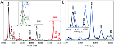
953 Da (theoretical mass of 15![[thin space (1/6-em)]](https://www.rsc.org/images/entities/char_2009.gif) 954 Da, Table S1, ESI†), as expected because the cluster cannot survive the acidic conditions of LC-MS sample preparation. Minor peaks at +32, +56 and occasionally +112 Da were also observed, corresponding to a low amount of a persulfide adduct, and one and two irons, respectively, most likely resulting from the loss of the cluster during sample preparation. Exposure to an excess of NO (8 NO per [4Fe–4S] cluster) prior to analysis by LC-MS resulted in a 2 Da decrease in the mass of the apo-protein to 15
954 Da, Table S1, ESI†), as expected because the cluster cannot survive the acidic conditions of LC-MS sample preparation. Minor peaks at +32, +56 and occasionally +112 Da were also observed, corresponding to a low amount of a persulfide adduct, and one and two irons, respectively, most likely resulting from the loss of the cluster during sample preparation. Exposure to an excess of NO (8 NO per [4Fe–4S] cluster) prior to analysis by LC-MS resulted in a 2 Da decrease in the mass of the apo-protein to 15![[thin space (1/6-em)]](https://www.rsc.org/images/entities/char_2009.gif) 951 Da, suggesting that NO-mediated oxidation of apo-NsrR, resulting in a disulfide bond, occurred upon reaction with NO (Fig. S1, ESI†). MS/MS analysis of digested NsrR revealed that the disulfide bond forms between Cys93 and Cys99 (Fig. S2, ESI†).
951 Da, suggesting that NO-mediated oxidation of apo-NsrR, resulting in a disulfide bond, occurred upon reaction with NO (Fig. S1, ESI†). MS/MS analysis of digested NsrR revealed that the disulfide bond forms between Cys93 and Cys99 (Fig. S2, ESI†).
In addition to this apo-protein mass shift, a number of additional peaks were observed at masses +232, +306, and +338 Da (relative to reduced apo-NsrR) upon reaction with NO, see Fig. 2A. Further low abundance peaks were also observed at +264 and +370 Da (Fig. 2B). The peak at +232 Da was consistent with the presence of an NsrR-bound RRE-type ([Fe2(NO)4]) species (Fig. 3A), while that at +306 Da corresponded to glutathionylated NsrR (NsrR covalently attached to a single glutathione, which was present in the protein sample buffer). Peaks at +338 and 370 Da could arise from glutathionylated NsrR with one and two additional sulfurs, respectively. The inclusion of glutathione was previously found to stabilize the solution against precipitation that occurred in the absence of a low molecular weight thiol at ratios of [NO]![[thin space (1/6-em)]](https://www.rsc.org/images/entities/char_2009.gif) :
:![[thin space (1/6-em)]](https://www.rsc.org/images/entities/char_2009.gif) [FeS] ≥ 2.12
[FeS] ≥ 2.12
To confirm these assignments and to help with those for other peaks, the reaction was repeated with 57Fe-substituted [4Fe–4S] NsrR and, separately, with 15NO. For the 57Fe NsrR experiment, the peak at +232 Da shifted to +234 Da, consistent with the two additional mass units associated with two 57Fe ions, see Fig. 2A. For 15NO, the peak at +232 Da shifted to +236 Da, consistent with the four additional mass units associated with four 15NO molecules (Fig. 2A and B). The peak at +264 Da was significantly increased in intensity and shifted to +268 Da in the 15NO spectrum, consistent with an RRE with one additional sulfur, i.e. a persulfide coordinated RRE, [[Fe2(NO)4](S)] (Fig. 3A). The peaks at +306, +338 and +370 Da were unaffected in the 15NO experiment (Fig. 2B), consistent with the absence of NO in glutathionylated NsrR containing zero, one and two additional sulfurs (i.e. NsrR-SG, NsrR-S-SG and NsrR-SS-SG), respectively.
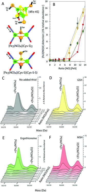
To further investigate the NO-mediated oxidation of NsrR and the formation of RRE and glutathionylated species, similar experiments were performed with increasing levels of NO, both in the absence and presence of GSH. In both cases, the mass of the apo-NsrR peak varied systematically with increasing NO, decreasing by 2 Da in the absence of GSH and by ∼1 Da in the presence, demonstrating that GSH protects NsrR from oxidation (Fig. S1, ESI†). As above, peaks due to RRE iron-nitrosyl adducts of NsrR were observed to form, but these remained at very low abundance until the ratio NO![[thin space (1/6-em)]](https://www.rsc.org/images/entities/char_2009.gif) :
:![[thin space (1/6-em)]](https://www.rsc.org/images/entities/char_2009.gif) [4Fe–4S] was >6 in the presence and absence of GSH (Fig. 3B–D). This indicated that RRE species are products of cluster nitrosylation in a non-concerted reaction.12
[4Fe–4S] was >6 in the presence and absence of GSH (Fig. 3B–D). This indicated that RRE species are products of cluster nitrosylation in a non-concerted reaction.12
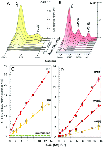
In the presence of GSH, glutathionylated NsrR was formed initially linearly with NO, suggesting that the formation of the mixed disulfide results from NO-dependent oxidation (Fig. 4A and C). The glutathionylated products with one or two additional sulfurs formed later in the titration, appearing only after the addition of ∼2 NO per cluster (Fig. 4A and D). The source of these additional sulfurs must be the bridging sulfides from the Fe–S cluster and therefore the data indicate that sulfide is not released from the cluster and oxidized until >2 NO per cluster are added. This is consistent with recent DNA-binding experiments showing that binding of [4Fe–4S] NsrR to the hmpA2 promoter (one of the three genes regulated by NsrR in S. coelicolor9) was abolished above a NO![[thin space (1/6-em)]](https://www.rsc.org/images/entities/char_2009.gif) :
:![[thin space (1/6-em)]](https://www.rsc.org/images/entities/char_2009.gif) [4Fe–4S] ratio of 2
[4Fe–4S] ratio of 2![[thin space (1/6-em)]](https://www.rsc.org/images/entities/char_2009.gif) :
:![[thin space (1/6-em)]](https://www.rsc.org/images/entities/char_2009.gif) 1.12
1.12
 | ||
| Fig. 4 Formation of GSH and MSH adducts of NsrR. (A) LC-MS spectra of [4Fe–4S] NsrR following additions of NO in (A) glutathione (GSH) and (B) mycothiol (MSH). (C and D) NO dependent formation of thiol adducts of apo-NsrR (as indicated) (C) and persulfide thiol adducts (D). Samples/data analyses as in Fig. 3. | ||
The cytoplasm of S. coelicolor does not contain GSH; instead, the low molecular weight thiols ergothioneine and mycothiol (MSH) are found, and so more physiologically relevant experiments were performed with ergothioneine and MSH.25,26 Like GSH, these protected NsrR from NO-mediated disulfide bond formation (Fig. S1C, ESI†). RRE iron-nitrosyl adducts of NsrR were observed, Fig. 3B, E and F, but these remained at very low abundance until the ratio NO![[thin space (1/6-em)]](https://www.rsc.org/images/entities/char_2009.gif) :
:![[thin space (1/6-em)]](https://www.rsc.org/images/entities/char_2009.gif) [4Fe–4S] was >8, indicating that ergothioneine/MSH inhibit the formation, or destabilize, NsrR-bound RREs. Mycothiolated NsrR was also observed, with the MSH adduct accumulating with increasing NO (Fig. 4B and C). The increase was more pronounced in the range 0–2 NO per cluster, compared to that above this ratio, and the extent of mycothiolation substantially greater than that observed for GSH. As for GSH, the formation of sulfur adducts of mycothiolated NsrR increased significantly above a ratio of 2 NO per cluster (Fig. 4D), again consistent with release/oxidation of cluster sulfide above this ratio. No covalent adducts were detected with ergothioneine (Fig. 4C). MS/MS analysis of digested NsrR revealed that GSH adducts are formed at Cys93 and Cys99 (see Fig. S3 and S4, ESI†), consistent with the structure of apo-NsrR, which indicates that these residues (but not Cys105) are surface exposed (Fig. S5, ESI†). Fragments containing Cys105 were not detected by MS/MS, so we cannot entirely rule out that GSH adducts are formed here also. Previous studies showed that nitrosylation of [4Fe–4S] NsrR in the presence of GSH resulted in complete loss of DNA binding,12 and so the function of thiol adducts is most likely the protection of Cys residues from NO-mediated oxidation of Cys93/Cys99 (Fig. S1 and S2, ESI†).
[4Fe–4S] was >8, indicating that ergothioneine/MSH inhibit the formation, or destabilize, NsrR-bound RREs. Mycothiolated NsrR was also observed, with the MSH adduct accumulating with increasing NO (Fig. 4B and C). The increase was more pronounced in the range 0–2 NO per cluster, compared to that above this ratio, and the extent of mycothiolation substantially greater than that observed for GSH. As for GSH, the formation of sulfur adducts of mycothiolated NsrR increased significantly above a ratio of 2 NO per cluster (Fig. 4D), again consistent with release/oxidation of cluster sulfide above this ratio. No covalent adducts were detected with ergothioneine (Fig. 4C). MS/MS analysis of digested NsrR revealed that GSH adducts are formed at Cys93 and Cys99 (see Fig. S3 and S4, ESI†), consistent with the structure of apo-NsrR, which indicates that these residues (but not Cys105) are surface exposed (Fig. S5, ESI†). Fragments containing Cys105 were not detected by MS/MS, so we cannot entirely rule out that GSH adducts are formed here also. Previous studies showed that nitrosylation of [4Fe–4S] NsrR in the presence of GSH resulted in complete loss of DNA binding,12 and so the function of thiol adducts is most likely the protection of Cys residues from NO-mediated oxidation of Cys93/Cys99 (Fig. S1 and S2, ESI†).
Recent studies of the reaction of NsrR with NO have provided clear evidence for the formation of RRE-like and RBS-like products, along with small amounts of DNIC species.24 That persulfide forms of these products might also be formed was postulated and DFT calculations supported this possibility. Along with the previous studies, the data presented here provide the first unequivocal proof that [Fe2(NO)4] and [[Fe2(NO)4](S)] adducts are formed following the NO reaction of NsrR. Persulfide adducts arise as a result of oxidation of cluster sulfide,18 and attachment of the resulting S0 species to Cys thiolate side chains, as observed following the O2 reaction of [4Fe–4S] FNR, the master anaerobic/aerobic metabolism regulator in E. coli.27,28 It is not clear why iron-nitrosyl species are able to remain bound to the protein under conditions of the LC-MS experiment, where non-covalent interactions are expected to be lost. Given that previous spectroscopic studies of these cluster nitrosylation reactions indicated that at least half the iron remained associated with the protein following gel filtration,18 a significant proportion of iron-nitrosyls must be lost under the conditions of the LC-MS experiment, as apo-protein is the most abundant species in the LC-MS spectra. Nevertheless, enough remains associated with the protein to permit detection and we suggest that this is a result of the relative chemical robustness of RRE compared to Fe–S clusters. RBS-type species, which were recently shown to be another major product of cluster nitrosylation in these proteins, apparently do not survive the conditions of the LC-MS experiment, possibly because any sulfide associated with such species would not survive the acidic conditions of sample preparation.24
We note that iron-nitrosyl species were previously observed by LC-MS attached to Clostridium botulinum HiPIP following reaction of the [4Fe–4S] cluster with NO.29 In that study, a peak at +232 Da was interpreted as arising from the association of two DNIC species. This was in part based on the observation of the typical DNIC S = ½ signal in the EPR spectrum. However, quantification of the EPR signal revealed sub-stoichiometric amounts of DNIC and so it is possible that both DNIC and RRE species were present. That a single DNIC adduct was not detected may suggest that the iron-nitrosyl detected was more likely to be the RRE species. Furthermore, in that study, additional peaks at +31–33 Da were also observed for apo-protein and iron-nitrosyl adducts, which were interpreted as representing nitrosated forms of the protein (predicted at +29–30 Da). Based on recent studies that have revealed the ease with which cluster sulfide is oxidized to form persulfide adducts18,19,28,30 it is perhaps more likely that these may represent persulfide (+32 Da) adducts. The iron-nitrosyl species observed here for NsrR are unlikely to be due to DNIC species because no single DNIC adduct was observed at +116 Da and it is known that DNIC species are only minor products of NsrR [4Fe–4S] cluster nitrosylation.12
In summary, here we have demonstrated the precise nature of the RRE-type species formed upon [4Fe–4S] cluster nitrosylation of NsrR as [Fe2(NO)4] and [[Fe2(NO)4](S)]. Reaction with NO also resulted in the formation of an oxidized (disulfide bonded) form of apo-NsrR, which was inhibited by the presence of low molecular weight thiols. The presence of GSH and MSH led to the formation of thiol adducts (representing glutathionylation and mycothiolation) at Cys93 and Cys99, while the presence of ergothioneine did not. Formation of thiol adducts most likely functions to protect Cys residues from NO-mediated oxidation that results in a disulfide bond between Cys93 and Cys99. Persulfide forms of GSH and MSH adducts, resulting from the release and oxidation of sulfide from the [4Fe–4S] cluster, were only observed to accumulate at significant levels at NO![[thin space (1/6-em)]](https://www.rsc.org/images/entities/char_2009.gif) :
:![[thin space (1/6-em)]](https://www.rsc.org/images/entities/char_2009.gif) [4Fe–4S] ratios >2, consistent with previous solution data showing the formation of a distinct intermediate at this ratio, and DNA-binding studies showing that NsrR dissociates from the hmpA2 promoter at the same ratio.12 This indicates that once two NO molecules are bound to the [4Fe–4S] core, the cluster begins to disassemble, leading to release of sulfide that can participate in further reactions.
[4Fe–4S] ratios >2, consistent with previous solution data showing the formation of a distinct intermediate at this ratio, and DNA-binding studies showing that NsrR dissociates from the hmpA2 promoter at the same ratio.12 This indicates that once two NO molecules are bound to the [4Fe–4S] core, the cluster begins to disassemble, leading to release of sulfide that can participate in further reactions.
This work was supported by Biotechnology and Biological Sciences Research Council Grant BB/P006140/1 to NLB & JCC, by UEA through the purchase of the mass spectrometer used in this work, and by the FeSBioNet COST Action CA15133. We thank Dr Gerhard Saalbach, John Innes Centre (Proteomics Facility), for assistance in acquiring and analysing MS/MS data.
Conflicts of interest
There are no conflicts to declare.Notes and references
- J. C. Crack, J. Green, A. J. Thomson and N. E. Le Brun, Acc. Chem. Res., 2014, 47, 3196–3205 CrossRef PubMed.
- R. Bruckdorfer, Mol. Aspects Med., 2005, 26, 3–31 CrossRef PubMed.
- A. F. Vanin, S. V. Kiladze and L. N. Kubrina, Biofizika, 1977, 22, 850–855 Search PubMed.
- J. C. Drapier, Methods, 1997, 11, 319–329 CrossRef PubMed.
- A. F. Vanin, Nitric oxide, 2009, 21, 1–13 CrossRef PubMed.
- S. Costanzo, S. Menage, R. Purrello, R. P. Bonomo and M. Fontecave, Inorg. Chim. Acta, 2001, 318, 1–7 CrossRef.
- R. K. Poole, Biochem. Soc. Trans., 2005, 33, 176–180 CrossRef PubMed.
- J. C. Crack, J. Green, A. J. Thomson and N. E. Le Brun, Curr. Opin. Chem. Biol., 2012, 16, 35–44 CrossRef PubMed.
- J. C. Crack, J. Munnoch, E. L. Dodd, F. Knowles, M. M. Al Bassam, S. Kamali, A. A. Holland, S. P. Cramer, C. J. Hamilton, M. K. Johnson, A. J. Thomson, M. I. Hutchings and N. E. Le Brun, J. Biol. Chem., 2015, 290, 12689–12704 CrossRef PubMed.
- N. P. Tucker, M. G. Hicks, T. A. Clarke, J. C. Crack, G. Chandra, N. E. Le Brun, R. Dixon and M. I. Hutchings, PLoS One, 2008, 3, e3623 Search PubMed.
- E. T. Yukl, M. A. Elbaz, M. M. Nakano and P. Moenne-Loccoz, Biochemistry, 2008, 47, 13084–13092 CrossRef PubMed.
- J. C. Crack, D. A. Svistunenko, J. Munnoch, A. J. Thomson, M. I. Hutchings and N. E. Le Brun, J. Biol. Chem., 2016, 291, 8663–8672 CrossRef PubMed.
- A. Volbeda, E. L. Dodd, C. Darnault, J. C. Crack, O. Renoux, M. I. Hutchings, N. E. Le Brun and J. C. Fontecilla-Camps, Nat. Commun., 2017, 8, 15052 CrossRef PubMed.
- S. Rajagopalan, S. J. Teter, P. H. Zwart, R. G. Brennan, K. J. Phillips and P. J. Kiley, Nat. Struct. Mol. Biol., 2013, 20, 740–747 Search PubMed.
- J. A. Santos, N. Alonso-Garcia, S. Macedo-Ribeiro and P. J. Pereira, Proc. Natl. Acad. Sci. U. S. A., 2014, 111, E2251–2260 CrossRef PubMed.
- W. Shepard, O. Soutourina, E. Courtois, P. England, A. Haouz and I. Martin-Verstraete, FEBS J., 2011, 278, 2689–2701 CrossRef PubMed.
- N. P. Tucker, N. E. Le Brun, R. Dixon and M. I. Hutchings, Trends Microbiol., 2010, 18, 149–156 CrossRef PubMed.
- J. C. Crack, L. J. Smith, M. R. Stapleton, J. Peck, N. J. Watmough, M. J. Buttner, R. S. Buxton, J. Green, V. S. Oganesyan, A. J. Thomson and N. E. Le Brun, J. Am. Chem. Soc., 2011, 133, 1112–1121 CrossRef PubMed.
- J. C. Crack, M. R. Stapleton, J. Green, A. J. Thomson and N. E. Le Brun, J. Biol. Chem., 2013, 288, 11492–11502 CrossRef PubMed.
- A. R. Butler, C. Glidewell and M. Li, Adv. Inorg. Chem., 1988, 32, 335–393 CrossRef.
- T. C. Harrop, Z. J. Tonzetich, E. Reisner and S. J. Lippard, J. Am. Chem. Soc., 2008, 130, 15602–15610 CrossRef PubMed.
- C. E. Tinberg, Z. J. Tonzetich, H. Wang, L. H. Do, Y. Yoda, S. P. Cramer and S. J. Lippard, J. Am. Chem. Soc., 2010, 132, 18168–18176 CrossRef PubMed.
- Z. J. Tonzetich, H. Wang, D. Mitra, C. E. Tinberg, L. H. Do, F. E. Jenney, Jr., M. W. Adams, S. P. Cramer and S. J. Lippard, J. Am. Chem. Soc., 2010, 132, 6914–6916 CrossRef PubMed.
- P. N. Serrano, H. Wang, J. C. Crack, C. Prior, M. I. Hutchings, A. J. Thomson, S. Kamali, Y. Yoda, J. Zhao, M. Y. Hu, E. E. Alp, V. S. Oganesyan, N. E. Le Brun and S. P. Cramer, Angew. Chem., 2016, 55, 14575–14579 CrossRef PubMed.
- D. S. Genghof and O. Van Damme, J. Bacteriol., 1968, 95, 340–344 Search PubMed.
- G. L. Newton, N. Buchmeier and R. C. Fahey, Microbiol. Mol. Biol. Rev., 2008, 72, 471–494 CrossRef PubMed.
- B. Zhang, J. C. Crack, S. Subramanian, J. Green, A. J. Thomson, N. E. Le Brun and M. K. Johnson, Proc. Natl. Acad. Sci. U. S. A., 2012, 109, 15734–15739 CrossRef PubMed.
- J. C. Crack, A. J. Thomson and N. E. Le Brun, Proc. Natl. Acad. Sci. U. S. A., 2017, 114, E3215–E3223 CrossRef PubMed.
- H. W. Foster and J. A. Cowan, J. Am. Chem. Soc., 1999, 121, 4093–4100 CrossRef.
- K. A. Johnson, M. F. J. M. Verhagen, P. S. Brereton, M. W. W. Adams and I. J. Amster, Anal. Chem., 2000, 72, 1410–1418 CrossRef PubMed.
Footnote |
| † Electronic supplementary information (ESI) available. See DOI: 10.1039/c8cc01339j |
| This journal is © The Royal Society of Chemistry 2018 |


