Non-natural amino acid peptide microarrays to discover Ebola virus glycoprotein ligands†
Joshua A.
Rabinowitz
 ,
John C.
Lainson
 ,
Stephen Albert
Johnston
and
Chris W.
Diehnelt
 *
*
Biodesign Institute Center for Innovations in Medicine, Arizona State University, Tempe, Arizona 85281, USA. E-mail: chris.diehnelt@asu.edu
First published on 3rd January 2018
Abstract
We demonstrate a platform to screen a virus pseudotyped with Ebola virus glycoprotein (GP) against a library of peptides that contain non-natural amino acids to develop GP affinity ligands. This system could be used for rapid development of peptide-based antivirals for other emerging or neglected tropical infectious diseases.
Ebola viruses and Marburg virus are a highly lethal group of filoviruses that are responsible for periodic outbreaks of hemorrhagic fever in Africa. There are five Ebola viruses (Ebola, Sudan, Reston, Bundibugyo, and Tai Forest) with Ebola Zaire (EBOV) and Sudan (SUDV) responsible for >70% of outbreaks and >90% of deaths.1,2 The EBOV outbreak in West Africa of 2014–2016 was the largest outbreak in history, killing over 11
![[thin space (1/6-em)]](https://www.rsc.org/images/entities/char_2009.gif) 000, and severely damaging healthcare systems of the affected countries. While there is no clinically approved therapeutic for EBOV, there are EBOV vaccines and therapeutics in clinical trials.2,3 Additionally, there are several monoclonal antibodies (mAbs) reported that target the EBOV glycoprotein (GP), including the ZMapp three mAb cocktail.4–9 While mAbs are promising candidates, many have low cross reactivity due to sequence variation of GPs from EBOV and SUDV10 although, cross-neutralizing mAbs were recently reported.11 A practical challenge for EBOV mAbs is that the resource limited settings of Ebola virus outbreaks pose challenges for both production cost and cold-chain, necessary for mAb therapeutics.9
000, and severely damaging healthcare systems of the affected countries. While there is no clinically approved therapeutic for EBOV, there are EBOV vaccines and therapeutics in clinical trials.2,3 Additionally, there are several monoclonal antibodies (mAbs) reported that target the EBOV glycoprotein (GP), including the ZMapp three mAb cocktail.4–9 While mAbs are promising candidates, many have low cross reactivity due to sequence variation of GPs from EBOV and SUDV10 although, cross-neutralizing mAbs were recently reported.11 A practical challenge for EBOV mAbs is that the resource limited settings of Ebola virus outbreaks pose challenges for both production cost and cold-chain, necessary for mAb therapeutics.9
In light of this ongoing need, others have reported peptides that target GP and block viral entry.12–14 However, peptide therapeutics composed of L-amino acids are sensitive to protease degradation and generally have poor in vivo stability. It is well known that incorporation of D-amino acids can increase stability but often in unpredictable ways. Many groups instead turn to discovery of D-amino acid containing peptides directly using techniques such as mirror-image phage display.15 In this method, a D-amino acid version of the target protein is screened against an L-amino acid phage display library to identify L-amino acid peptides, whose D-amino acid analogue binds to the L-amino acid protein target.15 This strategy has been successfully used for many years and has recently been demonstrated against PD-L116 and against the conserved N-trimer region of EBOV GP.14 However, this method is limited to small to medium sized protein targets due to the limitations of producing D-amino acid proteins through synthetic methods.
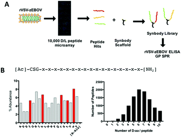
Our group has developed a peptide discovery system based on screening a protein, bacterial, or viral target against a peptide microarray to discover low affinity peptides that can be linked to produce high affinity bivalent peptides called synbodies.17–21 We have improved the peptide microarray platform through the production of a library of 10![[thin space (1/6-em)]](https://www.rsc.org/images/entities/char_2009.gif) 000 peptides of mixed L- and D-amino acid composition to enable the discovery of protease stabilized peptides in the primary screening step. This system is not limited to proteins of a particular size and in this work we show that it can be used to produce mixed L,D peptide ligands to the native EBOV GP through screening a vesicular stomatitis virus pseudotyped with EBOV Zaire GP (rVSV–zEBOV) against the mixed L,D peptide array. An added benefit of rVSV–zEBOV screening rather than using purified GP is that the replication competent rVSV–zEBOV preserves the secondary and tertiary structure of GP, increasing the probability that peptide hits will be functional in subsequent assays. Ten peptides containing D amino acids that bound rVSV–zEBOV in the peptide microarray screen were used to construct a library of synbodies through conjugation to a bi-functional peptide scaffold (Fig. 1A). The synbody library was screened for binding rVSV–zEBOV by ELISA and then screened by SPR against GP. Through this process, we identified multiple EBOV GP binding synbodies and characterized two with KD < 200 nM.
000 peptides of mixed L- and D-amino acid composition to enable the discovery of protease stabilized peptides in the primary screening step. This system is not limited to proteins of a particular size and in this work we show that it can be used to produce mixed L,D peptide ligands to the native EBOV GP through screening a vesicular stomatitis virus pseudotyped with EBOV Zaire GP (rVSV–zEBOV) against the mixed L,D peptide array. An added benefit of rVSV–zEBOV screening rather than using purified GP is that the replication competent rVSV–zEBOV preserves the secondary and tertiary structure of GP, increasing the probability that peptide hits will be functional in subsequent assays. Ten peptides containing D amino acids that bound rVSV–zEBOV in the peptide microarray screen were used to construct a library of synbodies through conjugation to a bi-functional peptide scaffold (Fig. 1A). The synbody library was screened for binding rVSV–zEBOV by ELISA and then screened by SPR against GP. Through this process, we identified multiple EBOV GP binding synbodies and characterized two with KD < 200 nM.
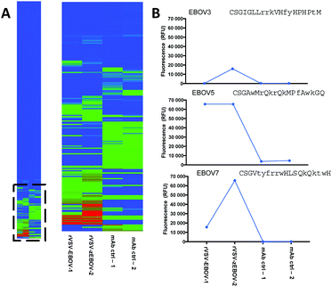
While live viruses can be screened against peptide microarrays to discover binding peptides,20 EBOV is a high biosafety pathogen and cannot be handled outside BSL4 level laboratories. However, the rVSV–zEBOV pseudotyped virus is antigenically similar to EBOV, has been demonstrated effective in producing neutralizing immunity when used as a vaccine3 and is compatible with BSL2 laboratories. Therefore, we used rVSV–zEBOV to select EBOV GP binding peptides. In this assay, rVSV–EBOV was screened at 106 PFU per mL against a peptide microarray containing 10![[thin space (1/6-em)]](https://www.rsc.org/images/entities/char_2009.gif) 000 random sequence 20-aa peptides. Peptides were composed of 17 variable positions and used 19 amino acids excluding Cys. Peptide sequences were produced using a random number generator with Arg, Lys, Trp, Phe, and Thr amino acids substituted with their D-entantiomers along with 10 peptides that contained β-Ala (Fig. 1B). This resulted in each peptide containing from 1 to 10 D-amino acids and with net charges that ranged from −5 to +15 with the median charge of +1 (Fig. 1B and Fig. S1, ESI†). Peptides were immobilized via thiol–maleimide conjugation with an N-terminal CSG linker. Binding to rVSV–zEBOV was detected using the ZMapp therapeutic mAb, 2G4.7 The mAb was incubated at 1 nM with rVSV–zEBOV along with 1 nM of a fluorescently labeled secondary antibody. In this way, the virus was effectively fluorescently labeled prior to incubation on the peptide microarray. A negative control composed of 2G4 with secondary antibody was also screened to eliminate antibody binding peptides. Hierarchical clustering was used to identify groups of peptides with distinct binding patterns across replicate arrays (Fig. 2A). The majority of the peptides do not bind the virus, however a number demonstrate high binding for rVSV–zEBOV and low mAb only binding. Analysis of the clusters demonstrate peptides with a number of binding profiles, with representative clusters in Fig. 2B (Fig. S2, ESI†). We chose peptides from different clusters (Fig. 2B, Fig. S3 and Table S1, ESI†) to use for synbody construction in order to increase the likelihood that the peptides would bind different sites on GP. These peptides were synthesized at the 10 mg scale with >90% purity by a vendor and used for synbody library production.
000 random sequence 20-aa peptides. Peptides were composed of 17 variable positions and used 19 amino acids excluding Cys. Peptide sequences were produced using a random number generator with Arg, Lys, Trp, Phe, and Thr amino acids substituted with their D-entantiomers along with 10 peptides that contained β-Ala (Fig. 1B). This resulted in each peptide containing from 1 to 10 D-amino acids and with net charges that ranged from −5 to +15 with the median charge of +1 (Fig. 1B and Fig. S1, ESI†). Peptides were immobilized via thiol–maleimide conjugation with an N-terminal CSG linker. Binding to rVSV–zEBOV was detected using the ZMapp therapeutic mAb, 2G4.7 The mAb was incubated at 1 nM with rVSV–zEBOV along with 1 nM of a fluorescently labeled secondary antibody. In this way, the virus was effectively fluorescently labeled prior to incubation on the peptide microarray. A negative control composed of 2G4 with secondary antibody was also screened to eliminate antibody binding peptides. Hierarchical clustering was used to identify groups of peptides with distinct binding patterns across replicate arrays (Fig. 2A). The majority of the peptides do not bind the virus, however a number demonstrate high binding for rVSV–zEBOV and low mAb only binding. Analysis of the clusters demonstrate peptides with a number of binding profiles, with representative clusters in Fig. 2B (Fig. S2, ESI†). We chose peptides from different clusters (Fig. 2B, Fig. S3 and Table S1, ESI†) to use for synbody construction in order to increase the likelihood that the peptides would bind different sites on GP. These peptides were synthesized at the 10 mg scale with >90% purity by a vendor and used for synbody library production.
To produce a library of synbodies to screen for binding, we used an approach where two peptides (A and B) are incubated with a bifunctional scaffold to produce a reaction mixture containing A–A, B–B, A–B, and B–A synbodies.20–22 The isomeric hetero-bivalent synbodies were not separated from one another, therefore we considered them as a single synbody (Fig. S3, ESI†). Synbodies were prepared through conjugation of purified peptides containing N-terminal Cys to the maleimide groups on the scaffold. We used the Sc0 scaffold,20,22 which contains a biotin-labeled Lys for streptavidin-based detection, for synbody construction. The synbody reactions were incubated overnight, purified by HPLC and the presence of each synbody was confirmed by MALDI. Using this scheme, we successfully produced 50 of 55 possible synbodies with an average yield of 0.5 mg for each hetero-bivalent synbody and 5 mg for each homo-bivalent synbody (Fig. S4, ESI†).
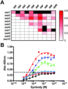
As a primary screen to select synbodies, we measured the relative binding of each synbody to rVSV–zEBOV and norovirus virus-like particle (VLP) coated wells via ELISA assay. Binding to the norovirus VLP was used as a negative control to filter out synbodies with poor specificity or those that bound the ELISA plate. Wells were coated with 105 PFU per well of rVSV–zEBOV and negative control wells were coated with 0.8 μg per well of norovirus VLP. Synbodies were incubated at 100 nM in duplicate wells and synbody binding was detected via streptavidin conjugated horseradish peroxidase (SA-HRP). A heat map of the differential binding as a function of the peptide components of each synbody is shown in Fig. 3A (Fig. S5, ESI†). From this data, a trend emerges in which synbodies composed of peptide 3, 5, or 7 have the highest differential binding while those containing peptide 9 or 10 had little to no differential binding. Synbodies with the highest differential binding (1–5, 2–3, 2–5, 2–7, 3–5, 3–7, 5–7, 7–7) were then tested across a range of concentrations (Fig. 3B). The assay format was the same as before, but with each synbody ranging in concentration from 1 nM to 1 μM. A mAb that targets GP, 13F6, was used as a positive control at 10 nM. Interestingly, the maximum binding of one group of synbodies (1–5, 2–3, 2–5) appears to saturate at a lower level than the synbodies containing peptide 7. It is unclear whether this is due to a limited number of binding sites or if peptide 7 binds multiple sites on rVSV–EBOV causing an increase in the maximum binding level above that of the 13F6 control. Regardless, these data demonstrate that EBOV synbodies bind rVSV–zEBOV in a concentration dependent manner.
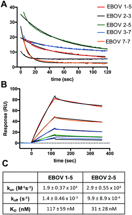
We then screened several rVSV–zEBOV binding synbodies against purified GP using surface plasmon resonance (SPR) to confirm that they bound GP rather than some other viral component. In this assay, synbodies 1–10, 1–5, 2–3, 2–5, 3–7 and 7–7 were injected over immobilized GP using a Biosensing Instruments 4500 Series SPR system. A carboxymethyldextran chip was immobilized with GP and synbodies were injected over the surface at 10 μM. Synbodies 2–3 and 1–10 had minimal binding while 1–5, 2–5, 3–7, and 7–7 bound immobilized GP (Fig. S6, ESI†). Due to the assay design, equilibrium dissociation constants (KD) were not determined, but the dissociation rates (koff) were estimated from the sensorgram dissociation phase (Fig. 4A). Synbodies 2–5, 1–5, and 3–7 had slower dissociation rates which were confirmed when these synbodies were screened a second time on a different immobilized GP surface using a Biacore T-200 SPR (Fig. S7, ESI†). We then characterized the binding affinity and kinetics of synbodies 1–5 and 2–5 by SPR to evaluate if there were any affinity differences of the synbodies that contained peptide 5. In this assay, each synbody was captured on either a streptavidin (SA chip) or a neutravidin (NA) coated CM5 chip and GP was injected over the captured synbody. EBOV 1–5 was captured on a SA chip and increasing concentrations of GP were injected at 30 μL min−1 over the surface. The sensorgrams were then fit to a 1![[thin space (1/6-em)]](https://www.rsc.org/images/entities/char_2009.gif) :
:![[thin space (1/6-em)]](https://www.rsc.org/images/entities/char_2009.gif) 1 binding model to determine the binding kinetics and KD (Fig. 4B). A similar analysis was performed on EBOV 2–5, which was captured on a NA chip, and replicate runs of GP over each synbody yielded a KD of 31 nM for EBOV 2–5 and 117 nM for EBOV 1–5 (Fig. 4C). These data indicate that D-amino acid peptides can be combined to produce high affinity synbodies with binding kinetics that suggest that these synbodies could function as EBOV GP inhibitors.
1 binding model to determine the binding kinetics and KD (Fig. 4B). A similar analysis was performed on EBOV 2–5, which was captured on a NA chip, and replicate runs of GP over each synbody yielded a KD of 31 nM for EBOV 2–5 and 117 nM for EBOV 1–5 (Fig. 4C). These data indicate that D-amino acid peptides can be combined to produce high affinity synbodies with binding kinetics that suggest that these synbodies could function as EBOV GP inhibitors.
In conclusion, we screened a pseudotyped version of EBOV, rVSV–zEBOV, on a peptide microarray to select D-amino acid containing peptide binders for EBOV GP. We then constructed a library of synbodies through conjugation of two peptides to a bivalent peptide scaffold and identified multiple synbodies that bound rVSV–zEBOV by ELISA. Further characterization demonstrated that several of the synbodies were specific binders to GP. This work demonstrates that peptides composed of both D- and L-amino acids can be easily discovered and combined to produce synbodies with KD < 200 nM for a large viral glycoprotein. Synbody binding affinity can be further improved if necessary by improving the peptide binding affinity prior to synbody conjugation.20,21,23 While the synbody scaffold used in this work undergoes thiol-exchange reactions due to the use of maleimide conjugation,22 there are other thiol-conjugation approaches that can be used to construct protease stable synbodies.24 Peptide microarray technologies are advancing rapidly and there are now in situ synthetic arrays with libraries of hundreds of thousands of random peptides.25 These methods can easily incorporate non-natural amino acids into peptides, greatly expanding the capability of discovering peptide binders containing non-natural amino acids in the primary screen. While the library sizes accessible with peptide microarrays are orders of magnitude smaller than those of display techniques, the ability to screen large proteins or whole viruses to quickly discover protease stable peptide ligands that can be further optimized is a powerful new technique for peptide drug discovery against targets as large and complex as viral glycoproteins.
We thank V. Gellar for her contribution to this work, B. Jacobs and K. Kiebler for providing rVSV–EBOV, C. Arntzen and H. Mason for providing 13F6, and L. Zeitlin from Mapp Biopharmaceutical, Inc. for providing 2G4. We thank B. Bratcher from Kentucky BioProcessing for providing the norovirus VLP. We thank A. Ueki and T. Jing (Biosensing Instruments) for the use of the BIA4500 SPR system. This work was funded by a grant from the Biodesign Institute at ASU to C. W. D., internal funds to C. W. D., from Barrett, The Honors College thesis funding to J. R., and by the Defense Advanced Research Projects Agency (W911NF-10-0299) to S. A. J.
Conflicts of interest
There are no conflicts to declare.Notes and references
- Center for Disease Control and Prevention Outbreaks Chronology: Ebola Virus Disease, http://www.cdc.gov/vhf/ebola/outbreaks/history/chronology.html.
- T. Zhang, M. Zhai, J. Ji, J. Zhang, Y. Tian and X. Liu, Bioorg. Med. Chem. Lett., 2017, 27, 2364–2368 CrossRef CAS PubMed.
- A. M. Henao-Restrepo, A. Camacho, I. M. Longini, C. H. Watson, W. J. Edmunds, M. Egger, M. W. Carroll, N. E. Dean, I. Diatta, M. Doumbia, B. Draguez, S. Duraffour, G. Enwere, R. Grais, S. Gunther, P.-S. Gsell, S. Hossmann, S. V. Watle, M. K. Kondé, S. Kéïta, S. Kone, E. Kuisma, M. M. Levine, S. Mandal, T. Mauget, G. Norheim, X. Riveros, A. Soumah, S. Trelle, A. S. Vicari, J.-A. Røttingen and M.-P. Kieny, Lancet, 2017, 389, 505–518 CrossRef CAS.
- D. Corti, J. Misasi, S. Mulangu, D. A. Stanley, M. Kanekiyo, S. Wollen, A. Ploquin, N. A. Doria-Rose, R. P. Staupe, M. Bailey, W. Shi, M. Choe, H. Marcus, E. A. Thompson, A. Cagigi, C. Silacci, B. Fernandez-Rodriguez, L. Perez, F. Sallusto, F. Vanzetta, G. Agatic, E. Cameroni, N. Kisalu, I. Gordon, J. E. Ledgerwood, J. R. Mascola, B. S. Graham, J.-J. Muyembe-Tamfun, J. C. Trefry, A. Lanzavecchia and N. J. Sullivan, Science, 2016, 351, 1339–1342 CrossRef CAS PubMed.
- Z. A. Bornholdt, H. L. Turner, C. D. Murin, W. Li, D. Sok, C. A. Souders, A. E. Piper, A. Goff, J. D. Shamblin, S. E. Wollen, T. R. Sprague, M. L. Fusco, K. B. J. Pommert, L. A. Cavacini, H. L. Smith, M. Klempner, K. A. Reimann, E. Krauland, T. U. Gerngross, D. K. Wittrup, E. O. Saphire, D. R. Burton, P. J. Glass, A. B. Ward and L. M. Walker, Science, 2016, 351, 1078–1083 CrossRef CAS PubMed.
- B. Wang, C. A. Kluwe, O. I. Lungu, B. J. DeKosky, S. A. Kerr, E. L. Johnson, J. Jung, A. B. Rezigh, S. M. Carroll, A. N. Reyes, J. R. Bentz, I. Villanueva, A. L. Altman, R. A. Davey, A. D. Ellington and G. Georgiou, Sci. Rep., 2015, 5, 13926 CrossRef PubMed.
- C. D. Murin, M. L. Fusco, Z. A. Bornholdt, X. Qiu, G. G. Olinger, L. Zeitlin, G. P. Kobinger, A. B. Ward and E. O. Saphire, Proc. Natl. Acad. Sci. U. S. A., 2014, 111, 17182–17187 CrossRef CAS PubMed.
- J. Audet, G. Wong, H. Wang, G. Lu, G. F. Gao, G. Kobinger and X. Qiu, Sci. Rep., 2014, 4, 6881 CrossRef CAS PubMed.
- X. G. Qiu, G. Wong, J. Audet, A. Bello, L. Fernando, J. B. Alimonti, H. Fausther-Bovendo, H. Y. Wei, J. Aviles, E. Hiatt, A. Johnson, J. Morton, K. Swope, O. Bohorov, N. Bohorova, C. Goodman, D. Kim, M. H. Pauly, J. Velasco, J. Pettitt, G. G. Olinger, K. Whaley, B. L. Xu, J. E. Strong, L. Zeitlin and G. P. Kobinger, Nature, 2014, 514, 47–53 CAS.
- J. Jin and G. Simmons, Clin. Vaccine Immunol., 2016, 23, 535–539 CrossRef CAS PubMed.
- A. Z. Wec, A. S. Herbert, C. D. Murin, E. K. Nyakatura, D. M. Abelson, J. M. Fels, S. He, R. M. James, M.-A. de La Vega, W. Zhu, R. R. Bakken, E. Goodwin, H. L. Turner, R. K. Jangra, L. Zeitlin, X. Qiu, J. R. Lai, L. M. Walker, A. B. Ward, J. M. Dye, K. Chandran and Z. A. Bornholdt, Cell, 2017, 169, 878–890 CrossRef CAS PubMed.
- C. D. Higgins, J. F. Koellhoffer, K. Chandran and J. R. Lai, Bioorg. Med. Chem. Lett., 2013, 23, 5356–5360 CrossRef CAS PubMed.
- E. K. Nyakatura, J. C. Frei and J. R. Lai, ACS Infect. Dis., 2015, 1, 42–52 CrossRef CAS PubMed.
- T. R. Clinton, M. T. Weinstock, M. T. Jacobsen, N. Szabo-Fresnais, M. J. Pandya, F. G. Whitby, A. S. Herbert, L. I. Prugar, R. McKinnon, C. P. Hill, B. D. Welch, J. M. Dye, D. M. Eckert and M. S. Kay, Protein Sci., 2015, 24, 446–463 CrossRef CAS PubMed.
- T. N. M. Schumacher, L. M. Mayr, D. L. Minor, M. A. Milhollen, M. W. Burgess and P. S. Kim, Science, 1996, 271, 1854–1857 CAS.
- H.-N. Chang, B.-Y. Liu, Y.-K. Qi, Y. Zhou, Y.-P. Chen, K.-M. Pan, W.-W. Li, X.-M. Zhou, W.-W. Ma, C.-Y. Fu, Y.-M. Qi, L. Liu and Y.-F. Gao, Angew. Chem., Int. Ed., 2015, 54, 11760–11764 CrossRef CAS PubMed.
- B. A. Williams, C. W. Diehnelt, P. Belcher, M. Greving, N. W. Woodbury, S. A. Johnston and J. C. Chaput, J. Am. Chem. Soc., 2009, 131, 17233–17241 CrossRef CAS PubMed.
- N. Gupta, P. E. Belcher, S. A. Johnston and C. W. Diehnelt, Bioconjugate Chem., 2011, 22, 1473–1478 CrossRef CAS PubMed.
- V. Domenyuk, A. Loskutov, S. A. Johnston and C. W. Diehnelt, PLoS One, 2013, 8, e54162 CAS.
- N. Gupta, J. Lainson, V. Domenyuk, Z.-G. Zhao, S. A. Johnston and C. W. Diehnelt, Bioconjugate Chem., 2016, 27, 2505–2512 CrossRef CAS PubMed.
- N. L. Gupta, J. C. Lainson, P. E. Belcher, L. Shen, H. S. Mason, S. A. Johnston and C. W. Diehnelt, Anal. Chem., 2017, 89, 7174–7181 CrossRef CAS PubMed.
- J. C. Lainson, M. F. Fuenmayor, S. A. Johnston and C. W. Diehnelt, Bioconjugate Chem., 2015, 26, 2125–2132 CrossRef CAS PubMed.
- M. P. Greving, P. E. Belcher, C. W. Diehnelt, M. J. Gonzalez-Moa, J. Emery, J. Fu, S. A. Johnston and N. W. Woodbury, PLoS One, 2010, 5, e15432 Search PubMed.
- S. C. Alley, D. R. Benjamin, S. C. Jeffrey, N. M. Okeley, D. L. Meyer, R. J. Sanderson and P. D. Senter, Bioconjugate Chem., 2008, 19, 759–765 CrossRef CAS PubMed.
- J. B. Legutki, Z.-G. Zhao, M. Greving, N. Woodbury, S. A. Johnston and P. Stafford, Nat. Commun., 2014, 5, 4785 CrossRef CAS PubMed.
Footnote |
| † Electronic supplementary information (ESI) available. See DOI: 10.1039/c7cc08242h |
| This journal is © The Royal Society of Chemistry 2018 |


