Cellular internalization of rod-like nano hydroxyapatite particles and their size and dose-dependent effects on pre-osteoblasts
Abstract
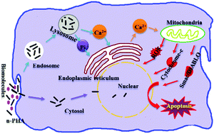
Nano hydroxyapatite particles (n-HA) have been reported to promote osteogenic activities of bone-related cells, while inhibiting tumor cell growth, and the biological effects of n-HA are related with the particle size, dose, culture time and cell type. In this work, we prepared n-HA with a strictly controlled rod-like shape and adjustable sizes without any surface chemical contaminations. Using the prepared n-HA, we investigated the size and dose effect of the nano particles on pre-osteoblasts for up to 7 days. We probed cell proliferation and gene expression in the presence of n-HA, the cellular uptake pathways of n-HA particles, as well as the extracellular and intracellular [Ca2+] ([Ca2+]i) changes caused by the particles, in order to get a better understanding of the biological effects of n-HA of various sizes. The n-HA exhibited size- and dose-dependent impacts on MC3T3-E1 proliferation, intracellular reactive oxygen species (ROS) generation, mitochondrial membrane potential, and osteogenic gene expression. 40 nm n-HA caused the slowest MC3T3-E1 growth, the highest intracellular ROS concentration, the largest mitochondrial membrane potential loss and the lowest level of osteogenic gene expression among the samples. The cytotoxicity of 40 nm n-HA increased with the dose and culture time. 70 nm n-HA showed beneficial effects on MC3T3-E1 growth, but the positive effect disappeared at the highest concentration on day 7. 100 nm n-HA promoted cell growth and the promoting effect increased with the dose. Cells cultured with 100 nm n-HA expressed the highest level of osteogenic gene expression among the experimental groups. We discovered that the presence of n-HA increased [Ca2+]i but did not elevate extracellular [Ca2+]. The [Ca2+]i increased as the n-HA size decreased. We also found that n-HA may enter cells through two pathways and that the amount of engulfed particles depended on the particle size. The internalized n-HA particles located in the cytosol, endosomes, lysosomes and nuclei. The particles dissolved in lysosomes and raised [Ca2+]i, which correlated with the cell death and osteogenic gene expression. In conclusion, the particle size, dose, and culture time influenced the biological effects of n-HA on ME3T3-E1 cells, probably by changing the [Ca2+]i in the cells instead of the extracellular [Ca2+].


 Please wait while we load your content...