
 Open Access Article
 This Open Access Article is licensed under a
 Creative Commons Attribution 3.0 Unported Licence
Stem cell therapies for ischemic stroke: current animal models, clinical trials and biomaterials
Hugh H. Chan
ab,
Connor A. Wathen
c,
Ming Ni
*a and
Shuangmu Zhuo
 *a
*a
aKey Laboratory of OptoElectronic Science and Technology for Medicine of Ministry of Education & Fujian Provincial Key Laboratory of Photonics Technology, Fujian Normal University, Fuzhou 350007, P. R. China. E-mail: shuangmuzhuo@gmail.com; mingni.sg@gmail.com
bDepartment of Neuroscience, Lerner Research Institute, Cleveland Clinic, Cleveland, Ohio, USA
cCenter for Neurological Restoration, Cleveland Clinic, Cleveland, Ohio, USA
First published on 28th March 2017
Abstract
Ischemic stroke is one of the most common causes of death. A brain that has suffered a stroke is significantly damaged due to the transient interruption of blood supply. Stroke survivors suffer from stroke-induced cognitive, motor and psychiatric problems that require increased use of the health care system, which results in a very poor quality of life and a heavy emotional and financial burden for patients and their families. Although rehabilitation can ameliorate some behavioral abnormalities, it does not significantly improve stroke symptoms. Neuroregeneration may be the principle process for replenishing of lost brain functions. Stem cell therapy is a novel approach to restore neurological function following ischemic stroke. Our goal in this review is to first briefly introduce the pathophysiology of stroke then document the progress of stem cell research, including clinical trials, towards the treatment of ischemic stroke. We highlight the promising applications of biomaterials to enhance the therapeutic effect of stem cell therapy in stroke treatment. As biomaterials provide efficient scaffolds for the growth and differentiation of stem cells, they may guarantee the quality and yield of stem cells for grafting. Furthermore, biomaterials also provide a convenient method for delivering the stem cells to be transplanted. This review provides direction for future research to combine tissue engineering approaches with stem cell therapy for the treatment of stroke.
 Hugh H Chan | Hugh H Chan received his PhD from Imperial College London in 2010. He is currently a postdoctoral fellow at the Neurosciences Department of the Lerner Research Institute of Cleveland Clinic. His current research focuses on rehabilitation and repair in experimental stroke models. |
 Connor A Wathen | Connor A Wathen received his BS from the University of Notre Dame in 2013. He is currently a medical student at the Cleveland Clinic Lerner College of Medicine of the Case Western Reserve University and will receive his MD in 2018. His current research focuses on deep cerebellar stimulation to improve post-stroke rehabilitation. |
 Ming Ni | Ming Ni received his Ph.D. in Chemical Engineering from the University of Washington in 2004. He is currently a research scientist at the Institute of Bioengineering and Nanotechnology, Agency for Science, Technology and Research in Singapore. His current research focuses on biomaterials and nanomaterials and their applications as probes for nonlinear optical microscopy and super high resolution microscopy. He is also interested in stem cell therapy and drug delivery. |
 Shuangmu Zhuo | Shuangmu Zhuo received his Ph.D. degree in Optics Engineering from the Fujian Normal University, China, in 2012. He then joined the Singapore-MIT Alliance for Research and Technology as a Postdoctoral Research Fellow. He is currently a Professor at the College of Photonic and Electronic Engineering, Fujian Normal University, China. His research interests include the development and applications of nonlinear optical microscopy in biological and biomedical research. |
1. Introduction
Stroke is one of the most common causes of death in developed countries,1 and occurs primarily in the elderly population with a higher risk in males.2 Ischemic stroke occurs when any of the many arteries perfusing the brain is occluded, resulting in anoxic damage to the portion of the brain supplied by the occluded artery.3 Even if there is reperfusion of the ischemic tissue, the presence of highly oxygenated blood allows for the production of reactive oxygen species which results in oxidative damage.4 The combination of ischemic and oxidative damage leads to neuronal death, including apoptosis,5 and loss of neural function. Because the location of the stroke is determined by the vessel that is occluded and frequently spans several functional domains, the resulting symptoms are usually a combination of motor, cognitive and psychiatric deficits.6–11Although there are emerging treatments that restore perfusion to the ischemic brain, including the administration of tissue plasminogen activator to dissolve thrombi and emboli as well as intra-arterial/endovascular procedures to re-canalize blood vessels,12,13 these types of treatment primarily serve to reduce stroke mortality. As neurological injury due to ischemia is largely irreversible, it is necessary to develop novel therapeutic approaches to restore lost neurological functions through the regeneration of neurons.14 A few studies that have provided evidence on the therapeutic effect of transplantation of different types of stem cells have been documented, in which the stroke symptoms were relieved and the lesion volumes were reduced.15,16 These studies suggested that the transplantation of stem cells could be a promising approach for the treatment of stroke. Currently, however, there are still problems associated with stem cell therapies for stroke. In this review, we will highlight the obstacles encountered during the development of such therapies. Transplanted stem cells often struggle to survive after injection, and biomaterials provide a perfect solution as scaffolds/delivery vehicles for cells to safely arrive at the injured sites. We suggest and discuss the applications of biomaterials to stem cell therapy in the treatment of stroke, which may provide a better understanding of the direction of research in the field of stem cell therapy for stroke.
2. Stem cell research in animal models
Although there is a small degree of spontaneous cellular regeneration following ischemic stroke, the amount of regeneration is not sufficient for functional neurological restoration. The administration of exogenous stem cells has the potential to overcome this limitation and provide neurological restoration in the compromised brain following stroke. The transplantation of stem cells from different sources has been evaluated in order to determine their efficacy and safety in the treatment of stroke. The goal of therapy is to develop stem cell therapy for (1) restoring compromised behavioral deficits and (2) restructuring the damaged brain regions. There are five primary stem cell types that are the focus of the development of stem cell therapy of stroke, namely embryonic stem cells (ESCs), neural stem cells (NSCs), mesenchymal stem cells (MSCs), hematopoietic stem cells (HSCs) and induced pluripotent stem cells (iPSCs). Table 1 summarizes the usage of different types of stem cells in stem cell therapy research for ischemic stroke.| Stem cell | Sources/example | Disadvantages | Advantages | |||
|---|---|---|---|---|---|---|
| Immune reaction | Tumorigenesis | Proliferativity | Differentiability | Efficacy | ||
| a When the MSCs and iPSCs are derived from the recipients’ somatic cells, there is hypothetically no immune response. | ||||||
| ESC32–37 | Fetus | ++ | +++ | +++ | +++ | ++ |
| NSC32,34,38,39 | Fetus | + | + | ++ | + | ++ |
| MSC15,40–50,55 | e.g. bone marrow | +a | + | ++ | ++ | ++ |
| HSC56–58 | + | ++ | + | ++ | ||
| iPSC59–61 | e.g. fibroblast | +a | + | ++ | +++ | + |
2.1 Endogenous stem cells
Although stroke induces brain tissue death, as compensation, there is a small degree of new cell formation, particularly neurons, after stroke.17 These endogenous stem cells, however, are not sufficient for recovery. In an experimental stroke model in rodents, young rats were shown to exhibit a higher level of new neuron formation, termed neurogenesis, than that in aged rats.18 To support neurogenesis after stroke, new blood vessels also form in order to reconstruct the compromised circulation, a process known as angiogenesis.19,20 Neurogenesis and angiogenesis are more prevalent in young brains, particularly at the postnatal to adolescent stages. Although most strokes occur in older individuals, limited degrees of neurogenesis and angiogenesis are still detected in adult brains.21,22 From the studies of rodents, the neurogenesis triggered by stroke occurs in the subventricular zone (SVZ) and subgranular zone (SGZ).23,24 Stem cells or neural progenitor cells (NPCs) divide and differentiate in the SVZ adjacent to the lateral ventricles.25 Guided by chemokines such as vascular endothelial growth factors, stem cell factor, and monocyte chemoattractant protein 1, NPCs migrate to the penumbra area that surrounds the ischemic lesion.26 NPCs are characterized by certain markers such as doublecortin, nestin, Sox2, and others.27,28 NPCs derived from the SGZ divide and develop into mature neurons then migrate to the hippocampus and other parts of the limbic system.29 Angiogenesis is primarily characterized by the formation of endothelial cells. New blood vessels are formed from pre-existing blood vessels, and elongate to access the sites at which neurogenesis occurs.30 De novo blood vessels can also form without the aid of pre-existing blood vessels, a process known as vasculogenesis.312.2 Exogenous stem cells
The therapeutic effect of bone marrow mononuclear cells (BMMNCs) in stroke has also been frequently studied in rodent models of ischemic stroke. In a rat MCAO model, transplantation of BMMNCs at the early stages of stroke reduced infarct volume, oxidative damage, and neuroinflammation, and the effects were enhanced by valproic acid.49 Furthermore, 5-fluorouracil pre-treated BMMNCs were also effective in reducing stroke lesion volume.50 Recently, MSCs from human dental pulp stem cells were found to have a more potent effect on the protection of ischemic human astrocyte and in the rodent stroke model,51,52 thus indicating that it could be a better source of MSCs.53
Current research now focuses on alternative administration routes of MSCs. Instead of focal administration of MSCs into stroked brains, systemic administration of MSCs may provide a less invasive approach for the delivery of stem cells. The safety of intra-arterial infusion of human bone marrow-derived MSCs into the external carotid artery in the MCAO rat brain model has been documented.54 Although there was only a transient peak distribution of transplanted MSCs in the brain, a significantly higher amount of MSCs was documented in the ipsilesional cerebral cortex. Another remarkable study investigating intravenous administration of allogenic MSCs derived from bone marrow and adipose tissue also demonstrated functional recovery following stroke.55 Furthermore, MSC transplants were also associated with a reduction in apoptosis and increase in cellular plasticity. These findings strongly suggested that peripheral administration of MSCs is possibly a safe and effective approach for stem cell transplantation.
3. Clinical application of stem cells towards stroke
3.1 Current trials
The clinical safety, including tumorigenesis and the immune response, of stem cells therapies for stroke has been reported in a series of studies.62,63 In one clinical study, autologous MSCs were intravenously delivered months after stroke.64 In this study, the lesion volume detected by MRI was reduced by 20%. Remarkably, there was no tumorigenesis, abnormal cell growth or venous embolism observed. The safety of this study and another similar pilot clinical trial demonstrated the encouraging potential of the application of MSCs in clinical settings.65 In another clinical study on stem cell therapy for acute stroke, mononuclear cells derived from bone marrow improved the clinical outcomes as shown by a reduction in the National Institute of Health Stroke Scale (NIHSS) score 6 months after grafting.66As mentioned previously, there are numerous studies on the efficacy of bone marrow-derived mononuclear cells in rodent models of stroke.49,50 Promising results have led to clinical trials of the BMMNCs. For instance, in a clinical study of autologous BMMNCs, the transplanted BMMNCs improved the prognosis of functional recovery in patients with chronic stroke (Table 2).67 The intravenous transplantation of autologous BMMNCs was also proven to be clinically safe in a pilot clinical trial;68 unfortunately there was no significant therapeutic effect in the patients (Table 2).69 In a later stage clinical study, autologous BMMNCs improved the cerebral blood flow and neurological recovery (Table 2).70 Although the restorative effect of BMMNCs only shows a small degree of therapeutic effect and remains controversial, clinical trials involving MSCs are the focus of development of stem cell therapies for stroke.62,63,65–67,69–71 Specifically, two meta-analyses of the efficacy of transplantation of autologous BMMNCs (one pre-clinical and another based on single arm proportional meta-analysis) in stroke patients have been reviewed.72,73
| Trial ID | Country | Cell type | Completion date | Outcome | |
|---|---|---|---|---|---|
| 1 | NCT00473057 | Brazil | MSC: BMSC | 2011 | No information |
| 2 | NCT01501773 | India | MSC: BMSC | 2011 | Clinical safety and efficacy68,69 |
| 3 | NCT00761982 | Spain | MSC: CD34+ | 2011 | Clinical safety74 |
| 4 | NCT00535197 | UK | MSC: CD34+ | 2012 | No information |
| 5 | NCT01297413 | USA | MSC: BMSC | 2013 | Pending |
| 6 | NCT01310114 | USA | MSC: PDA001 | 2013 | No information |
| 7 | NCT01453829 | Mexico | MSC: adipose derived cells | 2013 | Pending |
| 8 | NCT01678534 | Spain | MSC: adipose derived cells | 2013 | Clinical safety and efficacy75 |
| 9 | NCT00859014 | USA | MSC: mononuclear cells | 2013 | Clinical safety66 |
| 10 | NCT01028794 | Japan | Autologous BMMNC | 2013 | Clinical safety and efficacy70 |
| 11 | NCT01518231 | PRC | Hematopoietic SC | 2013 | No information |
| 12 | NCT01438593 | Taiwan | MSC: CD34+ | 2013 | Pending |
| 13 | NCT02065778 | India | Autologous BMMNC | 2014 | Clinical safety67 |
| 14 | NCT01151124 | UK | NSC: CTX0E03 | 2014 | Pending |
| 15 | NCT01832428 | India | MSC: BMSC | 2014 | Pending |
| 16 | NCT01461720 | Malaysia | MSC: BMSC | 2014 | Pending |
| 17 | NCT01273337 | USA | MSC: BMSC (ALD-401) | 2014 | Pending |
| 18 | NCT01436487 | USA & UK | Multistem cells | 2014 | Pending |
| 19 | NCT01287936 | USA | SB623 | 2015 | Pending |
| 20 | NCT01849887 | USA | MSC: BMSC | 2015 | Pending |
| 21 | NCT01714167 | PRC | MSC: BMSC | 2015 | Pending |
| 22 | NCT01468064 | PRC | BMSC and EPC | 2015 | Pending |
| 23 | NCT01716481 | Korea | MSC | 2016 | Pending |
| 24 | NCT00875654 | France | MSC | 2016 | Pending |
Currently, ReNeuron, a British based company, is sponsoring a phase I clinical trial investigating the clinical usage of a novel NSC, CTX0E03, in stroke treatment, and it has recently been announced that the study has been completed. Although the company has not disclosed any results, the phase II stage of the study of CTX0E03 has been registered. Table 2 contains a summary of the recent clinical trials of stem cell therapies for stroke.
3.2 Obstacles
Most of the systematic reviews cited the difficulties in conducting these analyses due to the significant heterogeneity in those clinical studies. For instance, the difference types of stem cells used, with or without genetic modifications, different numbers of stem cells transplanted, varied locations of grafting, diverse baseline patient characteristics, measurement of outcomes, etc. make a fair comparison difficult to conduct.87
Furthermore, several systematic reviews were critical of the fact that most of the stem cell studies did not report graft cell survival in the brain and generally reported that cell therapy-mediated adverse events were not observed. These findings raise further concerns regarding publication bias.86
4. Facilitation of stem cell therapy in stroke by tissue engineering and application of biomaterial
A biomaterial, as defined by Prof. David Williams, is “a substance that has been engineered to take a form which, alone or as part of a complex system, is used to direct, by control of interactions with components of living systems, the course of any therapeutic or diagnostic procedure.”89 Biomaterials have been used since the dawn of mankind. For example, nacre, also known as mother-of-pearl, was first used as a dental implant in the ancient Mayan civilization.90 The modern era of biomaterials started in the 1940s when Sir Harold Ridley used poly(methyl methacrylate) in intraocular lenses.91 Since that time, biomaterials have been applied successfully as scaffolds for tissue engineering and as delivery vehicles for drugs, proteins and genes. Since people have become aware of the problems associated with stem cell therapy, namely, low cell survival and inefficient neuronal cell differentiation, biomaterial-based scaffolds have been used to tackle such problems. To date, several biomaterials have been used (Table 3), including alginate,92 collagen,93 dextran,94 hyaluronan/methyl cellulose (HAMC),95,96 gelatin,97 and poly(lactic-co-glycolic acid) (PLGA).98–100 These biomaterials are either synthetic or natural polymers. Other types of biomaterials, such as ceramics and metals, are not suitable for brain tissue engineering due to the density and consistency of neural tissue. In this section, we will first address the desirable properties a biomaterial should possess for repairing or replacing brain tissue using six types of biomaterials as examples. Then we will look at the key properties of biomaterials that can influence stem cell proliferation and differentiation. Finally, we will describe the current strategy that combines two approaches, tissue engineering scaffolds and drug delivery vehicles, into one (Fig. 1).| Biomaterial | Origin | Biocompatible | Biodegradable | Major immune response | Neurotoxicity | Key reference |
|---|---|---|---|---|---|---|
| Alginate | Natural | Yes | Yes | No | No | 101 and 105 |
| Collagen | Natural | Yes | Yes | Yes | No | 106 and 108 |
| Dextran | Natural | Yes | Yes | No | No | 109 and 114 |
| Hyaluronan/methyl cellulose (HAMC) | Hybrid | Yes | Yes | No | No | 95 and 115 |
| Gelatin | Natural | Yes | Yes | Yes | No | 97 |
| Poly(lactic-co-glycolic acid) (PLGA) | Synthetic | Yes | Yes | Yes | No | 98 and 99 |
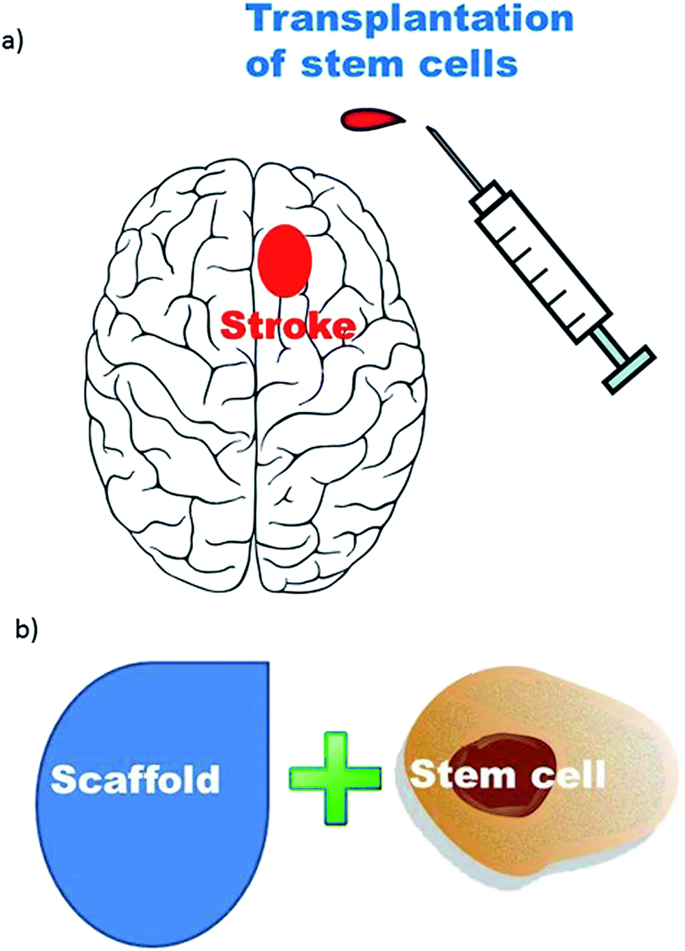 | ||
| Fig. 1 Facilitation of stem cell therapy in stroke by tissue engineering and the application of biomaterials. (a) Schematic showing the transplantation of stem cells into the brain; (b) stem cells can be either encapsulated into microcapsules or cultivated on microspheres before implantation. Both microcapsules and microspheres are tissue engineering scaffolds made of alginate, collagen or other biomaterials. | ||
4.1 Desired biomaterials for brain tissue engineering
The ideal biomaterial for use in brain tissue engineering should be biocompatible, biodegradable, should not generate an immune system reaction, and should not induce neurotoxicity.75 We will use six commonly used biomaterials as examples to illustrate these points.4.2 Key factors that influence stem cell proliferation and differentiation
We have discussed six major types of commonly used biomaterials for brain tissue engineering. Not only should the biomaterials facilitate the delivery of the substance for stroke treatment, but they should also provide a suitable medium for the growth and delivery of stem cells. To promote stem cell proliferation and differentiation, we can further adjust the properties of the materials. In the following section, we will cover four major properties of biomaterials: material size and shape, surface charge and hydrophobicity, surface chemistry and ligand/receptors, and mechano-regulation/material elasticity.4.3 Application of biomaterials in stem cell transplantation for stroke therapy
Stem cells have been transplanted into the brains of stroked animals, with stunning therapeutic effect,15,35,36,39,45,46 and mixed outcomes have also been observed in preliminary clinical cases.15,62 However, the survival rate and disorientation of stem cell differentiation are still major obstacles to guaranteeing the potential for clinical use. Applying biomaterials that have been shown to be beneficial for stem cell growth could increase the potency of stem cell treatment of stroke, as shown in a few successful examples. For instance, although there was no behavioral data to support the symptom-relieving effect, transplantation of neural stem cells supported with a PLGA scaffold reduced the ischemic lesion volume in the cerebral cortex in a rat MCAO model.98 As mentioned, Yu et al. successfully proved that administration of a complex containing collagen type I and neural stem cells can reduce the neurological stroke score in a rat MCAO model.93 Another phenomenal finding presented by Jin et al. showed that post-stroke administration of human neural precursor cells supported by matrigel not only reduced the infarct volume in the cerebral cortex following MCAO stroke, but also improved the preferred usage of the healthy limb only in the cylinder test and the learning and memory deficit in the Y-maze.131 Although there are only several successful reports on the therapeutic effect of combined usage of stem cells and biomaterials, it is expected that more reports will be published. These findings strongly suggest that the combination of biomaterials and stem cells can provide a better therapeutic effect for lesioned animal stroke models than stem cell therapies in isolation.4.4 Combining cell scaffolds and drug carriers into stem cell therapies for stroke
Pharmacologically active microcarrier (PAM) is an example of combining a cell scaffold and a drug carrier into one.100 In a recent study, mesenchymal stem cells and VEGF were loaded into PAM.100 It has been shown to be an effective strategy for repairing the brain damage caused by stroke. PAM was initially developed by Claudia N. Montero-Menei’s group at INSERM, France.132 It serves as a support for cell culture and a carrier for the controlled delivery of bioactive molecules. In addition to stroke, Claudia N. Montero-Menei’s group also investigated the use of PAM to treat Parkinson’s disease133 and osteoarthritis.134 PAM is based on PLGA microcarriers. Other types of biomaterials can be used and even better outcomes are expected to be achieved using the same strategy but with different types of microcarriers. In addition, a limited number of growth factors have been tested, such as VEGF104 and TGF-β.134 Further research into the therapeutic effects of a broader array of growth factors will be carried out in the near future. A combination of two or more growth factors can be used to achieve much better results for stem cell therapy.5. Conclusion
Stem cell therapy in stroke is in its infant stage. There are still numerous technical problems and obstacles yet to be solved. Biomaterial based tissue engineering is a promising approach for facilitation of the potency of transplanted stem cells in stroke treatment. However, the safety and biocompatibility of biomaterials are major issues to be considered in order to develop a more promising therapeutic approach for clinical use in stroke therapy. Furthermore, most of the research concerning the combination of biomaterials with stem cell transplantation in in vivo stroke models focuses mainly on cell survival, differentiation, and localization with limited data comparing behavioral outcomes, which have not been clinically reviewed. Another important issue that is worthy of attention is that transplanted stem cells cannot guarantee the formation of the desired neural circuit for functional neurorestoration following stroke. Further investigation is necessary in order to ensure that transplanted stem cells can re-integrate with functional circuitry. Undeniably, stem cell therapy will be developed into a safe and reliable treatment for restoration of brain functions from any form of brain injury, particularly stroke. We expect that more works on stem cell therapy development, including clinical trials, will be published in the near future.Acknowledgements
S. Z. thanks the National High Technology Research and Development Program of China (2015AA020508), the National Key Basic Research Program of China (2015CB352006), the National Natural Science Foundation of China (61335011), the Program for Changjiang Scholars and Innovative Research Team in University (Grant No. IRT_15R10), and the Fujian Provincial Youth Top-notch Talent Support Program.References
- V. L. Roger, A. S. Go and D. M. Lloyd-Jones, et al., Heart disease and stroke statistics – 2012 update: a report from the American Heart Association. Circulation, Jan 3 2012, vol. 125, 1, pp. e2–e220 Search PubMed.
- F. Gokcay, E. M. Arsava and T. Baykaner, et al., Age-dependent susceptibility to infarct growth in women, Stroke, 2011, 42(4), 947–951 CrossRef PubMed.
- M. Mott, K. Pahigiannis and W. Koroshetz, Small blood vessels: big health problems: National Institute of Neurological Disorders and Stroke update, Stroke, 2014, 45(12), e257–258 CrossRef PubMed.
- J. E. Jung, G. S. Kim and H. Chen, et al., Reperfusion and neurovascular dysfunction in stroke: from basic mechanisms to potential strategies for neuroprotection, Mol. Neurobiol., 2010, 41(2–3), 172–179 CrossRef CAS PubMed.
- A. Brassai, R. G. Suvanjeiev, E. G. Ban and M. Lakatos, Role of synaptic and nonsynaptic glutamate receptors in ischaemia induced neurotoxicity, Brain Res. Bull., 2015, 112, 1–6 CrossRef CAS PubMed.
- A. Pollock, S. E. Farmer and M. C. Brady, et al., Interventions for improving upper limb function after stroke, Cochrane Database of Systematic Reviews, 2014, 11, CD010820 Search PubMed.
- K. M. Langa and D. A. Levine, The diagnosis and management of mild cognitive impairment: a clinical review, J. Am. Med. Assoc., 2014, 312(23), 2551–2561 CrossRef CAS PubMed.
- J. H. Sun, L. Tan and J. T. Yu, Post-stroke cognitive impairment: epidemiology, mechanisms and management, Ann. Transl. Med., 2014, 2(8), 80 Search PubMed.
- A. De Ryck, R. Brouns, M. Geurden, M. Elseviers, P. P. De Deyn and S. Engelborghs, Risk factors for poststroke depression: identification of inconsistencies based on a systematic review, J. Geriatr. Psychiatr. Neurol., 2014, 27(3), 147–158 CrossRef PubMed.
- A. J. Wu, J. Radel and B. Hanna-Pladdy, Improved function after combined physical and mental practice after stroke: a case of hemiparesis and apraxia, Am. J. Occup. Ther., 2011, 65(2), 161–168 CrossRef PubMed.
- F. Malouin, S. Belleville, C. L. Richards, J. Desrosiers and J. Doyon, Working memory and mental practice outcomes after stroke, Arch. Phys. Med. Rehabil., 2004, 85(2), 177–183 CrossRef PubMed.
- R. M. Appireddy, A. M. Demchuk and M. Goyal, et al., Endovascular therapy for ischemic stroke, J. Clin. Neurol., 2015, 11(1), 1–8 CrossRef PubMed.
- S. Yaghi, A. Eisenberger and J. Z. Willey, Symptomatic intracerebral hemorrhage in acute ischemic stroke after thrombolysis with intravenous recombinant tissue plasminogen activator: a review of natural history and treatment, JAMA Neurol., 2014, 71(9), 1181–1185 CrossRef PubMed.
- K. Abe, T. Yamashita, S. Takizawa, S. Kuroda, H. Kinouchi and N. Kawahara, Stem cell therapy for cerebral ischemia: from basic science to clinical applications, J. Cereb. Blood Flow Metab., 2012, 32(7), 1317–1331 CrossRef CAS PubMed.
- T. Honma, O. Honmou and S. Iihoshi, et al., Intravenous infusion of immortalized human mesenchymal stem cells protects against injury in a cerebral ischemia model in adult rat, Exp. Neurol., 2006, 199(1), 56–66 CrossRef CAS PubMed.
- S. I. Savitz, M. Chopp, R. Deans, T. Carmichael, D. Phinney and L. Wechsler, Stem Cell Therapy as an Emerging Paradigm for Stroke (STEPS) II, Stroke, 2011, 42(3), 825–829 CrossRef PubMed.
- P. Thored, A. Arvidsson and E. Cacci, et al., Persistent production of neurons from adult brain stem cells during recovery after stroke, Stem Cells, 2006, 24(3), 739–747 CrossRef CAS PubMed.
- M. K. Tobin, J. A. Bonds, R. D. Minshall, D. A. Pelligrino, F. D. Testai and O. Lazarov, Neurogenesis and inflammation after ischemic stroke: what is known and where we go from here, J. Cereb. Blood Flow Metab., 2014, 34(10), 1573–1584 CrossRef PubMed.
- T. Talwar and M. V. Srivastava, Role of vascular endothelial growth factor and other growth factors in post-stroke recovery, Ann. Indian Acad. Neurol., 2014, 17(1), 1–6 CrossRef PubMed.
- M. Slevin, J. Krupinski and N. Rovira, et al., Identification of pro-angiogenic markers in blood vessels from stroked-affected brain tissue using laser-capture microdissection, BMC Genomics, 2009, 10, 113 CrossRef PubMed.
- J. Macas, C. Nern, K. H. Plate and S. Momma, Increased generation of neuronal progenitors after ischemic injury in the aged adult human forebrain, J. Neurosci., 2006, 26(50), 13114–13119 CrossRef CAS PubMed.
- S. L. Minger, A. Ekonomou, E. M. Carta, A. Chinoy, R. H. Perry and C. G. Ballard, Endogenous neurogenesis in the human brain following cerebral infarction, Regener. Med., 2007, 2(1), 69–74 CrossRef PubMed.
- J. M. Parent, Injury-induced neurogenesis in the adult mammalian brain, Neuroscientist, 2003, 9(4), 261–272 CrossRef PubMed.
- M. J. Zaben and W. P. Gray, Neuropeptides and hippocampal neurogenesis, Neuropeptides, 2013, 47(6), 431–438 CrossRef CAS PubMed.
- M. S. Kim, H. R. Park and M. Park, et al., Neurotoxic effect of 2,5-hexanedione on neural progenitor cells and hippocampal neurogenesis, Toxicology, 2009, 260(1–3), 97–103 CrossRef CAS PubMed.
- M. Komitova, B. Mattsson, B. B. Johansson and P. S. Eriksson, Enriched environment increases neural stem/progenitor cell proliferation and neurogenesis in the subventricular zone of stroke-lesioned adult rats, Stroke, 2005, 36(6), 1278–1282 CrossRef PubMed.
- H. Y. Zhou, Y. Katsman and N. K. Dhaliwal, et al., A Sox2 distal enhancer cluster regulates embryonic stem cell differentiation potential, Genes Dev., 2014, 28(24), 2699–2711 CrossRef PubMed.
- J. J. Ohab and S. T. Carmichael, Poststroke neurogenesis: emerging principles of migration and localization of immature neurons, Neuroscientist, 2008, 14(4), 369–380 CrossRef CAS PubMed.
- C. R. Tyler and A. M. Allan, Adult hippocampal neurogenesis and mRNA expression are altered by perinatal arsenic exposure in mice and restored by brief exposure to enrichment, PLoS One, 2013, 8(9), e73720 CAS.
- J. F. Arenillas, T. Sobrino, J. Castillo and A. Davalos, The role of angiogenesis in damage and recovery from ischemic stroke, Curr. Treat. Options Cardiovasc. Med., 2007, 9(3), 205–212 CrossRef PubMed.
- T. G. Liman and M. Endres, New vessels after stroke: postischemic neovascularization and regeneration, Cerebrovasc. Dis., 2012, 33(5), 492–499 CrossRef CAS PubMed.
- G. Bain, D. Kitchens, M. Yao, J. E. Huettner and D. I. Gottlieb, Embryonic stem cells express neuronal properties in vitro, Dev. Biol., 1995, 168(2), 342–357 CrossRef CAS PubMed.
- S. Okabe, K. Forsberg-Nilsson, A. C. Spiro, M. Segal and R. D. McKay, Development of neuronal precursor cells and functional postmitotic neurons from embryonic stem cells in vitro, Mech. Dev., 1996, 59(1), 89–102 CrossRef CAS PubMed.
- S. C. Zhang, M. Wernig, I. D. Duncan, O. Brustle and J. A. Thomson, In vitro differentiation of transplantable neural precursors from human embryonic stem cells, Nat. Biotechnol., 2001, 19(12), 1129–1133 CrossRef CAS PubMed.
- L. Wei, L. Cui and B. J. Snider, et al., Transplantation of embryonic stem cells overexpressing Bcl-2 promotes functional recovery after transient cerebral ischemia, Neurobiol. Dis., 2005, 19(1–2), 183–193 CrossRef CAS PubMed.
- D. Yanagisawa, M. Qi and D. H. Kim, et al., Improvement of focal ischemia-induced rat dopaminergic dysfunction by striatal transplantation of mouse embryonic stem cells, Neurosci. Lett., 2006, 407(1), 74–79 CrossRef CAS PubMed.
- D. Solter, From teratocarcinomas to embryonic stem cells and beyond: a history of embryonic stem cell research, Nat. Rev. Genet., 2006, 7(4), 319–327 CrossRef CAS PubMed.
- O. Lindvall and Z. Kokaia, Stem cell research in stroke: how far from the clinic?, Stroke, 2011, 42(8), 2369–2375 CrossRef PubMed.
- K. Takahashi, T. Yasuhara and T. Shingo, et al., Embryonic neural stem cells transplanted in middle cerebral artery occlusion model of rats demonstrated potent therapeutic effects, compared to adult neural stem cells, Brain Res., 2008, 1234, 172–182 CrossRef CAS PubMed.
- A. J. Friedenstein, S. Piatetzky and K. V. Petrakova II, Osteogenesis in transplants of bone marrow cells, J. Embryol. Exp. Morphol., 1966, 16(3), 381–390 CAS.
- Z. Zou, Y. Zhang and L. Hao, et al., More insight into mesenchymal stem cells and their effects inside the body, Expert Opin. Biol. Ther., 2010, 10(2), 215–230 CrossRef CAS PubMed.
- K. M. Sicard and M. Fisher, Animal models of focal brain ischemia, Exp. Transl. Stroke Med., 2009, 1, 7 CrossRef PubMed.
- G. Gilmour, S. D. Iversen, M. F. O’Neill and D. M. Bannerman, The effects of intracortical endothelin-1 injections on skilled forelimb use: implications for modelling recovery of function after stroke, Behav. Brain Res., 2004, 150(1–2), 171–183 CrossRef CAS PubMed.
- M. F. Pittenger, A. M. Mackay and S. C. Beck, et al., Multilineage potential of adult human mesenchymal stem cells, Science, 1999, 284(5411), 143–147 CrossRef CAS PubMed.
- K. Kurozumi, K. Nakamura and T. Tamiya, et al., BDNF gene-modified mesenchymal stem cells promote functional recovery and reduce infarct size in the rat middle cerebral artery occlusion model, Mol. Ther., 2004, 9(2), 189–197 CrossRef CAS PubMed.
- D. C. Ding, W. C. Shyu and M. F. Chiang, et al., Enhancement of neuroplasticity through upregulation of beta1-integrin in human umbilical cord-derived stromal cell implanted stroke model, Neurobiol. Dis., 2007, 27(3), 339–353 CrossRef CAS PubMed.
- Y. Omori, O. Honmou, K. Harada, J. Suzuki, K. Houkin and J. D. Kocsis, Optimization of a therapeutic protocol for intravenous injection of human mesenchymal stem cells after cerebral ischemia in adult rats, Brain Res., 2008, 1236, 30–38 CrossRef CAS PubMed.
- X. Bao, M. Feng and J. Wei, et al., Transplantation of Flk-1+ human bone marrow-derived mesenchymal stem cells promotes angiogenesis and neurogenesis after cerebral ischemia in rats, Eur. J. Neurosci., 2011, 34(1), 87–98 CrossRef PubMed.
- S. Suda, K. I. Katsura, M. Saito, N. Kamiya and Y. Katayama, Valproic acid enhances the effect of bone marrow-derived mononuclear cells in a rat ischemic stroke model, Brain Res., 2014, 1565, 74–81 CrossRef CAS PubMed.
- Y. Li, W. W. Mao and C. G. Zhang, et al., Neuroprotective effects of intravenous transplantation of bone marrow mononuclear cells from 5-fluorouracil pre-treated rats on ischemic stroke, Behav. Brain Res., 2016, 301, 287–292 CrossRef CAS PubMed.
- M. Song, S. S. Jue, Y. A. Cho and E. C. Kim, Comparison of the effects of human dental pulp stem cells and human bone marrow-derived mesenchymal stem cells on ischemic human astrocytes in vitro, J. Neurosci. Res., 2015, 93(6), 973–983 CrossRef CAS PubMed.
- M. Song, J. H. Lee, J. Bae, Y. Bu and E. C. Kim, Human dental pulp stem cells are more effective than human bone marrow-derived mesenchymal stem cells in cerebral ischemic injury, Cell Transplant., 2017 DOI:10.3727/096368916X694391.
- G. Varga and G. Gerber, Mesenchymal stem cells of dental origin as promising tools for neuroregeneration, Stem Cell Res. Ther., 2014, 5(2), 61 CrossRef PubMed.
- B. Mitkari, E. Kerkela and J. Nystedt, et al., Intra-arterial infusion of human bone marrow-derived mesenchymal stem cells results in transient localization in the brain after cerebral ischemia in rats, Exp. Neurol., 2013, 239, 158–162 CrossRef CAS PubMed.
- M. Gutierrez-Fernandez, B. Rodriguez-Frutos and J. Ramos-Cejudo, et al., Effects of intravenous administration of allogenic bone marrow- and adipose tissue-derived mesenchymal stem cells on functional recovery and brain repair markers in experimental ischemic stroke, Stem Cell Res. Ther., 2013, 4(1), 11 CrossRef CAS PubMed.
- E. M. Pietras, M. R. Warr and E. Passegue, Cell cycle regulation in hematopoietic stem cells, J. Cell Biol., 2011, 195(5), 709–720 CrossRef CAS PubMed.
- H. Felfly, A. Muotri, H. Yao and G. G. Haddad, Hematopoietic stem cell transplantation protects mice from lethal stroke, Exp. Neurol., 2010, 225(2), 284–293 CrossRef CAS PubMed.
- D. Doycheva, G. Shih, H. Chen, R. Applegate, J. H. Zhang and J. Tang, Granulocyte-colony stimulating factor in combination with stem cell factor confers greater neuroprotection after hypoxic-ischemic brain damage in the neonatal rats than a solitary treatment, Transl. Stroke Res., 2013, 4(2), 171–178 CrossRef CAS PubMed.
- H. Okano and S. Yamanaka, iPS cell technologies: significance and applications to CNS regeneration and disease, Mol. Brain., 2014, 7, 22 CrossRef PubMed.
- S. J. Chen, C. M. Chang and S. K. Tsai, et al., Functional improvement of focal cerebral ischemia injury by subdural transplantation of induced pluripotent stem cells with fibrin glue, Stem Cells Dev., 2010, 19(11), 1757–1767 CrossRef CAS PubMed.
- M. Jiang, L. Lv and H. Ji, et al., Induction of pluripotent stem cells transplantation therapy for ischemic stroke, Mol. Cell. Biochem., 2011, 354(1–2), 67–75 CrossRef CAS PubMed.
- D. Kalladka and K. W. Muir, Brain repair: cell therapy in stroke, Stem Cells Cloning., 2014, 7, 31–44 CAS.
- F. Bifari, L. Pacelli and M. Krampera, Immunological properties of embryonic and adult stem cells, World J. Stem Cells., 2010, 2(3), 50–60 CrossRef PubMed.
- O. Honmou, K. Houkin and T. Matsunaga, et al., Intravenous administration of auto serum-expanded autologous mesenchymal stem cells in stroke, Brain., 2011, 134(Pt 6), 1790–1807 CrossRef PubMed.
- A. Bhasin, M. V. Srivastava, S. Mohanty, R. Bhatia, S. S. Kumaran and S. Bose, Stem cell therapy: a clinical trial of stroke, Clin Neurol Neurosurg., 2013, 115(7), 1003–1008 CrossRef PubMed.
- S. I. Savitz, V. Misra and M. Kasam, et al., Intravenous autologous bone marrow mononuclear cells for ischemic stroke, Ann Neurol., 2011, 70(1), 59–69 CrossRef PubMed.
- A. Sharma, H. Sane and N. Gokulchandran, et al., Autologous bone marrow mononuclear cells intrathecal transplantation in chronic stroke, Stroke Res. Treat., 2014, 2014, 234095 Search PubMed.
- K. Prasad, S. Mohanty and R. Bhatia, et al., Autologous intravenous bone marrow mononuclear cell therapy for patients with subacute ischaemic stroke: a pilot study, Indian J. Med. Res., 2012, 136(2), 221–228 Search PubMed.
- K. Prasad, A. Sharma and A. Garg, et al., Intravenous autologous bone marrow mononuclear stem cell therapy for ischemic stroke: a multicentric, randomized trial, Stroke., 2014, 45(12), 3618–3624 CrossRef CAS PubMed.
- A. Taguchi, C. Sakai and T. Soma, et al., Intravenous Autologous Bone Marrow Mononuclear Cell Transplantation for Stroke: Phase1/2a Clinical Trial in a Homogeneous Group of Stroke Patients, Stem Cells Dev., 2015, 24(19), 2207–2218 CrossRef CAS PubMed.
- G. H. Petit, T. T. Olsson and P. Brundin, The future of cell therapies and brain repair: Parkinson’s disease leads the way, Neuropathol Appl Neurobiol., 2014, 40(1), 60–70 CrossRef CAS PubMed.
- Q. Vu, K. Xie, M. Eckert, W. Zhao and S. C. Cramer, Meta-analysis of preclinical studies of mesenchymal stromal cells for ischemic stroke, Neurology., 2014, 82(14), 1277–1286 CrossRef PubMed.
- H. Jeong, H. W. Yim and Y. S. Cho, et al., Efficacy and safety of stem cell therapies for patients with stroke: a systematic review and single arm meta-analysis, Int J Stem Cells., 2014, 7(2), 63–69 CrossRef PubMed.
- F. Moniche, A. Gonzalez and J. R. Gonzalez-Marcos, et al., Intra-arterial bone marrow mononuclear cells in ischemic stroke: a pilot clinical trial, Stroke., 2012, 43(8), 2242–2244 CrossRef PubMed.
- E. Diez-Tejedor, M. Gutierrez-Fernandez and P. Martinez-Sanchez, et al., Reparative therapy for acute ischemic stroke with allogeneic mesenchymal stem cells from adipose tissue: a safety assessment: a phase II randomized, double-blind, placebo-controlled, single-center, pilot clinical trial, J. Stroke Cerebrovasc Dis., 2014, 23(10), 2694–2700 CrossRef PubMed.
- Y. Imanishi, A. Saito and H. Komoda, et al., Allogenic mesenchymal stem cell transplantation has a therapeutic effect in acute myocardial infarction in rats, J. Mol. Cell. Cardiol., 2008, 44(4), 662–671 CrossRef PubMed.
- J. R. Evans, S. L. Mason and R. A. Barker, Current status of clinical trials of neural transplantation in Parkinson’s disease, Prog. Brain Res., 2012, 200, 169–198 Search PubMed.
- N. Lenoir, Europe confronts the embryonic stem cell research challenge, Science, 2000, 287(5457), 1425–1427 CrossRef CAS PubMed.
- I. Kalaszczynska and K. Ferdyn, Wharton’s jelly derived mesenchymal stem cells: future of regenerative medicine? Recent findings and clinical significance, Biomed Res Int., 2015, 2015, 430847 Search PubMed.
- A. Hilfiker, C. Kasper, R. Hass and A. Haverich, Mesenchymal stem cells and progenitor cells in connective tissue engineering and regenerative medicine: is there a future for transplantation?, Langenbecks Arch. Surg., 2011, 396(4), 489–497 CrossRef PubMed.
- S. Kelly, T. M. Bliss and A. K. Shah, et al., Transplanted human fetal neural stem cells survive, migrate, and differentiate in ischemic rat cerebral cortex, Proc. Natl. Acad. Sci. U. S. A., 2004, 101(32), 11839–11844 CrossRef CAS PubMed.
- J. W. Jung, M. Kwon and J. C. Choi, et al., Familial occurrence of pulmonary embolism after intravenous, adipose tissue-derived stem cell therapy, Yonsei Med J., 2013, 54(5), 1293–1296 CrossRef PubMed.
- T. Makela, R. Takalo and O. Arvola, et al., Safety and biodistribution study of bone marrow-derived mesenchymal stromal cells and mononuclear cells and the impact of the administration route in an intact porcine model, Cytotherapy., 2015, 17(4), 392–402 CrossRef CAS PubMed.
- A. Popa-Wagner, A. M. Buga, T. R. Doeppner and D. M. Hermann, Stem cell therapies in preclinical models of stroke associated with aging, Front Cell Neurosci., 2014, 8, 347 Search PubMed.
- E. Enwere, T. Shingo, C. Gregg, H. Fujikawa, S. Ohta and S. Weiss, Aging results in reduced epidermal growth factor receptor signaling, diminished olfactory neurogenesis, and deficits in fine olfactory discrimination, J. Neurosci., 2004, 24(38), 8354–8365 CrossRef CAS PubMed.
- J. S. Lees, E. S. Sena and K. J. Egan, et al., Stem cell-based therapy for experimental stroke: a systematic review and meta-analysis, Int J Stroke., 2012, 7(7), 582–588 CrossRef PubMed.
- W. Cao and P. Li, Effectiveness and Safety of Autologous Bone Marrow Stromal Cells Transplantation After Ischemic Stroke: A Meta-Analysis, Med. Sci. Monit., 2015, 21, 2190–2195 CrossRef PubMed.
- G. B. Boncoraglio, A. Bersano, L. Candelise, B. A. Reynolds and E. A. Parati, Stem cell transplantation for ischemic stroke, Cochrane Database of Systematic Reviews, 2010,(9), CD007231 Search PubMed.
- D. F. Williams, On the mechanisms of biocompatibility, Biomaterials, 2008, 29(20), 2941–2953 CrossRef CAS PubMed.
- M. Ni and B. D. Ratner, Nacre surface transformation to hydroxyapatite in a phosphate buffer solution, Biomaterials, 2003, 24(23), 4323–4331 CrossRef CAS PubMed.
- B. D. Ratner and S. J. Bryant, Biomaterials: where we have been and where we are going, Annu. Rev. Biomed. Eng., 2004, 6, 41–75 CrossRef CAS PubMed.
- D. F. Emerich, E. Silva and O. Ali, et al., Injectable VEGF hydrogels produce near complete neurological and anatomical protection following cerebral ischemia in rats, Cell Transplant., 2010, 19(9), 1063–1071 Search PubMed.
- H. Yu, B. Cao and M. Feng, et al., Combinated transplantation of neural stem cells and collagen type I promote functional recovery after cerebral ischemia in rats, Anat. Rec., 2010, 293(5), 911–917 CrossRef PubMed.
- B. D. Cherksey, V. S. Sapirstein and A. L. Geraci, Adrenal chromaffin cells on microcarriers exhibit enhanced long-term functional effects when implanted into the mammalian brain, Neuroscience, 1996, 75(2), 657–664 CrossRef CAS PubMed.
- Y. Wang, M. J. Cooke, C. M. Morshead and M. S. Shoichet, Hydrogel delivery of erythropoietin to the brain for endogenous stem cell stimulation after stroke injury, Biomaterials, 2012, 33(9), 2681–2692 CrossRef CAS PubMed.
- M. J. Cooke, Y. Wang, C. M. Morshead and M. S. Shoichet, Controlled epi-cortical delivery of epidermal growth factor for the stimulation of endogenous neural stem cell proliferation in stroke-injured brain, Biomaterials, 2011, 32(24), 5688–5697 CrossRef CAS PubMed.
- N. P. Stover and R. L. Watts, Spheramine for treatment of Parkinson’s disease, Neurotherapeutics, 2008, 5(2), 252–259 CrossRef CAS PubMed.
- E. Bible, D. Y. Chau, M. R. Alexander, J. Price, K. M. Shakesheff and M. Modo, The support of neural stem cells transplanted into stroke-induced brain cavities by PLGA particles, Biomaterials, 2009, 30(16), 2985–2994 CrossRef CAS PubMed.
- E. Bible, O. Qutachi, D. Y. Chau, M. R. Alexander, K. M. Shakesheff and M. Modo, Neo-vascularization of the stroke cavity by implantation of human neural stem cells on VEGF-releasing PLGA microparticles, Biomaterials, 2012, 33(30), 7435–7446 CrossRef CAS PubMed.
- M. S. Quittet, O. Touzani and L. Sindji, et al., Effects of mesenchymal stem cell therapy, in association with pharmacologically active microcarriers releasing VEGF, in an ischaemic stroke model in the rat, Acta Biomater., 2015, 15, 77–88 CrossRef CAS PubMed.
- K. Y. Lee and D. J. Mooney, Alginate: properties and biomedical applications, Prog. Polym. Sci., 2012, 37(1), 106–126 CrossRef CAS PubMed.
- A. D. Augst, H. J. Kong and D. J. Mooney, Alginate hydrogels as biomaterials, Macromol. Biosci., 2006, 6(8), 623–633 CrossRef CAS PubMed.
- U. Zimmermann, G. Klock and K. Federlin, et al., Production of mitogen-contamination free alginates with variable ratios of mannuronic acid to guluronic acid by free flow electrophoresis, Electrophoresis, 1992, 13(5), 269–274 CrossRef CAS PubMed.
- M. Otterlei, K. Ostgaard, G. Skjak-Braek, O. Smidsrod, P. Soon-Shiong and T. Espevik, Induction of cytokine production from human monocytes stimulated with alginate, J. Immunother., 1991, 10(4), 286–291 CrossRef CAS PubMed.
- B. Eftekharzadeh, F. Khodagholi, A. Abdi and N. Maghsoudi, Alginate protects NT2 neurons against H2O2-induced neurotoxicity, Carbohydr. Polym., 2010, 79(4), 1063–1072 CrossRef CAS.
- L. Cen, W. Liu, L. Cui, W. Zhang and Y. Cao, Collagen tissue engineering: development of novel biomaterials and applications, Pediatr. Res., 2008, 63(5), 492–496 CrossRef CAS PubMed.
- C. Soo, G. Rahbar and R. L. Moy, The immunogenicity of bovine collagen implants, Dermatol. Surg., 1993, 19(5), 431–434 CrossRef CAS.
- A. K. Lynn, I. V. Yannas and W. Bonfield, Antigenicity and immunogenicity of collagen, J. Biomed. Mater. Res., Part B, 2004, 71(2), 343–354 CrossRef CAS PubMed.
- J. A. Cadee, M. J. van Luyn and L. A. Brouwer, et al., In vivo biocompatibility of dextran-based hydrogels, J. Biomed. Mater. Res., 2000, 50(3), 397–404 CrossRef CAS PubMed.
- C. Gebb, B. Lundgren, J. Clark and U. Lindskog, Harvesting and subculturing cells growing on denatured-collagen coated microcarriers (Cytodex 3), Dev. Biol. Stand., 1983, 55, 57–65 CAS.
- C. J. De Groot, M. J. Van Luyn and W. N. Van Dijk-Wolthuis, et al., In vitro biocompatibility of biodegradable dextran-based hydrogels tested with human fibroblasts, Biomaterials, 2001, 22(11), 1197–1203 CrossRef CAS PubMed.
- E. M. Bachelder, T. T. Beaudette, K. E. Broaders, J. Dashe and J. M. Frechet, Acetal-derivatized dextran: an acid-responsive biodegradable material for therapeutic applications, J. Am. Chem. Soc., 2008, 130(32), 10494–10495 CrossRef CAS PubMed.
- J. Gilroy, M. I. Barnhart and J. S. Meyer, Treatment of acute stroke with dextran 40, J. Am. Med. Assoc., 1969, 210(2), 293–298 CrossRef CAS.
- N. Lennard, J. Smith and J. Dumville, et al., Prevention of postoperative thrombotic stroke after carotid endarterectomy: the role of transcranial Doppler ultrasound, J. Vasc. Surg., 1997, 26(4), 579–584 CrossRef CAS PubMed.
- D. Gupta, C. H. Tator and M. S. Shoichet, Fast-gelling injectable blend of hyaluronan and methylcellulose for intrathecal, localized delivery to the injured spinal cord, Biomaterials, 2006, 27(11), 2370–2379 CrossRef CAS PubMed.
- B. D. Ratner, A. S. Hoffman, F. J. Schoen and J. E. Lemons, Biomaterials Science: An Introduction to Materials in Medicine, Elsevier Academic Press, Amsterdam, Boston, 2004 Search PubMed.
- W. S. Hu and D. I. Wang, Selection of microcarrier diameter for the cultivation of mammalian cells on microcarriers, Biotechnol. Bioeng., 1987, 30(4), 548–557 CrossRef CAS PubMed.
- E. K. Yim, S. W. Pang and K. W. Leong, Synthetic nanostructures inducing differentiation of human mesenchymal stem cells into neuronal lineage, Exp. Cell Res., 2007, 313(9), 1820–1829 CrossRef CAS PubMed.
- J. Yoo, M. Noh, H. Kim, N. L. Jeon, B. S. Kim and J. Kim, Nanogrooved substrate promotes direct lineage reprogramming of fibroblasts to functional induced dopaminergic neurons, Biomaterials, 2015, 45, 36–45 CrossRef CAS PubMed.
- S. E. Gratton, P. A. Ropp and P. D. Pohlhaus, et al., The effect of particle design on cellular internalization pathways, Proc. Natl. Acad. Sci. U. S. A., 2008, 105(33), 11613–11618 CrossRef CAS PubMed.
- M. Ni, W. H. Tong, D. Choudhury, N. A. Rahim, C. Iliescu and H. Yu, Cell culture on MEMS platforms: a review, Int. J. Mol. Sci., 2009, 10(12), 5411–5441 CrossRef CAS PubMed.
- K. Fairman and B. S. Jacobson, Unique morphology of HeLa cell attachment, spreading and detachment from microcarrier beads covalently coated with a specific and non-specific substratum, Tissue Cell, 1983, 15(2), 167–180 CrossRef CAS PubMed.
- E. Walter, D. Dreher and M. Kok, et al., Hydrophilic poly(DL-lactide-co-glycolide) microspheres for the delivery of DNA to human-derived macrophages and dendritic cells, J. Controlled Release, 2001, 76(1–2), 149–168 CrossRef CAS PubMed.
- L. T. Allen, E. J. Fox and I. Blute, et al., Interaction of soft condensed materials with living cells: phenotype/transcriptome correlations for the hydrophobic effect, Proc. Natl. Acad. Sci. U. S. A., 2003, 100(11), 6331–6336 CrossRef CAS PubMed.
- S. Zhuo, M. Ni, L. Bi, L. Xia, J. Fan and H. Chan, Stem Cell-Biomaterial Interactions for Tissue Engineering, Stem Cells Int., 2015, 126710 Search PubMed.
- G. Klein, M. Langegger, R. Timpl and P. Ekblom, Role of laminin A chain in the development of epithelial cell polarity, Cell, 1988, 55(2), 331–341 CrossRef CAS PubMed.
- K. Yang, J. S. Lee and J. Kim, et al., Polydopamine-mediated surface modification of scaffold materials for human neural stem cell engineering, Biomaterials, 2012, 33(29), 6952–6964 CrossRef CAS PubMed.
- D. S. Benoit, M. P. Schwartz, A. R. Durney and K. S. Anseth, Small functional groups for controlled differentiation of hydrogel-encapsulated human mesenchymal stem cells, Nat. Mater., 2008, 7(10), 816–823 CrossRef CAS PubMed.
- A. J. Engler, S. Sen, H. L. Sweeney and D. E. Discher, Matrix elasticity directs stem cell lineage specification, Cell, 2006, 126(4), 677–689 CrossRef CAS PubMed.
- S. Ilkhanizadeh, A. I. Teixeira and O. Hermanson, Inkjet printing of macromolecules on hydrogels to steer neural stem cell differentiation, Biomaterials, 2007, 28(27), 3936–3943 CrossRef CAS PubMed.
- K. Jin, X. Mao and L. Xie, et al., Transplantation of human neural precursor cells in Matrigel scaffolding improves outcome from focal cerebral ischemia after delayed postischemic treatment in rats, J. Cereb. Blood Flow Metab., 2010, 30(3), 534–544 CrossRef PubMed.
- V. M. Tatard, M. C. Venier-Julienne and P. Saulnier, et al., Pharmacologically active microcarriers: a tool for cell therapy, Biomaterials, 2005, 26(17), 3727–3737 CrossRef CAS PubMed.
- N. Daviaud, E. Garbayo, L. Sindji, A. Martinez-Serrano, P. C. Schiller and C. N. Montero-Menei, Survival, differentiation, and neuroprotective mechanisms of human stem cells complexed with neurotrophin-3-releasing pharmacologically active microcarriers in an ex vivo model of Parkinson’s disease, Stem Cells Transl. Med., 2015, 4(6), 670–684 CrossRef CAS PubMed.
- C. Bouffi, O. Thomas and C. Bony, et al., The role of pharmacologically active microcarriers releasing TGF-beta3 in cartilage formation in vivo by mesenchymal stem cells, Biomaterials, 2010, 31(25), 6485–6493 CrossRef CAS PubMed.
| This journal is © The Royal Society of Chemistry 2017 |