Permittivity imaged at the nanoscale using tip-enhanced Raman spectroscopy
Julien
Plathier
 a,
Andrey
Krayev
b,
Vasili
Gavrilyuk
b,
Alain
Pignolet
a and
Andreas
Ruediger
*a
aINRS-EMT, 1650 Boul. Lionel-Boulet, Varennes, Québec, J3X1S2, Canada. E-mail: ruediger@emt.inrs.ca
bAIST-NT, 359 Bel Marin Keys Blvd. #20, Novato, California 94949, USA
First published on 11th August 2017
Abstract
Localized surface plasmon resonances are the dominating contribution to the optical enhancement and the lateral resolution in tip-enhanced Raman spectroscopy. This well studied phenomenon may give access to more information about the sample than the enhanced Raman spectra alone due to its sensitivity to the permittivity of the tip environment. In this work, the effects of the permittivity of the sample on the properties of localized surface plasmon resonance are studied through the amplified signal of the luminescence of gold tips.
Conceptual insightsOptical characterization at the nanoscale is a key for the development of new ultra-compact optical devices. Already established, surface plasmon resonance sensing yields a global quantification of the refractive indices of molecules and thin films. Now, apertureless scanning near-field optical microscopy is demonstrated to identify materials, at the nanoscale, using the relative change in the refractive index over the sample. This communication presents the possibility of combining nanoscale resolution with quantitative measurements of the permittivity using tip-enhanced Raman spectroscopy, thus enabling the mapping of the optical properties of isolated nano-objects. |
Introduction
Since their prediction in 1957,1 surface plasmon resonances (SPRs) have been studied and used to measure the optical properties of materials.2 When, in metals, an electromagnetic field induces a collective oscillation of the electron cloud, this oscillation generates a surface plasmon. This surface plasmon is characterized by its resonance frequency and its ability to very efficiently convert propagating electromagnetic waves to an optical near field.3 Due to the sensitivity of the SPR frequency to the permittivity of its direct environment, surface plasmons are used as sensors to probe the permittivities of molecules and thin films.4,5 When localized at the apex of a nano-antenna, an SPR can also be used as an optical amplifier, allowing the enhancement of the far field incoming electromagnetic radiation.6 This enhancement is used in surface-enhanced Raman spectroscopy (SERS) to push the sensitivity of Raman spectroscopy to the single molecule detection limit.7 Tip-enhanced Raman spectroscopy (TERS) uses the same enhancement mechanism as SERS but the nano-antenna is a metallic scanning probe microscope (SPM) tip. It allows both improving the Raman spectroscopy detection limit and overcoming the diffraction limit, enabling the investigation of isolated molecules and nano-objects.8Tip-enhanced Raman spectroscopy exploits the optical amplification of an SPR by focusing a laser on the extremity of a metallic tip to generate a localized surface plasmon resonance (LSPR) at its apex. The illuminated tip, generally part of an atomic force microscope (AFM) or scanning tunneling microscope (STM), is then used to scan surfaces by conventional SPM. Most TERS experiments use the LSPR only as an optical amplifier to study nanostructures with an optical spatial resolution of few nanometers.6,8,9 However, the sensitivity of the LSPR frequency to the sample permittivity is too often neglected. A change in the sample permittivity in the vicinity of the tip is measurable by monitoring the change in the resonance condition of the system including the nano-sized sample under investigation, the localized surface plasmon at the apex of the tip, and possibly the image surface plasmon generated in the substrate, if metallic.5 Yet, a shift of the LSPR frequency has also been observed by increasing the tip-to-sample distance.10 So, in order to be able to extract the sample permittivity from a TERS map, one has to be able not only to identify the shifts in the resonance condition of the system but also their different causes.
In this communication, we present an analysis of the resonance frequency shifts recorded during the TERS mapping of carbon nanotubes and graphene oxide nanosheets deposited on gold. We show that the study of the amplified signal of the luminescence of the gold tip can be used to estimate the local permittivity of the object investigated at the nanoscale. For the sake of simplicity, although it is physically correct to describe the shift in the frequency of the whole system composed of the tip (and its surface plasmon), the sample, and the substrate's surface (with possibly an image of the surface plasmon, if metallic, generating itself a secondary image surface image at the tip apex), we will use the commonly used expression “shift of the LSPR frequency”.
Experimental
TERS setup
Experimental data are acquired using an AIST-NT OmegaScope 1000 scanning probe microscope coupled to a Horiba XploRA confocal Raman spectrometer. The TERS probe consists of an etched gold tip glued on a 32.768 kHz tuning fork from Abracon Corporation. To maintain the tip-to-sample distance close to one nanometer, the AFM, in non-contact mode, uses the resonance frequency of the tuning fork as a feedback signal. To excite the LSPR at the tip apex, the latter is side-illuminated using a He–Ne laser (632.8 nm, TEM 00) using a Mitutoyo M Plan Apo 100× objective (0.7 NA) mounted on a piezoelectric stage. The laser beam and the optical axis of the objective make an angle of 65° with the normal to the surface of the substrate and sample. The laser polarization is set to be along the tip axis to maximize the EM field enhancement,11 and the laser power is adjusted to approximately 600 μW to prevent any heat-induced effects that might damage the tip (melting). The focal spot of the objective is pre-aligned at the apex of the tip using a video camera placed in the optical path. Subsequently, a fine alignment is performed by imaging the emission of the tip apex with the objective.12 Finally, a 600 l mm−1 grating, coupled to a thermoelectrically cooled Andor iDus 420 BU CCD detector, spectrally resolves the backscattered light.Tip etching
Annealed gold wires with a diameter of 100 μm are electrochemically etched using fuming hydrochloric acid.13 The electric signal is composed of 30 μs pulses at 3 kHz (4.5 V peak to peak, +0.5 V DC bias) delivered by a Keithley 3390 arbitrary waveform generator. The counter electrode is a ring made of the same wire as the tips. This process gives reproducible smooth tips with a radius of curvature of about 30 nm.Sample preparation
The samples are prepared by functionalizing a gold-coated glass slide with amino-terminated polyamidoamine (PAMAM) dendrimers, generation 4. Then, a water dispersion of graphene oxide from Graphene Supermarket is placed on top of the functionalized substrate for one minute and, eventually, the excess is removed by spinning the sample. Next, 100 μL of a suspension of individual NanoIntegris metallic carbon nanotubes is drop-cast on top of the sample, held there for 3 minutes, with the excess being removed in a spin-coater. Eventually, 40 μL of a 10−5 M toluene solution of fullerenes (C60) is spin-coated. The samples are rinsed with distilled water and dried while spinning. The final stage of the sample preparation is the removal of the majority of the organic residue by drop-casting acidic piranha solution on top of the sample for five seconds with subsequent rinsing with distilled water.Data treatment
For each measurement, the far field signal is subtracted from the TERS spectrum. The far field signal is acquired without the tip under the same conditions and for the same area as the associated TERS measurements. Then, the tip emission is extracted using a non-linear regression of a Lorentzian function10 in spectral regions without visible Raman signal and is removed from the data. The frequency scale of Raman spectra is calibrated using a silicon (100) crystal.Simulations
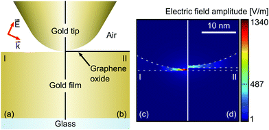
LSPR simulations are performed using Maxwell's equations and the finite element method. This method allows using a triangle-based mesh to sample the geometry of the system and to increase the triangle density in areas of interest. This mesh provides the ability to study complex configurations with high accuracy in a short amount of time. The system simulated consists of the apex of a gold tip with 30 nm curvature radius, based on SEM micrographs of etched tips, on top of a flat 50 nm gold surface on a 1 μm glass substrate. An electromagnetic wave p-polarized, with an electric field amplitude of 1 V m−1, propagates through the air medium to reach the tip-sample system with an angle of incidence of 65°. A layer of graphene oxide is added between the tip and the gold surface to simulate its effects on the electric field at the apex of the tip, as visible in Fig. 1. Experimental data are used to set the tip-to-sample distance.Results and discussion
LSPRs are sensitive to their direct environment, especially to their permittivity.4 In order to be able to observe the effects of the environment on the properties of the tip localized surface plasmon, the sample is scanned. During scanning, the (enhanced) Raman signal, the tip emission and the sample topography are simultaneously mapped. The mapping also allows checking the repeatability of the LSPR properties and its correlation with the sample topography and spatial distribution.Fig. 2a shows the topography of a 2.5 μm × 2.5 μm area obtained by AFM simultaneously with the TERS map presented in Fig. 2b, both having a pixel size of about 10 nm. The AFM image shows a set of carbon nanotubes and a piece of graphene oxide. The apparent sizes of those objects are wider than expected due to a double apex tip,14 a characteristic artifact that is visible by looking carefully at the bottom left nanotube. The TERS map reveals more nanotubes than those visible in the topography picture and a lateral resolution twice as high, breaking the diffraction limit by two orders of magnitude. Despite the double apex tip, only one hot spot enhances the Raman signal, giving a sharp spectral map.
 | ||
| Fig. 2 (a) Topography (256 × 256 px) of carbon nanotubes and graphene oxide deposited on a gold surface. Several carbon nanotubes and a piece of graphene oxide are scattered across the visible area. (b) TERS map (100 ms px−1) simultaneously recorded. The signal intensity is integrated over the 2D band of the carbon nanotubes (2600–2800 cm−1). Gray profile lines give the locations of the line scans presented in Fig. 4. Red dots show the locations of the spectra presented in Fig. 3. (c) Tip emission intensity extracted from the TERS map. A dark contrast is visible at the locations of carbon nanotubes revealed by the topography. The graphene oxide piece is also distinguishable from the gold substrate. | ||
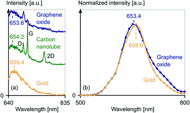
The enhanced emission, visible in Fig. 3a, shows a clear enhancement of the D, G and 2D Raman modes of the carbon nanotubes at 1350 cm−1 (691.9 nm), 1630 cm−1 (705.5 nm) and 2700 cm−1 (763.2 nm), respectively, and an enhancement of the D and G Raman modes of the graphene oxide compared with the far field signal obtained over the gold film. The enhancement factor, defined as the fourth root of the ratio of the near field intensity to the far field intensity corrected by the interaction surface of each field,15 is estimated to be 6.5.
 | ||
| Fig. 3 (a) Emission spectra of the gold film (yellow), a carbon nanotube (green) and the graphene oxide piece (dark blue). The position of the enhanced gold luminescence is indicated for each spectrum. The locations of the measurement points are shown in Fig. 2b. (b) Normalized simulated electric field intensity at the apex of the tip on top of gold (yellow dots) and graphene oxide (dark blue diamonds). The wavelength giving the maximum intensity is indicated in both cases. | ||
Those spectra also exhibit a broad emission signal, interpreted as the gold luminescence, arising from interband electronic transition (5d-6sp),16 amplified by the LSPR of the tip. This amplified luminescence can be used to perform near-field optical imaging.17 Therefore, the gold emission intensity, extracted from TERS data as described previously, is presented in Fig. 2c. It exhibits a net contrast at the locations of some carbon nanotubes and of the graphene oxide sheet. A comparison of Fig. 2a–c reveals that this contrast is correlated with the topography. This is visible at the bottom left nanotube, which is only partially resolved on the topography and tip emission images, but clearly visible on the TERS map. This large variation in the tip emission intensity for a small change in the tip-to-substrate distance and the spatial resolution of the map suggests a near field origin. Therefore, the gold luminescence signal has to be enhanced by the LSPR. The frequency of the maximum of the enhanced luminescence is plotted across graphene oxide and carbon nanotubes, as presented in Fig. 4a and b. A blue shift of the maximum of the enhanced luminescence, also visible in Fig. 3a, occurs when the tip travels across the graphene oxide and the nanotube. This shift is clearly correlated with the topography at the same location, showing that both the intensity and peak shift of the enhanced luminescence are correlated with the tip-position on the sample.
 | ||
| Fig. 4 Enhanced tip luminescence peak shift (turquoise diamonds) and topography (brown dots) over graphene oxide (a) and carbon nanotubes (b). Positions of line scans are visible in Fig. 2b. | ||
A similar blue shift and an intensity decrease of TERS signals with the tip-to-sample distance have been reported on perchlorate and guanine coadsorbed on gold, the enhanced luminescence being explained as reflecting the LSPR properties.10 In our case, the distance between the tip and the gold film increases when the tip is on top of a nanotube and on top of the graphene oxide sheet. But at the same time, the permittivity in the vicinity of the tip also changes due to the presence of those carbon nanostructures.
Therefore, both the permittivity distribution in the vicinity of the tip and the tip-to-substrate distance act on the LSPR frequency. The permittivity dependence comes from the discontinuity of the normal component of the electric displacement field, across an interface between two media.18 The tip-to-substrate distance dependence comes from the coupling between the electric field in the vicinity of the tip and the electric field of the image dipole supported by the gold film and can be described by the Dipolar-Coupling Model.19,20 If the enhanced luminescence reflects the LSPR properties, this emission and the surface plasmon resonance should shift by the same amount for a given variation in the permittivity of the sample, assuming a constant sample thickness and a constant tip-to-substrate distance. To check this hypothesis, the LSPR is simulated as previously described. The tip-to-sample distance and the graphene oxide sheet thickness are set at 0.4 nm and 0.8 nm, respectively, based on AFM measurements. The permittivities of gold and graphene oxide are based on the work of Rakić et al.21 and Jung et al.22 respectively. The electric field enhancement from the surface plasmon exhibits a maximum around 660 nm as visible in Fig. 3b. By comparing the LSPR frequencies averaged over the gold substrate (659.4 nm) and the graphene oxide sheet (653.6 nm) to the simulated ones (659.6 nm and 653.4 nm, respectively) we notice that the shift of the resonance frequency is similar in both cases (5.8 nm and 6.2 nm). The average value over carbon nanotubes (654.2 nm) also qualitatively agrees with our hypothesis, considering that the exact value of their permittivity is not known but should be close to that of graphene, and also considering that carbon nanotubes’ contribution to the permittivity distribution in the vicinity of the tip should be smaller than that of graphene due to their smaller volume.
In order to be able to compare the experimental results to the simulations, the value of the enhanced luminescence position is an average of many spectra taken over the gold film, carbon nanotubes and graphene oxide sheet and discriminated using the presence of Raman peaks and the topography. This averaging limits the influence of the tip-to-sample distance due to the feedback loop of the AFM and the roughness of the sample. This influence is visible in Fig. 2c, where the enhanced luminescence intensity is directly impacted by the tip-to-substrate distance. The near field contribution to the intensity being inversely proportional to the 10th power of the tip-to-substrate distance,23 the enhanced luminescence intensity decreases rapidly for a small increase of the tip-to-substrate distance. Variation in the coupling between the tip and the gold film also contributes significantly to the noise visible in the peak shift signal of Fig. 4a and b.
Conclusions
In this study, we have shown that the gold tip luminescence, enhanced by the LSPR, can be used to obtain the permittivity of nano-objects. By simulating the electric field enhancement from the LSPR of a gold tip on gold and graphene, we were able to demonstrate that the LSPR frequency shift, measured experimentally, is due to the difference in the permittivities of gold and graphene. Although more investigations are needed, the experimental shift induced by the nanotube also qualitatively supports our hypothesis. Further investigations will be performed in order to determine quantitatively the permittivity of the nanotube.Conflicts of interest
There are no conflicts to declare.Acknowledgements
A. R. is grateful for an NSERC discovery grant. We thank Gitanjali Kolhatkar for fruitful discussions.Notes and references
- R. H. Ritchie, Phys. Rev., 1957, 106, 874–881 CrossRef CAS.
- J. Homola, S. S. Yee and G. Gauglitz, Sens. Actuators, B, 1999, 54, 3–15 CrossRef CAS.
- A. V. Zayats and I. I. Smolyaninov, J. Opt. A: Pure Appl. Opt., 2003, 5, S16 CrossRef CAS.
- K. A. Willets and R. P. Van Duyne, Annu. Rev. Phys. Chem., 2007, 58, 267–297 CrossRef CAS PubMed.
- G. Kolhatkar, et al. , Phys. Chem. Chem. Phys., 2016, 18, 30546–30553 RSC.
- R. Zhang, et al. , Nature, 2013, 498, 82–86 CrossRef CAS PubMed.
- K. Kneipp, et al. , Phys. Rev. Lett., 1997, 78, 1667–1670 CrossRef CAS.
- C. Chen, N. Hayazawa and S. Kawata, Nat. Commun., 2014, 5, 3312 Search PubMed.
- V. Deckert, et al. , Faraday Discuss., 2015, 177, 9–20 RSC.
- B. Pettinger, et al. , Surf. Sci., 2009, 603, 1335–1341 CrossRef CAS.
- A. L. Demming, F. Festy and D. Richards, J. Chem. Phys., 2005, 122, 184716 CrossRef CAS PubMed.
- M. Nicklaus, et al. , Rev. Sci. Instrum., 2012, 83, 066102 CrossRef PubMed.
- S. S. Kharintsev, et al. , Nanotechnology, 2011, 22, 025202 CrossRef PubMed.
- D. Ricci and P. C. Braga, Recognizing and avoiding artifacts in AFM imaging, Press, Humana, Totowa, New Jersey, 2004 Search PubMed.
- S. S. Kharintsev, et al. , J. Phys. D: Appl. Phys., 2013, 46, 145501 CrossRef.
- J. Zheng, et al. , Nanoscale, 2012, 4, 4073–4083 RSC.
- A. Merlen, J. Plathier and A. Ruediger, Phys. Chem. Chem. Phys., 2015, 17, 21176–21181 RSC.
- J. D. Jackson, Classical Electrodynamics, John Wiley & Sons, Inc., 3rd edn, 1999 Search PubMed.
- J. H. Jeans, The mathematical theory of electricity and magnetism, Cambridge University Press, 2009 Search PubMed.
- P. K. Jain, W. Huang and M. A. El-Sayed, Nano Lett., 2007, 7, 2080–2088 CrossRef CAS.
- A. D. Rakić, et al. , Appl. Opt., 1998, 37, 5271–5283 CrossRef.
- I. Jung, et al. , J. Phys. Chem. C, 2008, 23, 8499–8506 Search PubMed.
- J. W. Suk, et al. , ACS Nano, 2010, 4, 6557–6564 CrossRef CAS PubMed.
| This journal is © The Royal Society of Chemistry 2017 |

