
Reproducible in vitro model for dystrophic calcification of cardiac valvular interstitial cells: insights into the mechanisms of calcific aortic valvular disease†

Abstract
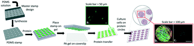
Calcific aortic valvular disease (CAVD) is the most prevalent valvular pathology in the United States. Development of a pharmacologic agent to slow, halt, or reverse calcification has proven to be unsuccessful as still much remains unknown about the mechanisms of disease initiation. Although in vitro models of some features of CAVD exist, their utility is limited by the inconsistency of the size and time course of the calcified cell aggregates. In this study, we introduce and verify a highly reproducible in vitro method for studying dystrophic calcification of cardiac valvular interstitial cells, considered to be a key mechanism of clinical CAVD. By utilizing micro-contact printing, we were able to consistently reproduce cell aggregation, myofibroblastic markers, programmed cell death, and calcium accumulation within aggregates of 50–400 μm in diameter on substrates with moduli from 9.6 to 76.8 kPa. This method is highly repeatable, with 70% of aggregates staining positive for Alizarin Red S after one week in culture. Dense mineralized calcium-positive nanoparticles were found within the valvular interstitial cell aggregates as shown by scanning electron microscopy (SEM) and energy dispersive spectrometry (EDS). The area of micro-contact printed aggregates staining positive for caspase 3/7 activity increased from 5.9 ± 0.9% to 12.6 ± 4.5% over one week in culture. Z-VAD-FMK reduced aggregates staining positive for Alizarin Red S by 60%. The state of cell stress is hypothesized to play a role in the disease progression; traction force microscopy indicates high substrate stresses along the aggregate periphery which can be modulated by altering the size of the aggregates and the modulus of the substrate. Micro-contact printing is advantageous over the currently used in vitro model as it allows the independent study of how cytokines, substrate modulus, and pharmacologic agents affect calcification. This controlled method for aggregate creation has the potential to be used as an in vitro assay for the screening of promising therapeutics to mitigate CAVD.
 Please wait while we load your content...

