
Handheld XRF and Raman equipment for the in situ investigation of Roman finds in the Villa dei Quintili (Rome, Italy)
R.
Alberti
a,
V.
Crupi
b,
R.
Frontoni
c,
G.
Galli
c,
M. F.
La Russa
d,
M.
Licchelli
e,
D.
Majolino
b,
M.
Malagodi
f,
B.
Rossi
g,
S. A.
Ruffolo
d and
V.
Venuti
*b
aXGLab srl, Via Francesco D'Ovidio 3, 20131 Milano, Italy
bDipartimento di Scienze Matematiche e Informatiche, Scienze Fisiche e Scienze della Terra, Università degli Studi di Messina, Viale Ferdinando Stagno D'Alcontres 31, 98166 Messina, Italy. E-mail: vvenuti@unime.it; Fax: +39 090395004; Tel: +39 0906765299
cSoprintendenza Speciale per i Beni Archeologici di Roma, Villa dei Quintili, Via Appia Nuova 1092, 00197 Roma, Italy
dDipartimento di Biologia, Ecologia e Scienze della Terra (DiBEST), Università degli Studi della Calabria, Via Pietro Bucci, 87036 Arcavacata di Rende, Cs, Italy
eDipartimento di Chimica, Università di Pavia, Via Taramelli 12, 27100 Pavia, Italy
fDipartimento di Musicologia e Beni Culturali, Università di Pavia, Corso Garibaldi 178, 26100 Cremona, Italy
gElettra – Sincrotrone Trieste, Strada Statale 14 km 163.5, Area Science Park, 34149 Trieste, Italy
First published on 4th November 2016
Abstract
In the present work, a variety of fragments of frescoes coming from the Villa dei Quintili in Rome (Italy) and dating back to the II century A.D. were subjected to, first of all, an X-ray fluorescence (XRF) analysis by optimizing a portable spectrometer for non-destructive investigation in the field of cultural heritage. The innovative aspect is the ability to obtain coloured maps referring to the distribution and concentration of elements present in the sample. Other than characterization, the aim was to improve the technique for non-invasive and fast in situ analysis. It has been performed, in conjunction with portable Raman analysis, at the ruins of the Villa dei Quintili on samples of different typologies including pottery, statues and frescoes, dating back to the II/III century A.D., different in colour, support and shape. From the results, the identification of the main pigmenting agents is attempted, providing the group of archaeologists in the villa with extremely valuable information for their work too. In particular, by comparing the XRF results for frescoes analysed in the laboratory, taken from the warehouse of the villa, and XRF and Raman data of frescoes analysed in situ, for which the provenance area inside the villa was known, the context of excavation and the manufacture process for some of the former has been hypothesized.
Introduction
Nowadays, cultural heritage is very strictly interconnected with scientific methodologies. A modern view of archaeometry, conservation science and, generally, art analysis cannot be separated from the use of spectroscopic and imaging techniques such as, among the others, X-ray fluorescence spectroscopy (XRF),1,2 inductively coupled plasma mass spectrometry (ICP-MS),3 scanning electron microscopy with energy dispersive X-ray spectroscopy (SEM-EDS),2,4,5 high performance liquid chromatography (HPLC),6 Fourier transform infrared spectroscopy (FT-IR)2,7 and Raman spectroscopy.8Archaeometric studies deal with valuable artifacts. So, their fundamental prerequisite remains an investigation that could be as informative as possible, but, at the same time, non-invasive or, at least, micro-destructive.9,10 In this context, in particular, the combined use of XRF and Raman spectroscopies represents a successful approach for a non-invasive study that allows for complementary compositional information, the first one at the elemental level, and the second one on a molecular scale. It has been already applied in a variety of findings including potteries,11 frescoes,12 glasses,13 gemstones14 and paintings.15 In fact, the elemental (qualitative and/or semi-quantitative) analysis performed by XRF becomes necessary for the unambiguous phase identification of both organic and inorganic materials by the Raman technique, and vice versa. Starting from such a characterization, a reliable hypothesis on the provenance, manufacturing technology, and state of preservation of the artefact can be made.
Recently, mobile instruments have contributed significantly to the in situ investigation of large and/or immovable objects. They have been successfully applied in the case of historical buildings,16 sculptures,17 rock art,18 and stained glass windows.19 Recently, very interesting reviews and books of the progress of mobile spectrometers in archaeometry have been published.20,21
The present work is mainly aimed at the development of a portable instrument (ELIO spectrometer produced by GLAB s.r.l.) for a non-destructive XRF analysis in the field of cultural heritage. The innovative aspect is the ability to obtain, with the same tool used for XRF analysis, colored maps referring to the distribution and concentration of elements present in a given sample. In this sense, the first part of the study, performed in the laboratory, allowed us to test the instrumental set-up and its software. The final aim was to optimize the instrumental performances for a fast and non-invasive in situ analysis. As a case study, a variety of frescoes, potteries and statues coming from the archaeological site of the Villa dei Quintili in Rome (Italy), dating back to the II–III century A.D., have been considered, and precious information for the archaeometric characterization of the fragments has been obtained.
The Villa dei Quintili [Fig. 1], extending between the Via Appia and the Via Appia Nuova, was the largest villa of the Roman suburbs.22 In 1985 the State acquired 23 hectares of the ancient complex, also developed in neighboring properties. The site is known in the ancient cartography as Statuary (for the richness of the sculptures accumulated through the centuries) and Old Rome (for the grandeur of monumental ruins).
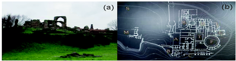 | ||
| Fig. 1 (a) A panoramic view of the Villa dei Quintili. (b) Plan of the Villa dei Quintili and details of the sampled areas. Notes: A = representation area; B = private residences; C = basis villae; D = frigidarium; E = calidarium; F = Ludus-Viridarium; M = small bathroom; R = hippodrome; S = stadium; T = arcades. | ||
The owners, the brothers Sixth Quintilius Condianus and Sixth Quintilius Valerio Massimo, consuls in 151 A.D., were killed by the Emperor Commodus in 182 A.D., and all their properties were confiscated, including the villa at the V mile of the Via Appia which remained an imperial property until the third century A.D.23,24
The excavations, carried out between the end of the XVIII and the beginning of the XIX century, have yielded numerous works of art, now preserved in various museums and private collections in Italy and abroad (the Louvre, the Hermitage, the Vatican Museums, and the Torlonia Collection). Additional findings in 1925 and 1929 have been occasional: the works discovered are now exhibited in the National Museum of Palazzo Massimo and in the Villa dei Quintili itself, along with some findings of the most recent excavations.
The residential complex underwent over the centuries a sequence of different building phases until the fourth century AD, and it became a center of recycling and production of different materials, such as marble, bricks, tuffs, and metallic and glassy materials. Several materials, especially frescos fragments, were removed from their original location and reused. From the archeological point of view, the site is thus a complex case study and the analytical characterization of the pigments can support the conservators and restorers to (i) increase the knowledge on the original position of the frescos and (ii) rewrite a correct map of the different areas of the villa.
This study allowed us to identify, easily and quickly thanks to the use of portable spectrometers, the elemental constituents, by XRF, of the pigmenting agents of the analysed samples, and, as an innovative aspect, their distribution and concentration in a given sample. Nevertheless, as is well known, in the case of pigments with the same or similar colour and same main elements, or pigments with the same colour and same formula, or mixtures of pigments, any conclusion on the nature of pigments coming only from the elemental composition as obtained from XRF remains a reasonable hypothesis. So, the unambiguous identification of the pigments, starting from their elemental composition, was also attempted by a complementary inspection, by Raman spectroscopy, of their molecular nature. The obtained results are crucial in archaeological research, since the knowledge of materials and preparation methods of pigmenting agents, peculiar to a specific population and technology development, is extremely helpful for a correct historical-geographical classification of the artworks. In particular, in our case, starting from similarities observed in the elemental XRF composition of frescoes analysed in the laboratory, taken from the warehouse of the villa and hence of an unknown provenance area and for which a Raman investigation of the molecular nature of the pigments was impossible because of the organic layers that covered them, and some of the frescoes investigated in situ, of a certain provenance area and for which the Raman technique allowed the identification of pigmenting agents, a hypothesis concerning the context of excavation and the manufacturing technology of some of the former has been for the first time attempted.
The reported investigation is carried out in the framework of well-established multi-technique archaeometric research on this renowned site.25–27 The sample selection is performed according to the NORMAL 3/80 (ref. 28) with the support of archaeologists from the Archaeological Superintendence of Rome.
Experimental
Materials
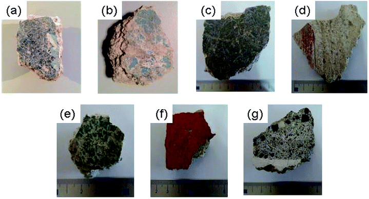 | ||
| Fig. 2 Images of the samples analysed in the laboratory. (a) Fresco Cod. r19d, (b) Fresco Cod. r19e, (c) Fresco Cod. r19b, (d) Fresco Cod. r19c, (e) Fresco Cod. r19f, (f) Fresco Cod. r63, and (g) Fresco Cod. section. | ||
We want to also add that since these aforementioned samples came from the warehouse of the Villa dei Quintili, they have, in the course of time, accumulated layers of organic materials on the pictorial film. So, even if Raman spectroscopy on these samples was also attempted, the presence of these layers generated a high fluorescence that completely covered the signal coming from the pigment, making the Raman results unreliable.
Investigated samples are earthenwares, statues and frescoes; for each category, chosen samples differ in colour, shape and support.
All the information relative to the investigated samples is reported in Table 1.
| Sample | Information | Performed analysis |
|---|---|---|
| Earthenwares | ||
| Brick with red writing | Not inventoried | XRF |
| Figured tile terminal | Not inventoried | XRF |
| Head of a man with a stucco tiara | II century A.D. | XRF |
| From the area (R) | ||
![[thin space (1/6-em)]](https://www.rsc.org/images/entities/char_2009.gif) |
||
| Statues | ||
| Statue of Zeus sitting on a rock | First half of the II century A.D. | XRF, Raman |
| From the Villa, 1925–1926 | ||
| Memorial, votive support to Zeus Brontoon dedicated by the matron Arrulia Hygia for the happy return from a trip | III century A.D. | XRF |
| From the VII km of the Via Appia Nuova cd. “Oriental sanctuary”, 1929 | ||
| Relief with the Phoenician goddess Astarte | II century A.D. | XRF |
| From the VII km of the Via Appia Nuova cd. “Oriental sanctuary”, 1929 | ||
| Alabaster bustier of Isis | II–III century A.D. | XRF |
| From the VII km of the Via Appia Nuova cd. “Oriental sanctuary”, 1929 | ||
| Headless statue of Heracles | II/III century A.D. | XRF |
| From the VII km of the Via Appia Nuova cd. “Oriental sanctuary”, 1929 | ||
|
||
| Frescoes | ||
| Fragment of a wall painting with floral elements and gems | II century A.D. | XRF |
| From the Ludus Viridarium area | ||
| Fragment of a wall painting with vegetable traces | II century A.D. | XRF, Raman |
| From the area between the residential sector (B) and the basis villae (C) | ||
| Fragment of a wall painting with vegetable festoon | II century A.D. | XRF, Raman |
| From the area between the residential sector (B) and the basis villae (C) | ||
| Fragment of a wall painting with geometric and plant decorations | II century A.D. | XRF, Raman |
| From the area between the residential sector (B) and the basis villae (C) | ||
Some representative images are reported in Fig. 3, as examples.
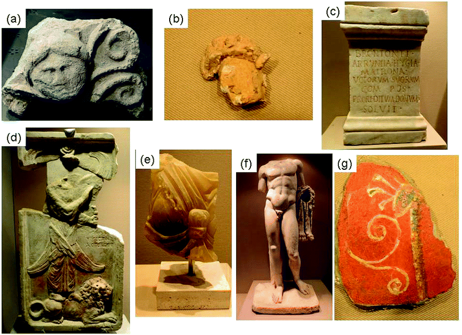 | ||
| Fig. 3 Some representative images of the samples analysed in situ. (a) Figured tile terminal, (b) head of a man with a stucco tiara, (c) memorial, votive support to Zeus Brontoon dedicated by the matron Arrulia Hygia for the happy return from a trip, (d) relief with the Phoenician goddess Astarte, (e) alabaster bustier of Isis, (f) headless statue of Heracles, and (g) fragment of a wall painting with vegetable traces. | ||
Methods
000. The instrument is equipped with a large area Silicon Drift X-ray Detector (SDD), with an active area of 25 mm2 and a resolution energy less than 135 eV, measured at the MnKα line (5.9 eV). As an extremely innovative aspect of this instrument, the elemental 2D mapping of the sample surface can be achieved through automatic XY raster scanning to perform elemental mapping. The translator stage can cover a maximum area of 10 × 10 cm2 with an accuracy of 10 μm. With respect to other XRF mapping devices,29 the compact head and the absence of any X-ray optics make it easily usable for in situ studies and ensure a good sensitivity even to lighter elements.30
Raman measurements have been performed by using a portable ‘BTR 111 MiniRamTM’ (B&W TEK, USA) spectrometer with an excitation wavelength of 785 nm (diode laser), ∼280 mW maximum laser power at the excitation port, and a Charge Coupled Device (CCD) detector (Thermoelectric Cooled, TE). In addition, the laser output power can be continuously adjusted in order to obtain the best signal-to-noise ratio in the minimum integration time. The system is equipped with a fibre optic interface for convenient sampling. The excitation laser emits from the probe's end where the Raman signal is then collected from the sample. The spot size is 85 μm at a working distance of 5.90 mm.
After that, an ELIO spectrometer has also been used for 2D or 1D mapping of the elements. This is an innovative aspect: in this way it is possible to obtain the mapping of the elements present in a precise area of the sample. In our case, the following measurement conditions were used: measurement time per point: 1 s, tube voltage: 50 kV, tube current: 80 μA, movement delay: 100 ms, number of rows: 36, number of columns: 36, row distance: 0.5 mm, and column distance: 0.5 mm. The maps have been elaborated by using PyMCA software.
As far as Raman measurements are concerned, the maximum power at the samples for our measurements was ∼55 mW. Spectra have been registered in the 60–3150 cm−1 wavenumber range, by using an acquisition time of 40 s and a resolution of 8 cm−1, adding several scans for each spectrum in order to improve the signal-to-noise ratio. An accurate wavelength calibration was performed by using an instrument from B&W Tek. Anyway, the optimal performance of the instrument was guaranteed by a calibration procedure before each measurement using the peak at 520.6 cm−1 of a silicon chip. Each spectrum has been processed by subtracting the blank spectrum; in addition, a baseline correction and a smoothing process have been performed by using the BWSpec 3.27 software. The collected Raman spectra have been compared with data from databases and the literature.
Results and discussion
XRF laboratory analysis
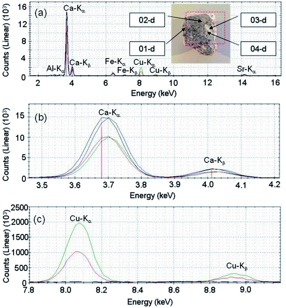 | ||
| Fig. 4 (a) XRF spectrum of the fresco Cod. r19d. In the inset, a photograph of the sample together with the analysed points is shown. (b) Magnification of the XRF spectrum corresponding to the Ca-Kα and Ca-Kβ peaks. (c) Magnification of the XRF spectrum corresponding to the Cu-Kα and Cu-Kβ peaks. 01-d: black line, 02-d: blue line, 03-d: red line, and 04-d: green line. | ||
The points 01-d and 02-d (points of the base) show similar counts of Ca, higher than that of the point 03-d (white pigment) and of the point 04-d (blue pigment). In the point 04-d (blue pigment) Cu is observed, with higher counts than that of the point 03-d (white point). Instead, in the points of the base Cu is completely absent. In Fig. 5 we report the maps obtained by the XRF mapping analysis.
 | ||
| Fig. 5 XRF maps of the fresco Cod. r19d, together with a photo of the analysed part. | ||
The mapping confirms what was observed by point analysis: calcium and copper are, in fact, complementary. Calcium is primarily present in the ground of the fresco, whereas corresponding to the blue pigment its concentration is much lower. Conversely, copper is present only in the blue pigment and completely absent in the ground. Strontium is present in a negligible amount, while the iron map might be the result of a change in the thickness of the sample. From the results, it is reasonable to hypothesize that the used blue pigment consisted mainly of copper. The ground of the fresco is instead a stone material mainly containing calcium.31
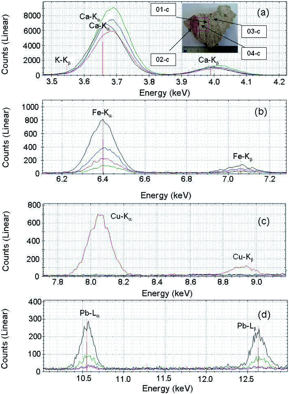 | ||
| Fig. 6 Magnification of the XRF spectrum of the fresco Cod. r19c corresponding to (a) the Ca-Kα and Ca-Kβ peaks, (b) the Fe-Kα and Fe-Kβ peaks, (c) the Cu-Kα and Cu-Kβ peaks, and (d) the Pb-Lα and Pb-Lβ peaks. In the inset, a photograph of the sample together with the analysed points is given. 01-c: blue line, 02-c: black line, 03-c: red line, and 04-c: green line. | ||
 | ||
| Fig. 7 XRF total spectrum of the fresco Cod. r19c. 01-c: blue line, 02-c: black line, 03-c: red line, and 04-c: green line. | ||
In all the analysed points, Ca was identified. The points 01-c (without pigment) and 02-c (red pigment) have a very similar Ca amount, the point 04-c (base) has a slightly greater Ca amount, while the point 03-c (blue pigment) has a slightly minor Ca amount. According to the Fe concentration, it is possible to classify the points in the following ascending order: 04-c (base), 03-c (blue pigment), 01-c (without pigment) and finally 02-c (red pigment). The point with traces of the blue pigment (03-c) shows the presence of Cu, which is completely absent in all other analysed points. The point analysed corresponding to the red pigment (02-c) shows the presence of Pb, observed in a much lesser extent in the point of the base (04-c) and completely absent in the other two points. The results of the XRF mapping are reported in Fig. 8.
 | ||
| Fig. 8 XRF maps of the fresco Cod. r19c together with a photo of the analysed part. | ||
It is possible to observe how calcium is primarily present in the base of the fresco, and corresponding to the red pigment, in fact, its concentration decreases. Again, the red pigment reveals the presence of calcium, iron and manganese. The study of the elemental composition of the pigments is important just in order to compare the fragments of frescos with each other and to obtain information about the technical methodology of Romans to preparing frescos. Strontium is non-uniformly distributed over the whole sample and in a negligible amount compared to the other elements.32 Cu, observed in the point analysis corresponding to the trace of blue pigment, was not identified in the XRF mapping due to the reduced amount.
XRF and Raman in situ analysis
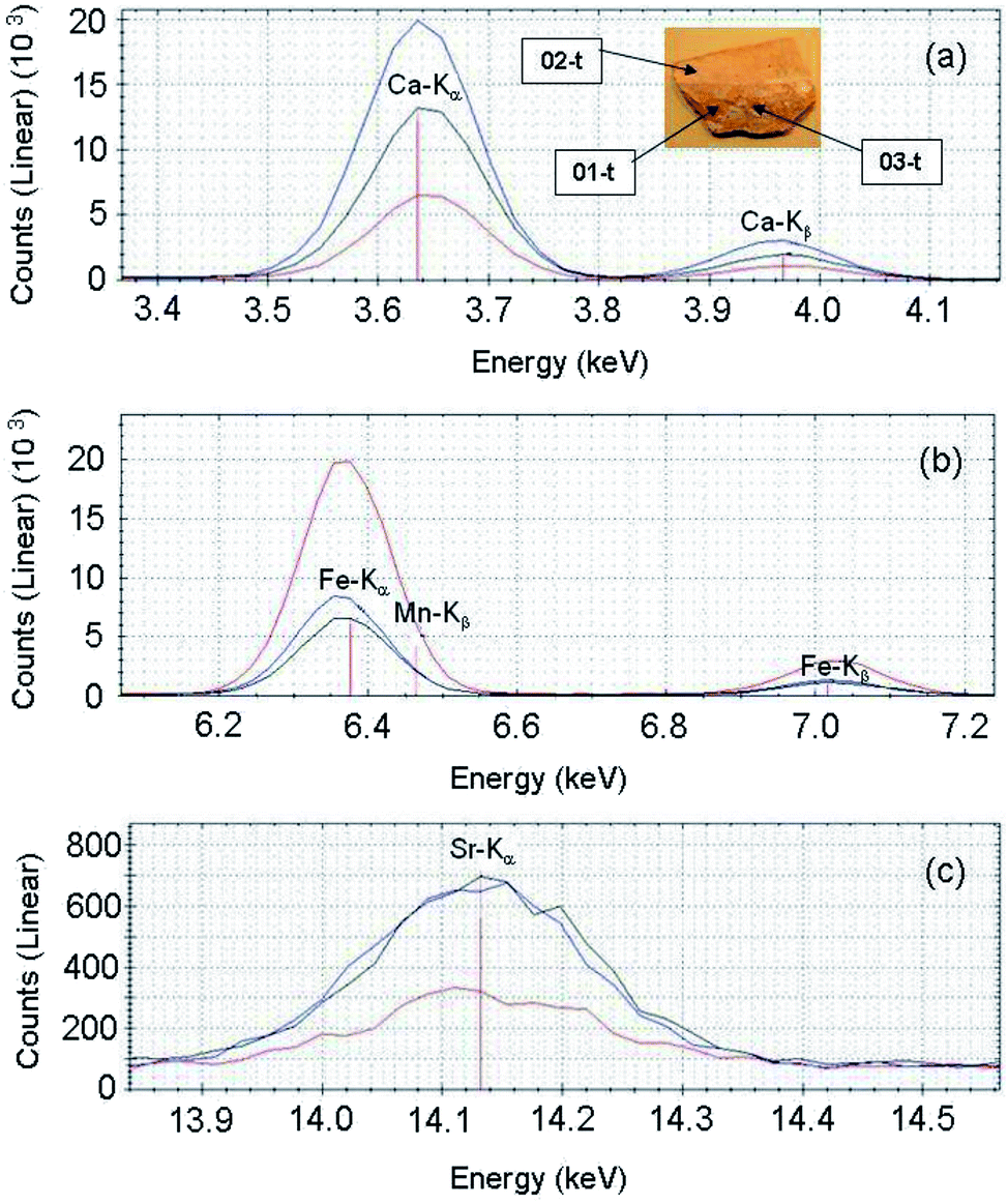 | ||
| Fig. 9 Magnification of the XRF spectrum of the brick with red writing corresponding to (a) the Ca-Kα and Ca-Kβ peaks, (b) the Fe-Kα and Fe-Kβ peaks, and (c) the Sr-Kα peak. In the inset, a photograph of the sample together with the analysed points is given. 01-t: blue line, 02-t: black line, and 03-t: red line. | ||
 | ||
| Fig. 10 XRF total spectrum of the brick with red writing for the three analysed points. 01-t: blue line, 02-t: black line, and 03-t: red line. | ||
From the analysis of the XRF spectra, Ca is mainly observed in the 01-t point (red pigment), in a lower quantity in the 02-t point (pigment absent), and, finally, it is not revealed in the 03-t point (more intense red pigment). Again, the points 01-t (red pigment) and 02-t (pigment absent) have a very similar amount of Fe, and the point 03-t (more intense red pigment) has a greater Fe amount than the other two points. In contrast, Sr is mainly observed, in a similar amount, in 01-t (red pigment) and 02-t points (pigment absent), and in a lower quantity in the 03-t point (more intense red pigment). These observations allowed us to affirm that the used red pigment was an earth (inorganic pigment of differently hydrated iron oxide). The support is rather a Ca-based stone material.
:1.
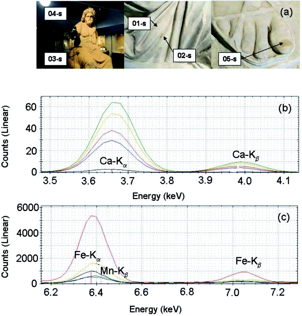 | ||
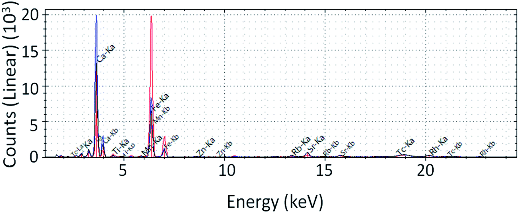
| Fig. 11 (a) Photograph of the analysed points of the sample, together with a magnification of the XRF spectrum corresponding to (b) the Ca-Kα and Ca-Kβ peaks and (c) the Fe-Kα and Fe-Kβ peaks. 01-s: blue line, 02-s: black line, 03-s: red line, 04-s: green line, and 05-s: yellow line. | ||
 | ||
| Fig. 12 XRF total spectrum of the statue of Zeus sitting on a rock for the five analysed points. 01-s: blue line, 02-s: black line, 03-s: red line, 04-s: green line, and 05-s: yellow line. | ||
Based on the increasing concentration of Ca, the analyzed points can be ordered as follows: 02-s (details of the dress, right leg with a red pigment), 01-s (details of the dress, right leg without any pigment), 03-s (details of the dress, right leg without pigment_1), 05-s (right big toe toenail), and 04-s (right shoulder). Again, based on the increasing concentration of Fe, the analyzed points can be ordered as follows: 04-s (right shoulder) and 01-s (details of the dress, right leg without any pigment) have a concentration of Fe almost identical, followed by 02-s (details of the dress, right leg with a red pigment), 05-s (right big toe toenail) and 03-s (especially details of the dress, right leg without pigment_1). From a general point of view, one can note that the concentrations of calcium and iron are inversely proportional: points having a high Fe-concentration exhibit a low Ca-concentration and vice versa. The iron traces corresponding to the red pigment may suggest the use of a clay (inorganic pigment based on differently hydrated iron oxide).
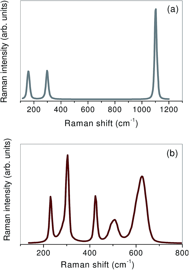
Raman measurements performed on different reddish areas of the statue allowed us to clearly identify the peaks centred at ∼166 cm−1, ∼311 cm−1, and ∼1116 cm−1, typical of calcite, as shown in Fig. 13.
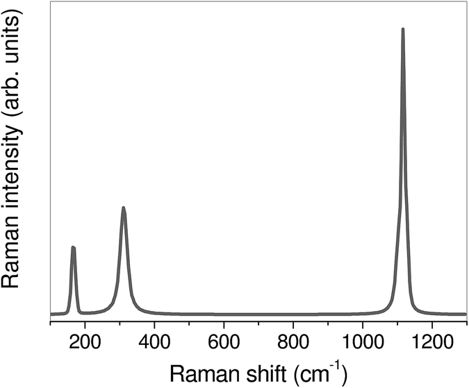 | ||
| Fig. 13 Raman spectrum collected on a reddish area of the Statue of Zeus sitting on a rock. | ||
:1.
 | ||
| Fig. 14 Photograph of the analysed part of the fragment of a wall painting with floral elements and gems, together with the XRF maps of the sample. | ||
Furthermore, Fig. 14 shows the XRF maps obtained for this sample.
Taking into account both the point XRF analysis and XRF mapping we can hypothesize that the red pigment used was vermilion. The presence of Fe in the yellow pigment allows us to hypothesize the use of yellow ocher (natural earths, yellow coloured by hydrated iron oxides, the most important of which is Fe2O3·H2O). The presence of Pb corresponding to the brown pigment could be the consequence of an alteration. Minium, an inorganic red pigment having formula Pb2O3, in the murals and in the case of humid environments oxidizes to brown PbO2. Today the pigment appears brown in colour, but in the past it could have been red.
The observations by XRF analysis are clearly supported by Raman data collected on white, green and red pigments and reported in Fig. 15(a)–(c), respectively. In particular, the white pigment turned out to be mainly composed of calcite. Going on, for the green pigment the Raman peaks centred at ∼149 cm−1, 310 cm−1, 550 cm−1, 640 cm−1, 680 cm−1, 850 cm−1, 1040 cm−1 and 1094 cm−1 can be recognized, typically attributable to green earth. Finally, the Raman spectrum of the red pigment clearly reveals the contributions at ∼268 cm−1, ∼305 cm−1 and ∼365 cm−1, usually ascribed to vermilion.
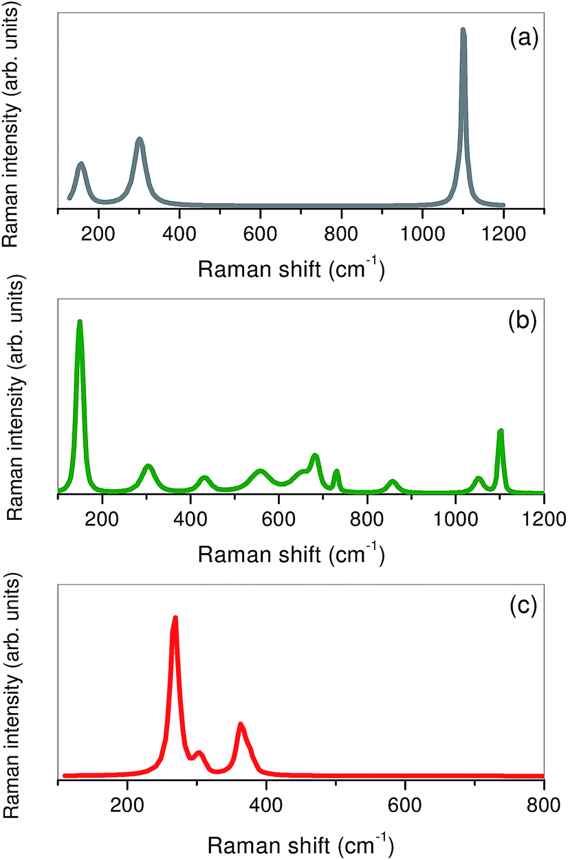 | ||
| Fig. 15 Raman spectra of (a) white, (b) green, and (c) red pigments of the fragment of a wall painting with vegetable traces. | ||
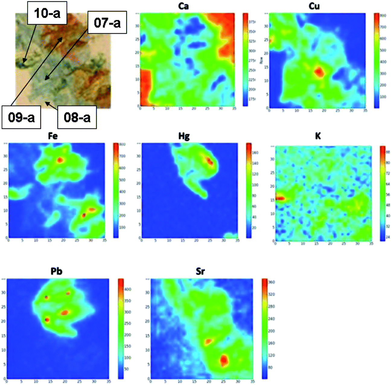 | ||
| Fig. 16 Photograph of the analysed part of the fragment of a wall painting with vegetable festoon, together with the XRF maps of the sample. | ||
Combining the point XRF analysis and XRF mapping it is possible to deduce that a Ca-based pigment (perhaps simple calcite, also used in the preparatory layer) has been used as the white pigment.33 The blue pigment, the only one presenting Cu traces, could be azurite (pigment consisting of basic copper carbonate); while the green pigment, which shows the presence of Fe, could be green earth. Finally, the presence of Pb only in the red pigment might suggest the use of vermilion to which, however, a clay (inorganic pigment containing impure anhydrous ferric oxide) was added. Calcium is the main element of the material used as the support.
Raman spectra collected on white and red pigments (Fig. 17(a) and (b) respectively) support the experimental XRF observations. On the one side, the observation of the typical peaks of calcite allows us to classify the white pigment as calcite. On the other side, the spectrum of the red pigment exhibits peaks at ∼228 cm−1, and ∼302 cm−1, which are attributable to vermilion and/or red ochre, and ∼425 cm−1, ∼504 cm−1 and ∼625 cm−1 that can be ascribed to red ochre.
 | ||
| Fig. 17 Raman spectra of (a) white and (b) red pigments of the fragment of a wall painting with vegetable festoon. | ||
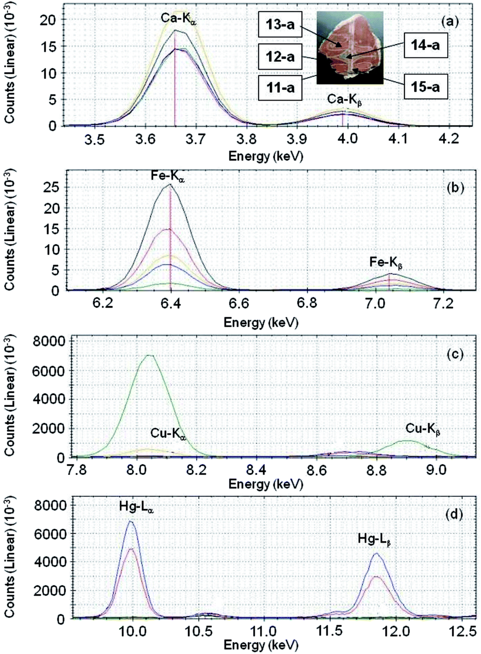 | ||
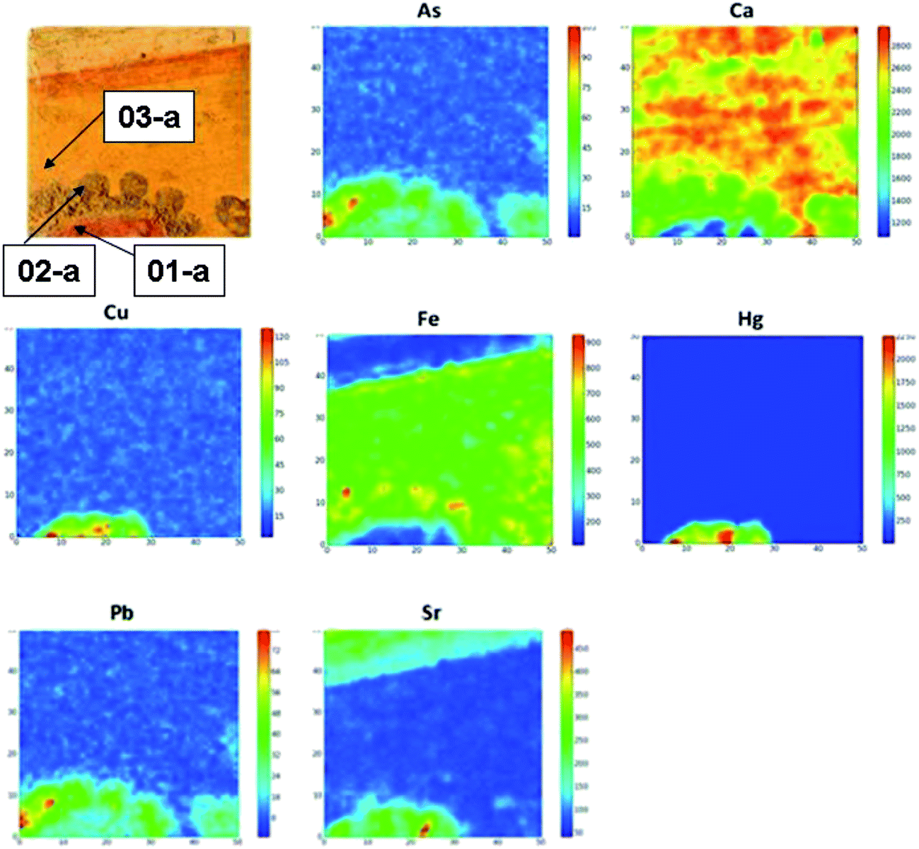
| Fig. 18 Magnification of the XRF spectrum of the fragment of a wall painting with geometric and plant decorations corresponding to (a) the Ca-Kα and Ca-Kβ peaks, (b) the Fe-Kα and Fe-Kβ peaks, (c) the Cu-Kα and Cu-Kβ peaks, and (d) the Hg-Lα and Hg-Lβ peaks. In the inset, a photograph of the sample together with the analysed points is given. 11-a: blue line, 12-a: black line, 13-a: red line, 14-a: green line, and 15-a: yellow line. | ||
Ca is observed in increasing amount passing from red pigments of the decorations and blue pigments (11-a, 13-a and 14-a, respectively), to the red pigment in the base (12-a), to, finally, the white pigment (15-a). As far as the Fe concentration is concerned, it is observed to increase going from the blue pigment (14-a), the red pigment in the decoration at the bottom (11-a), the white pigment (15-a), the red pigment of the decoration at the top (13-a), and the red pigment of the base (12-a). The presence of Cu is detected in the blue pigment (14-a) and, in traces, in the white pigment (15-a). Hg was detected in the red pigments used for decorations (11-a and 13-a). Observing the results of the punctual XRF analysis it is possible to say that a Ca-based pigment (perhaps simple calcite, also used for the realization of the preparatory layer) has been used as the white pigment. The presence of Cu in the blue pigment suggests the use of azurite (inorganic pigment mainly consisting of Cu). The red support and the red decoration show two different pigments. For the red pigment of the base red clay (inorganic Fe-based pigment) was used, while for the red pigment of the decoration vermilion (more precious than the red earth) has been used. The support is a Ca-based stone material. The XRF mapping results, not reported here, confirm the results obtained by punctual XRF analysis. Furthermore, the identification of lead corresponding to the red decorations allowed us to hypothesize that the red decorations have been made using vermilion, to which a small amount of red lead was added in order to obtain a specific shade.
Once again, the use of Raman spectroscopy supported the XRF results, allowing us to unambiguously identify the use of calcite (Fig. 19(a)) for the white pigment, and vermilion (Fig. 19(b)) for the red pigment of the decoration.
 | ||
| Fig. 19 Raman spectra of (a) the white and (b) red pigments of the decoration of the fragment of a wall painting with geometric and plant decorations. | ||
Now, the XRF results for the frescoes analysed in situ “wall painting with vegetable traces”, “wall painting with vegetable festoon” and “wall painting with geometric and plant decorations”, excavated from specific areas of the villa, as reported in Table 1, and for which Raman spectroscopy allowed for the unambiguous identification at the molecular level of the pigmenting agents, have been compared with the XRF data for the frescoes analysed in the laboratory, of an unknown provenance area inside the villa and for which the molecular identification of the pigments was made impossible by Raman analysis because of their covering organic layer that gave rise to a too high fluorescence. The comparison revealed remarkable similarities in the elemental composition between the fresco “wall painting with vegetable traces” and the fresco Cod. r63, and between the fresco “wall painting with vegetable festoon” and the fresco Cod. r19c. It is then reasonable to hypothesize, for frescoes Cod. r63 and r19c, a context of excavation from the area between the residential sector (B) and the basis villae (C) (as in the case of the two aforementioned frescoes analysed in situ). Again, taking into account the Raman results observed for the frescoes investigated in situ, we can make some hypotheses on the nature of pigments. In the case of fresco Cod. r63 a red pigment vermillion, HgS precisely, could have been used for the accomplishment of the work. As far as fresco Cod. r19c is concerned, red ochre (anhydrous ferric oxide impure of clays) could have been used for the realization of the red pigment, with a certain amount of red lead added in order to obtain a particular shade of colour. The trace of the blue pigment could be azurite.
Conclusions
In the present work, an innovative use of a portable spectrometer for non-invasive X-ray fluorescence (XRF) analysis of art objects, consisting in the compositional mapping of the sample's surface, other than the usual punctual analysis, is proposed. It has been applied, as the first step, in the laboratory, on a series of fresco fragments coming from the archaeological site of the Villa dei Quintili in Rome (Italy) and dating back to the II century A.D. Other than the compositional characterization at the elemental level, the research was devoted to an improvement of the performance of the instrument for a non-destructive and fast in situ analysis.34After that, the performance of the XRF portable set-up in identifying the key-elements of a variety of colouring matters has been tested in situ on samples of different nature including potteries, statues and frescoes, dating back to the II–III century A.D. The limits of this technique in mapping surfaces not perfectly flat and concentrations of the pigments were considered. The hypothesized nature of pigmenting agents is achieved by molecular characterization non-invasively performed by means of a handheld Raman spectrometer.
The relevance of this preliminary and non-invasive approach is also due to the comparison of several samples to each other, that allowed us to ascribe the right positioning of some frescos in a specific area of the villa, a historical task requested from the conservators of the archaeological site.
Acknowledgements
The authors acknowledge Dr Rita Paris, Archaeologist Director of the Soprintendenza Speciale per I Beni Archeologici di Roma, for supplying the investigated fragments of painted plasters, withdrawn from the archaeological site of the Villa dei Quintili (Rome, Italy), and for allowing the in situ analysis. We thank Dr Michele Gironda and the staff of the XGLab company.Notes and references
- F. Bardelli, G. Barone, V. Crupi, F. Longo, D. Majolino, P. Mazzoleni and V. Venuti, Anal. Bioanal. Chem., 2011, 339, 3147 CrossRef PubMed.
- E. Aquilia, G. Barone, V. Crupi, F. Longo, D. Majolino, P. Mazzoleni and V. Venuti, J. Anal. At. Spectrom., 2011, 26, 977 RSC.
- B. Li, J. Zhao, K. D. Collerson and A. Greig, Chin. Sci. Bull., 2003, 48, 1219 CrossRef CAS.
- G. Montana, E. Tsantini, L. Randazzo and A. Burgio, Archaeometry, 2013, 55, 591 CrossRef CAS.
- E. Basso, C. Invernizzi, M. Malagodi, M. F. La Russa, D. Bersani and P. P. Lottici, J. Raman Spectrosc., 2014, 45, 238 CrossRef CAS.
- I. Karapanagiotis, J. Theologou, A. Lakka, A. Ozoline and C. Panayiotou, Archaeometry, 2011, 53, 587 CrossRef CAS.
- D. Barilaro, G. Barone, V. Crupi, D. Majolino, P. Mazzoleni, M. Triscari and V. Venuti, Vib. Spectrosc., 2006, 42, 381 CrossRef CAS.
- G. Barone, D. Bersani, V. Crupi, F. Longo, U. Longobardo, P. P. Lottici, I. Aliatis, D. Majolino, P. Mazzoleni, S. Raneri and V. Venuti, J. Raman Spectrosc., 2014, 45, 1309 CrossRef CAS.
- D. Miriello, M. Malagodi, S. A. Ruffolo, M. F. La Russa, G. M. Crisci, A. Pezzino, R. Galluccio, D. Barca and E. Marasco, Environ. Earth Sci., 2010, 60, 829 CrossRef CAS.
- M. F. La Russa, S. A. Ruffolo, M. Malagodi, D. Barca, R. Cirrincione, A. Pezzino, G. M. Crisci and D. Miriello, Appl. Phys. A: Mater. Sci. Process., 2010, 100, 865 CrossRef CAS.
- R. Scarpelli, R. J. Clark and A. M. De Francesco, Spectrochim. Acta, Part A, 2014, 120, 60 CrossRef CAS PubMed.
- L. Appolonia, D. Vaudan, V. Chatel, M. Aceto and P. Mirti, Anal. Bioanal. Chem., 2009, 395, 2005 CrossRef CAS PubMed.
- D. Lauwers, A. Candeias, A. Coccato, J. Mirao, L. Moens and P. Vandenabeele, Spectrochim. Acta, Part A, 2016, 157, 146 CrossRef CAS PubMed.
- Z. Petrová, J. Jehlička, T. Čapoun, R. Hanus, T. Trojek and V. Goliáš, J. Raman Spectrosc., 2012, 43, 1275 Search PubMed.
- G. Casuccio, K. Bunker, S. Kennedy, M. Sparrow, B. Pacolay, P. Ioannidis and R. Foulke, Microsc. Microanal., 2012, 18, 958 CrossRef.
- N. Prieto-Taboada, I. Ibarrondo, O. Gómez-Laserna, I. Martinez-Arkarazo, M. A. Olazabal and J. M. Madariaga, J. Hazard. Mater., 2013, 248–249, 451 CrossRef CAS PubMed.
- C. Colombo, F. Bevilacqua, L. Brambilla, C. Conti, M. Realini, J. Striova and G. Zerbi, Anal. Bioanal. Chem., 2011, 401, 757 CrossRef CAS PubMed.
- L. C. Prinsloo, A. Tournié, P. Colomban, C. Paris and S. T. Bassett, J. Archaeol. Sci., 2013, 40, 2981 CrossRef CAS.
- P. Colomban and A. Tournié, Journal of Cultural Heritage, 2007, 8, 242 CrossRef.
- P. Colomban, J. Raman Spectrosc., 2012, 43, 1529 CrossRef CAS.
- D. N. Papadopoulou, G. A. Zachariadis, A. N. Anthemidis, N. C. Tsirliganis and J. A. Stratis, Talanta, 2006, 68, 1692 CrossRef CAS PubMed.
- A. Rotondi, Forma Urbis-Itinerari nascosti di Roma Antica, ANNO XVII(2), Roma, 2012 Search PubMed.
- R. Frontoni, Forma Urbis-Itinerari nascosti di Roma Antica, ANNO V(7/8), Roma, 2000 Search PubMed.
- R. Frontoni and G. Galli, Arkos, Scienza e Restauro, 2010, vol. 25, p. 66 Search PubMed.
- C. M. Belfiore, G. V. Fichera, M. F. La Russa, A. Pezzino, S. A. Ruffolo, G. Galli and D. Barca, Archaeometry, 2015, 57, 269 CrossRef CAS.
- V. Crupi, G. Galli, M. F. La Russa, F. Longo, G. Maisano, D. Majolino, M. Malagodi, A. Pezzino, M. Ricca, B. Rossi, S. A. Ruffolo and V. Venuti, Appl. Surf. Sci., 2015, 349, 924 CrossRef CAS.
- V. Crupi, V. Allodi, C. Bottari, F. D'Amico, G. Galli, A. Gessini, M. F. La Russa, F. Longo, D. Majolino, G. Mariotto, C. Masciovecchio, A. Pezzino, B. Rossi, S. A. Ruffolo and V. Venuti, Vib. Spectrosc., 2016, 83, 78 CrossRef CAS.
- NORMAL 3/80, Materiali lapidei: campionamento e conservazione dei campi-oni, Roma, 1980 Search PubMed.
- S. Mosca, T. Frizzi, M. Pontone, R. Alberti, L. Bombelli, V. Capogrosso, A. Nevin, G. Valentini and D. Comelli, Identification of pigments in different layers of illuminated manuscripts by X-ray fluorescence mapping and Raman spectroscopy, Microchem. J., 2016, 124, 775–784, DOI:10.1016/j.microc.2015.10.038.
- M. Alfeld, J. V. Pedroso, M. van Eikema Hommes, G. Van der Snickt and K. Janssens, et al., A mobile instrument for in situ scanning macro-XRF investigation of historical paintings, J. Anal. At. Spectrom., 2013, 28, 760, 10.1039/c3ja30341a . s.l.: Microchem. J. 124(2016) 775–784.
- A. Zielińska, W. Dąbrowski, T. Fiutowski, B. Mindur, P. Wiącek and P. Wróbel, X-ray fluorescence imaging system for fast mapping of pigment distributions in cultural heritage paintings, J. Instrum., 2013, 8, P10011, DOI:10.1088/1748-0221/8/10/p10011.
- A. Deneckere, B. Vekemans, L. Van de Voorde, P. De Paepe, L. Vincze, L. Moens and P. Vandenabeele, Feasibility study of the application of micro-Raman imaging as complement to micro-XRF imaging, Appl. Phys. A, 2012, 116, 363–376, DOI:10.1007/s00339-011-6693-5.
- A. Deneckere, M. De Reu, M. P. J. Martens, K. De Coene, B. Vekemans, L. Vincze, P. De Maeyerd, P. Vandenabeelee and L. Moensa, The use of a multi-method approach to identify the pigments in the 12th century manuscript Liber Floridus, Spectrochim. Acta, Part A, 2011, 80, 125–132, DOI:10.1016/j.saa.2011.03.005.
- E. Ravaud, L. Pichon, E. Laval, V. Gonzalez, M. Eveno and T. Calligaro, Development of a versatile XRF scanner for the elemental imaging of paintworks, Appl. Phys. A: Mater. Sci. Process., 2016, 122, 1–7, DOI:10.1007/s00339-015-9522-4.
| This journal is © The Royal Society of Chemistry 2017 |