
Highlights from Faraday Discussion FDSERS17: Surface Enhanced Raman Scattering – SERS, Glasgow, UK, 30th August–1st September 2017
G.
Di Martino†
 a,
H.
Fleming†
b,
M.
Kamp†
*c and
F.
Lussier†
d
a,
H.
Fleming†
b,
M.
Kamp†
*c and
F.
Lussier†
d
aNanoPhotonics Centre, Cavendish Laboratory, University of Cambridge, CB3 0HE, UK
bEaStCHEM, School of Chemistry, University of Edinburgh, EH9 3FJ Edinburgh, UK
cMelville Laboratory for Polymer Synthesis, University of Cambridge, CB2 1EW, UK. E-mail: mk841@cam.ac.uk
dDepartment of Chemistry, Université de Montréal, H3C 3J7, Canada
First published on 16th November 2017
Abstract
The 2017 Faraday Discussion on Surface Enhanced Raman Scattering (SERS) attracted more than a hundred delegates from a broad spectrum of backgrounds and experience levels, bringing together leading scientists involved in the long living field of SERS. The meeting gave an overview of the liveliness of the topic, characterised by open questions and fascinating science still to discover. In the following, we discuss the topics covered during this meeting and briefly highlight the content of each presentation.
On 30th August 2017, around 140 delegates representing 30 different countries gathered in a remarkably sunny Glasgow, at the University of Strathclyde, to attend the 2017 Faraday Discussion on Surface Enhanced Raman Scattering (Fig. 1). “The discussion meeting brought together a real mix of SERS researchers embracing chemistry, physics, and engineering. It mixed theory, modelling and experimental approaches together to produce a holistic view of the state of the art in SERS”, observed Prof. D. Graham (University of Strathclyde, UK).
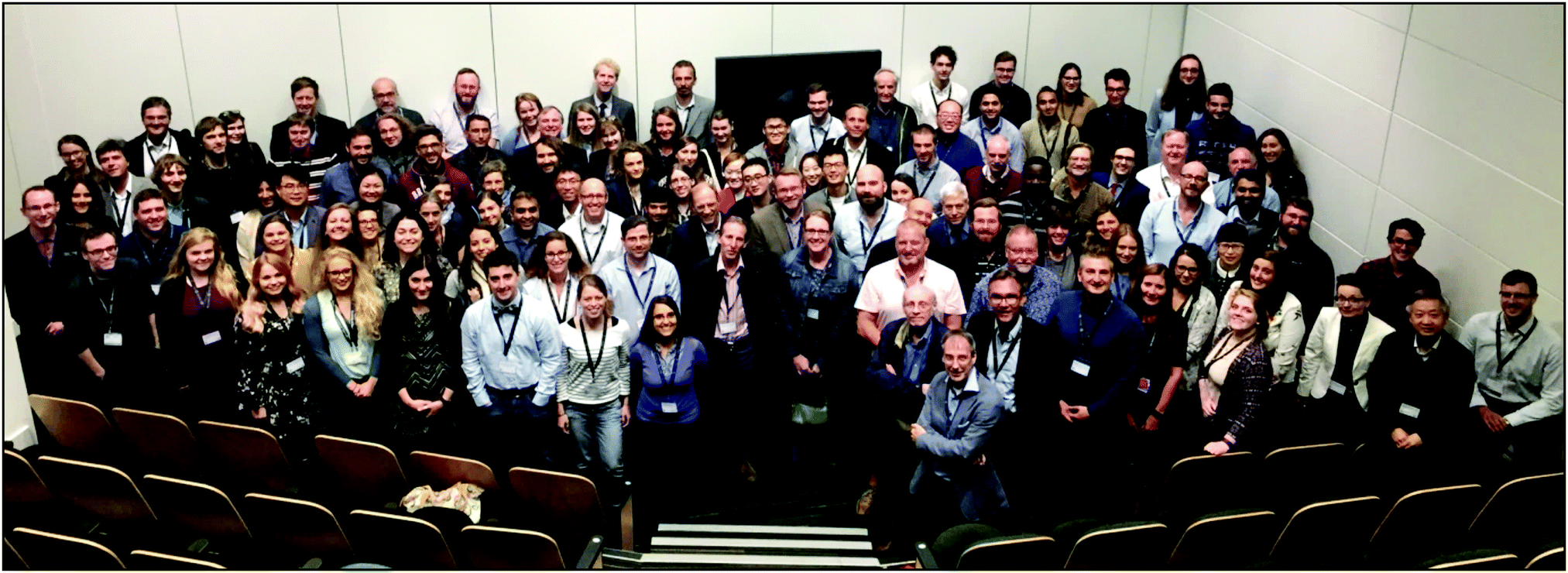 | ||
| Fig. 1 All of the delegates participating in the Faraday Discussion: Surface-Enhanced Raman Scattering – SERS held on 30th August – 1st September in Glasgow, UK. Photo taken by Mr Y. Li, Royal Society of Chemistry. | ||
The Faraday Discussions are unique international discussion meetings that focus on rapidly developing areas of chemistry and their interfaces with other scientific disciplines. The Discussions were founded in 1902; it was an idea conceived by Frederick S. Spiers who later became the Faraday Discussions Secretary of the Faraday Society. In line with the format of Faraday Discussions, all of the speakers submitted a paper prior to this meeting, which was made available for all of the participants to read before the event. During their respective sessions, the presenters were given 5 minutes each to briefly summarize the main findings of their work, followed by an open discussion with 30 minutes allotted for public and live peer review of each article contributed.
The discussions were preceded by a brief introduction from Prof. D. Graham on the topic of the meeting (Fig. 2a) and an introduction to the Faraday Discussions' format by the RSC Publishing Editors for the event, Sarah Sharp and Alexander Whiteside. The president of the Faraday Society Prof. E. Campbell (University of Edinburgh, UK) conferred the Spiers Memorial Award to Prof. R. Van Duyne (Northwestern University, USA) for his outstanding contribution to the SERS community (Fig. 2b).
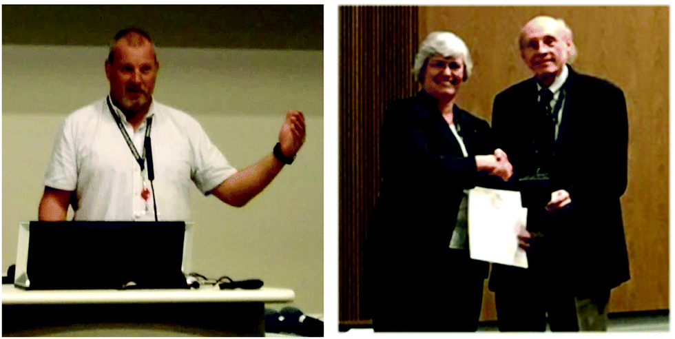 | ||
| Fig. 2 (a) Prof. D. Graham gives a short introduction to the meeting. (b) Prof. R. Van Duyne receives the Spiers Memorial Award from the president of the Faraday Society, Prof. E. Campbell. | ||
The meeting was divided into four sessions, which focused on particular aspects of SERS: (1) the theory of SERS enhancement, (2) ultrasensitive and towards single molecule SERS, (3) SERS in biology/biomedical SERS and (4) Analytical SERS. The opening lecture was given by Prof. R. Van Duyne, while the concluding remarks were given by Prof. M. Porter (University of Utah, USA).
Opening lecture
With his 45 minute opening lecture, Prof. R. Van Duyne set the scene for the discussion, providing a stimulating and wide-ranging introduction to the field of SERS. Prof. Van Duyne noted that the previous Faraday Discussions on SERS date back to 2005, demonstrating how this topic is still vibrant after more than ten years. In spite of SERS being an established field, there are still several dynamics to understand and interesting new directions to investigate, from strong coupling to the interconnection between physics and chemistry to name a few. Prof. R. Van Duyne ended his lecture with some open questions: “Is the metal altering the molecule's charge transfer? Can we deal with things that do not easily bind to surfaces?” The discussions could then start.Session 1 – The theory of SERS enhancement
Understanding the SERS from nanostructures is still an area of intense interest, as experiments become more robust and evolve to show discrepancies. This session covered a fascinating range, from the fundamentals of the SERS and TERS process using a quantum mechanical description via calculations of real EM enhancements, to the effects of applied voltages and semiconductor surfaces in SERS. The session started with Prof. J. Aizpurua (Centre for Materials Physics, San Sebastian, Spain), who discussed the different levels of approximation for the methodological solution of the optomechanical Hamiltonian of a generic SERS configuration, underlining their different approaches to the phonon population (DOI: 10.1039/C7FD00145B). The following talk presented by L. Velleman (Imperial College London, UK) focused on improving the understanding of nanoparticle assembly processes at liquid–liquid interfaces, with the aim of working towards finely controlling their structure and producing tailored optical and enhanced Raman signals (DOI: 10.1039/C7FD00162B). Prof. S. Reich (Freie Universität Berlin, Germany) presented a very interesting study where organic dyes encapsulated in single-walled carbon nanotubes become ideal probes for quantifying plasmonic enhancement in a Raman experiment (Fig. 3). Having the molecules chemically protected through the nanotube wall and spatially isolated from the metal prevents enhancement by chemical means and through surface roughness, making a step forward in distinguishing the different components contributing to such enhancement (DOI: 10.1039/C7FD00127D). The theory of SERS on semiconductor nanoparticles was then discussed by Prof. J. Lombardi (City College of New York, USA) with the aim of the optimization of SERS sensors (DOI: 10.1039/C7FD00138J). Dr P. Dawson (Queens University Belfast, UK) addressed the two systems of a tip–substrate system yielding tip-enhanced Raman scattering (TERS) and structures supporting hybrid plasmon-waveguide (HPWG) modes for SERS, offering schematic modelling for these systems (DOI: 10.1039/C7FD00128B). The last talk of the session faced the still strongly debated contribution of chemical mechanisms to the SERS enhancement. Here Prof. G. Schatz (Northwestern University, USA) presented theoretical modelling of voltage effects and the chemical mechanism in SERS, investigating the role of charge transfer (DOI: 10.1039/C7FD00122C).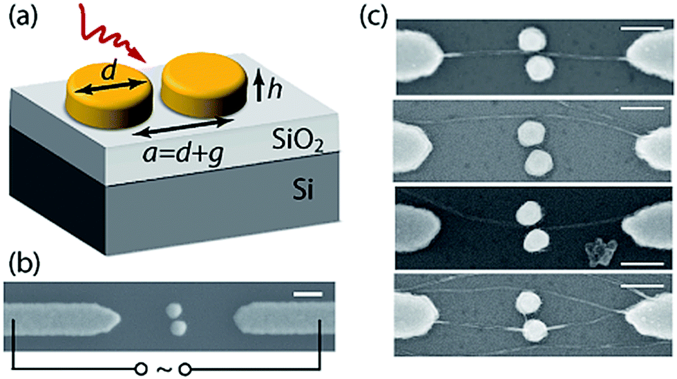 | ||
| Fig. 3 (a) The geometry of a plasmonic nanodimer on a SiO2/Si substrate. (b) Exemplary nanodimer between the electrodes used for the dielectrophoresis. (c) SEM images of 6T@CNT after deposition. The scale bars in (b) and (c) are 200 nm. Reproduced from DOI: 10.1039/C7FD00127D with permission from The Royal Society of Chemistry. | ||
According to Prof. J. Baumberg (University of Cambridge, UK), who chaired this session, “getting the quantum description correct is a major advance and the realisation that this mirrors the optomechanical descriptions which emerged in physics within the last 5 years provides important insights which will help develop the field of coherent Raman spectroscopies. Modelling of specific systems is also of much interest. Using carbon nanotubes as micro-vessels attached to plasmonics resonators, or semiconductor surfaces, or tip geometries under bias, all take better-understood theories and for us to try and apply them in new contexts.” From his point of view, “the CNT loading is just at a first step and more work will be very valuable. Experimental data for this and the other systems is crucial to provide verification of some of the predictions made and this is at an early stage. The theories will have to be modified in light of this. Finally, the introduction of further tuneable plasmonics at liquid surfaces is starting to become viable and is of much intrigue.” The very large number and range of questions in this session shows the liveliness of the interest and the emerging science that continues to develop.
Session 2 – Ultrasensitive and towards single molecule SERS
The second session of this Faraday Discussion focused on how the great sensitivity of SERS can be pushed towards the single molecule detection regime. Prof. V. Deckert (Friedrich Schiller University Jena, Germany) opened the session by describing the advantage of tip enhanced Raman scattering (TERS) to enable the localized plasmon-mediated polymerization of D3ATP by directing the tip to a specific location. TERS was also used to simultaneously catalyse the reaction and detect structural changes (DOI: 10.1039/C7FD00157F). Prof. K. Hewitt (Dalhousie University, Canada) then demonstrated the possibility of using a low-power continuous wave (CW) laser source for surface enhanced-stimulated Raman Scattering (SE-SRS) using metallic nanoparticles (DOI: 10.1039/C7FD00137A). SE-SRS was successfully used to probe commercial Raman-active nanoparticles, suggesting the potential application of stimulated Raman to probe the cellular environment. The influence of different scanning tunnelling microscopy (STM) parameters (i.e. current set point and bias voltage between the tip and sample) on the TERS response was then discussed by Miss N. Martín Sabanés (Max Planck Institute for Polymer Research, Germany; DOI: 10.1039/C7FD00164A). She evidenced how current literature failed to show the impact of both fundamental parameters and their synergistic effect on reaching the single molecule regime. Compared to typical AFM-based TERS set-ups, STM-TERS coupling provided a higher enhancement (a couple of orders of magnitude), allowing single molecule detection in liquids. Prof. K. Willets (Temple University, USA) presented how the SERS emission patterns of nanoparticles on a mirror (Fig. 4) provide insight into the position of a molecule within the plasmonic hot-spot (DOI: 10.1039/C7FD00163K). Moreover, the quality of the substrate strongly impacted how the gap plasmon-mediated emission couples to the far field and this is becoming an interesting tool to investigate the heterogeneity of a SERS substrate. Prof. K. Murakoshi (Hokkaido University, Japan) then described how electrochemical control over a plasmonic nanostructure can be used to enhance the SERS response of an adsorbed dye molecule (DOI: 10.1039/C7FD00126F). By changing the redox state of the dye, the coupling strength between the localized surface plasmon and the dye can be optimized, leading to an increased Raman intensity. The session was closed by Prof. F. Giorgis (Politecnico di Torino, Italy) presenting novel SERS-active metal–dielectric nanostructures integrated in microfluidic devices for the label-free quantitative detection of miRNA (DOI: 10.1039/C7FD00140A). The described SERS-based biosensor was optimized in two different ways: (1) direct one-step detection of the modified target miRNA and (2) two-step capturing and labelled DNA probes for label-free miRNA sensing. The direct quantification of miRNA-222 in RNA cellular extract was then demonstrated, showing the feasibility of the detection and quantification of miRNAs in real biological samples for clinical applications.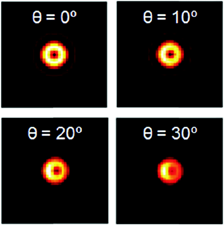 | ||
| Fig. 4 Theoretical calculations of the emission from a single dipole at a glass–air interface with φ = 0° and θ varying from 0° to 30°. Adapted from DOI: 10.1039/C7FD00163K with permission from the Royal Society of Chemistry. | ||
According to Prof. J. Edel (Imperial College London, UK), chair of the session, one of the biggest challenges is related to the selectivity of SERS at low concentrations, since single molecule detection usually implies the presence of spectral features, e.g. blinking (on/off signal), variation in the Raman band relative intensities and also variation in the frequency of specific Raman bands. Another important point discussed during the session was how efficiently we can screen for specific target analytes in complex mixtures with highly sensitive sensors. In addition to high selectivity, SERS also has a great capability for multiplexing on account of its intrinsic small bandwidth. However, although SERS has a high multiplexing potential compared to its most renowned competitor, fluorescence, few examples of true multiplexing SERS are currently present in the literature. Finally, an essential requirement for quantitative results is the use of reproducible and highly sensitive SERS substrates in order to ensure routinely quantitative measurements over a large dynamic range.
Session 3 – SERS in biology/biomedical SERS
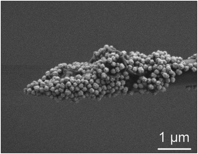
The third session of the meeting explored the use of SERS in biological and biomedical applications. Covering a range of interesting areas, from the in situ synthesis of nanoparticles in bacteria, approaches to measuring enzyme activity and the quantification of proteins, and sensing near neurons using dynamic-SERS (D-SERS), to topics on the variation of SERS measurements in biological systems, which lead to an involved discussion on the reproducibility of SERS experiments. Starting the session, Prof. R. Goodacre (University of Manchester, UK) presented a technique in which E. coli bacteria were quantitatively detected through isotopically enriching the bacteria (DOI: 10.1039/C7FD00150A). Through exposure to 12C/13C-glucose and 14N/15N-ammonium chloride, coupled with the in situ synthesis of silver nanoparticles (forming on the outside of the bacteria), the isotope levels of the enriched E. coli could be quantified using SERS (Fig. 5). The following talk by Mr P. C. Wuytens (Ghent University, Belgium) described a label-free approach to measuring trypsin activity by monitoring the cleavage of a peptide decorated on gold nanodomes, with a view to being able to conduct both single and multiplexed measurements of protease activity (DOI: 10.1039/C7FD00124J).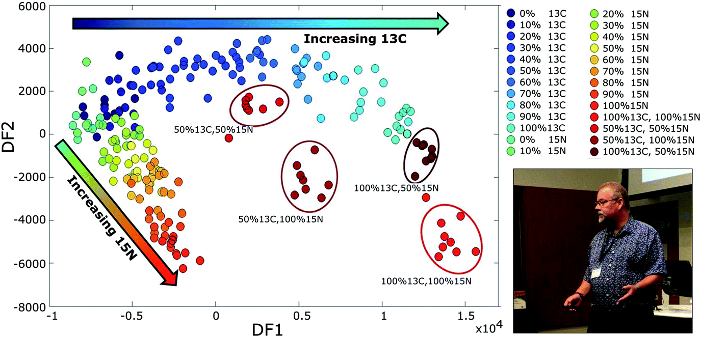 | ||
| Fig. 5 Prof. R. Goodacre speaks in the third session: SERS in biology/biomedical SERS (Photo taken by Prof. D. Graham). Graph: PC-DFA scores plot of the pre-processed SERS spectral data of E. coli cells cultivated on different ratios of unlabelled (12C and 14N) and isotopically labelled 13C and/or 15N growth substrates. Adapted from DOI: 10.1039/C7FD00150A with permission from the Royal Society of Chemistry. | ||
After a short break, the session resumed with a method of detecting galectin in real-time using glycan-decorated gold nanoparticles, presented by J. Langer (CIC biomaGUNE, Spain). By altering the densities of the Raman reporters on the surface of the gold nanoparticles, the aggregation dynamics of the particles could be tuned (DOI: 10.1039/C7FD00123A). Prof. J.-F. Masson (University of Montreal, Canada) followed up by presenting an interesting semi-quantitative SERS approach for neurotransmitter sensing near neurons throughout multiple stimulated dopamine secretion cycles (DOI: 10.1039/C7FD00131B and Fig. 6). This novel technique, referred to as dynamic SERS optophysiology (D-SERS), allowed the multiplex sensing of five neurotransmitters under physiological conditions and constitutes a strong proof-of-concept for the potential application of SERS to study normal and pathological cellular functions. The ability to acquire SERS data without altering the biological sample in any way has been a difficult feat, and therefore reproducibility has been a problem for SERS on biological samples. Using immuno-Raman microspectroscopy (iSERS), the repeated imaging of a single cell was demonstrated by Prof. S. Schlücker (University of Duisburg-Essen, Germany; DOI: 10.1039/C7FD00135E). In the closing paper of the session, the focus was on the highly debated issue of “What do we actually see in intracellular SERS?” (DOI: 10.1039/C7FD00156H). Prof. S. Mahajan (University of Southampton, UK) explored the extent of how experimental conditions can affect gold nanoparticle internalisation, which in turn affect cell metabolism and induce changes. The discussion on the topic afforded the consensus that there is a need to move toward a standard methodology of nanoparticle treatments in order to validate intracellular SERS experiments.
 | ||
| Fig. 6 Scanning electron microscopy (SEM) image of the D-SERS nanosensor coated with nanoraspberries. Adapted from DOI: 10.1039/C7FD00131B with permission from The Royal Society of Chemistry. | ||
Prof. K. Faulds (University of Strathclyde, UK), chair of the session, remarked: “Biological and biomedical SERS has advanced greatly in the last 20 years and, in particular, since the first Faraday SERS meeting in 2005. The applications covered at the meeting ranged from bacterial and cell imaging to in vitro assays for enzyme activity, small molecule and protein detection. One of the greatest achievements in the field is that we are now able to routinely make sensitive, quantitative measurements and obtain rapid, high resolution images from single cells/bacteria that give us a huge amount of information about a system. One of the biggest challenges in biological SERS is standardisation of approaches. Differences between research groups in synthesising and functionalising nanoparticles result in slightly different surface chemistry, SERS response and toxicity. This affects the reproducibility of experiments between different laboratories and as a community we need to ensure that all conditions, the appropriate characterisation techniques utilized (particle size, zeta potential, extinction), and that the synthesis methods, buffers/media used, and measuring cellular uptake and toxicity, are all carefully reported. These issues, as well as the use of robust and reliable data analysis methods, were discussed during the meeting.”
Session 4 – Analytical SERS
The final session of the conference demonstrated the broad potential for SERS to also be used as an analytical tool outside of biomedical SERS, and investigated the challenges to progress SERS from a lab-based technique to the markets. Prof. R. Van Duyne remembered the exact episode of Crime Scene Investigation (CSI) which contains a SERS-based instrument, however such SERS-based devices are not yet widely available. Potential applications brought forward in the session were: the detection of methanol in alcoholic beverages (Dr B. de Nijs), the study of processes in electrochemical cells (Dr G. Di Martino) and batteries (Prof. L. Hardwick), and the analysis of the composition of DNA (Prof. S. Bell).The morning part of the session was opened by Prof. Z.-Q. Tian (Xiamen University, China) with a fascinating talk on expanding the use of SERS to non-traditional (weakly SERS-active) substrates and even to non-SERS-active substrates such as aluminium oxide (DOI: 10.1039/C7FD00144D, see Fig. 7). In the second talk, Prof. L. Hardwick (University of Liverpool, UK) showed how SHINERS can be exploited to study chemical processes at the surfaces of battery electrode materials, both on the lithium metal anode and carbon cathode of a LiO2 cell (DOI: 10.1039/C7FD00151G). Prof. P. Vikesland (Virginia Tech, USA) discussed how to use the Rayleigh band intensity as a parameter for the normalization of SERS intensity (DOI: 10.1039/C7FD00125H). Finally, Dr B. de Nijs (University of Cambridge, UK) demonstrated that SERS is able explore the local environment in a nano-gap as it is sensitive to the different configurational states of molecules and even allows for the detection of hydrogen bonding (DOI: 10.1039/C7FD00147A). In his talk, Prof. Z.-Q. Tian remarked on the importance of translating SERS to markets: “The field of SERS has a very low entry level because nanoparticles can now be produced easily following published literature. This reflects in a large body of work and citations on SERS. However, the actual number of user products based on SERS is quite low. We can infer that translating SERS to markets is not easy.” A frequently suggested reason for this discrepancy is the problem with reproducibility between SERS studies, but Prof. R. Goodacre reminded the delegates that “SERS is often ‘undersold’ as not reproducible. However, in general it is very reproducible. The issue is that different applications require different platforms and substrates.” In several presentations, experimental and computational techniques were mentioned which can indeed handle such sources of variability, among which are principle component analysis (Prof. R. Goodacre, Dr B. de Nijs), the Rayleigh band intensity (Prof. P. Vikesland) and internal standards (Prof. R. Van Duyne).
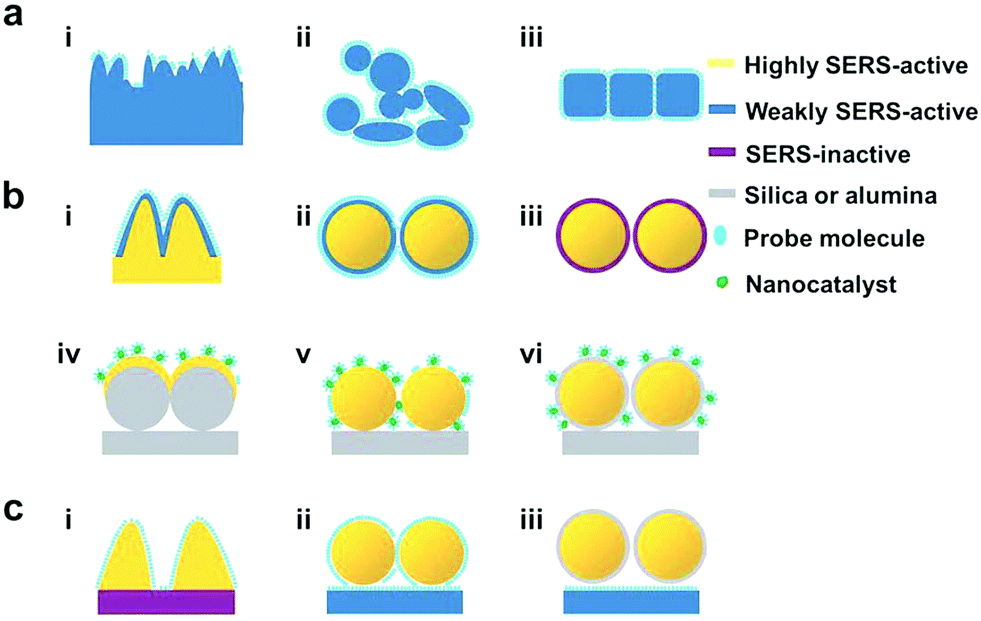 | ||
| Fig. 7 Different strategies to probe SERS-active materials and inactive materials. Adapted from DOI: 10.1039/C7FD00144D with permission from the Royal Society of Chemistry. | ||
After a coffee break, the session on Analytical SERS resumed with Prof. S. Bell (Queen's University Belfast, UK) who presented exciting work on the quantitative detection of oligonucleotides using SERS (DOI: 10.1039/C7FD00134G). He showed that even though spectral changes when adding a single nucleotide to the 3′ terminus are small (Fig. 8), the signal-to-noise levels in these SERS spectra are low enough that difference spectra can be used to detect the nucleotide by comparing them with nucleobase reference spectra. Moreover, the SERS DNA chain signal is also influenced by the secondary structure (coiling) of the chain, an effect which was eliminated by thermal pre-treatment, which uncoils the chains. Dr G. Di Martino (University of Cambridge, UK) presented an exciting study into the optical response of individual nm-wide plasmonic nanocavities, created by fabricating nanoparticle-on-mirror geometry (NPoM) inside an electrochemical cell (DOI: 10.1039/C7FD00130D, see Fig. 9). She showed that the SERS response (peak intensity, resonance full width at half maximum (FWHM), and the spectral position of the coupled plasmon mode) is influenced by the bias voltage and discussed four scenarios which potentially induce these changes. The scenarios are ion penetration into the SAM, the potential driven movement of the double layer changing the local refractive index, electronic coulombic scattering from individual ionic charges in the double layer, and H2 gas formation around the AuNP. Finally, J. Guicheteau (RDECOM Edgewood Chemical Biological Center, USA) discussed the influence of various synthesis protocols on SERS enhancement, as well as the role of thermodynamics during substrate formation (DOI: 10.1039/C7FD00141J). The protocols were drop and dry on a substrate, leave the substrate in solution for a constant time and volume, and immerse until equilibrium is reached. To express the advantage of SERS over normal Raman sensing for a given protocol and analyte, a figure of merit termed the SERS enhancement value (SEV) was proposed.
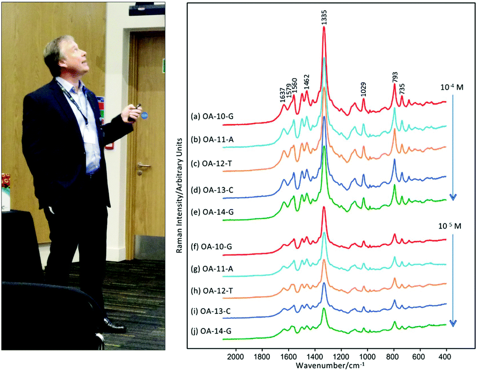 | ||
| Fig. 8 Prof. S. Bell speaks in the fourth session: Analytical SERS. Graphs: raw SERS spectra of ODN sequences showing the effect of adding nucleobases at the 3′ end on the spectra. Adapted from DOI: 10.1039/C7FD00134G with permission from the Royal Society of Chemistry. | ||
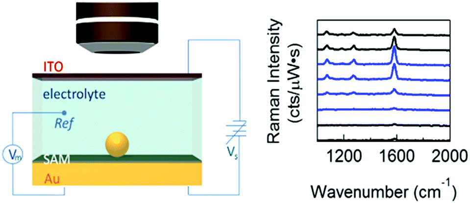 | ||
| Fig. 9 Left panel: Optically transparent thin (sub-mm) electrochemical cell for spectroscopy of single Au NPs on molecular layer on Au. Right panel: SERS intensity time evolution under an applied voltage of 0 V (black) to −1.2 V (blue). Adapted from DOI: 10.1039/C7FD00130D with permission from The Royal Society of Chemistry. | ||
Chair of the session Prof. J. Popp (Leibniz-IPHT, Germany) gave his perspective on Analytical SERS: “The key for moving SERS into routine analytics is to improve the reproducibility of nanostructured SERS surfaces as well as the ability to perform quantitative SERS analysis. In this context, shell-isolated nanoparticle-enhanced Raman spectroscopy (SHINERS) will be an important step towards the reproducible characterization of the chemical composition of surfaces having inorganic, organic or biological origins. The quantification of analyte molecules can be significantly improved by employing internal standards to address variations in the SERS activity. Moreover, the direct label-free SERS method is best suited for the analysis/detection of low-molecular weight substances with high affinity toward the metallic surface. Thus, an intrinsic sample preparation step is included to enrich molecules with high affinity from complex matrices such as environmental samples or human body fluids. To detect molecules with less affinity toward silver or gold surfaces, the design of special recognition molecules changing their Raman signal while interacting with the analyte of interest due to conformational changes will be the solution. Finally, medical and in vivo SERS applications via smart SERS tags modified with antibodies or aptamers allowing for a specific interaction of biomarkers to detect e.g. tumour cells in blood or tissue will be an important SERS topic within the next years.”
Several themes recurred in all four sessions (Fig. 10). First, the use of the NPoM geometry as a versatile platform for SERS was present transversally in all of the sessions. Examples include opto-mechanics studies in NPoM geometry (Prof. J. Aizpurua) in session 1, dynamic heterogeneity on account of molecular motion inside the hot spots (Prof. K. Willets) in session 2, NPoMs of weakly and strongly SERS-active materials (Prof. Z. Q. Tian) and NPoM in electrochemical cells (Dr G. Di Martino) in session 4. Tip-enhanced Raman Spectroscopy or TERS frequently recurred as a versatile platform as well, sparking a discussion on whether (coupled) plasmons on the tip of a TERS probe are affected by the tip's shape. Prof. R. Van Duyne pinpointed the advantage of TERS in his opening lecture as “TERS combines the sensitivity of Raman with the spatial resolution of AFM/STM”. Finally, the chemical and plasmonic contributions to SERS signals were another recurring point of interest (e.g. Prof. S. Reich).
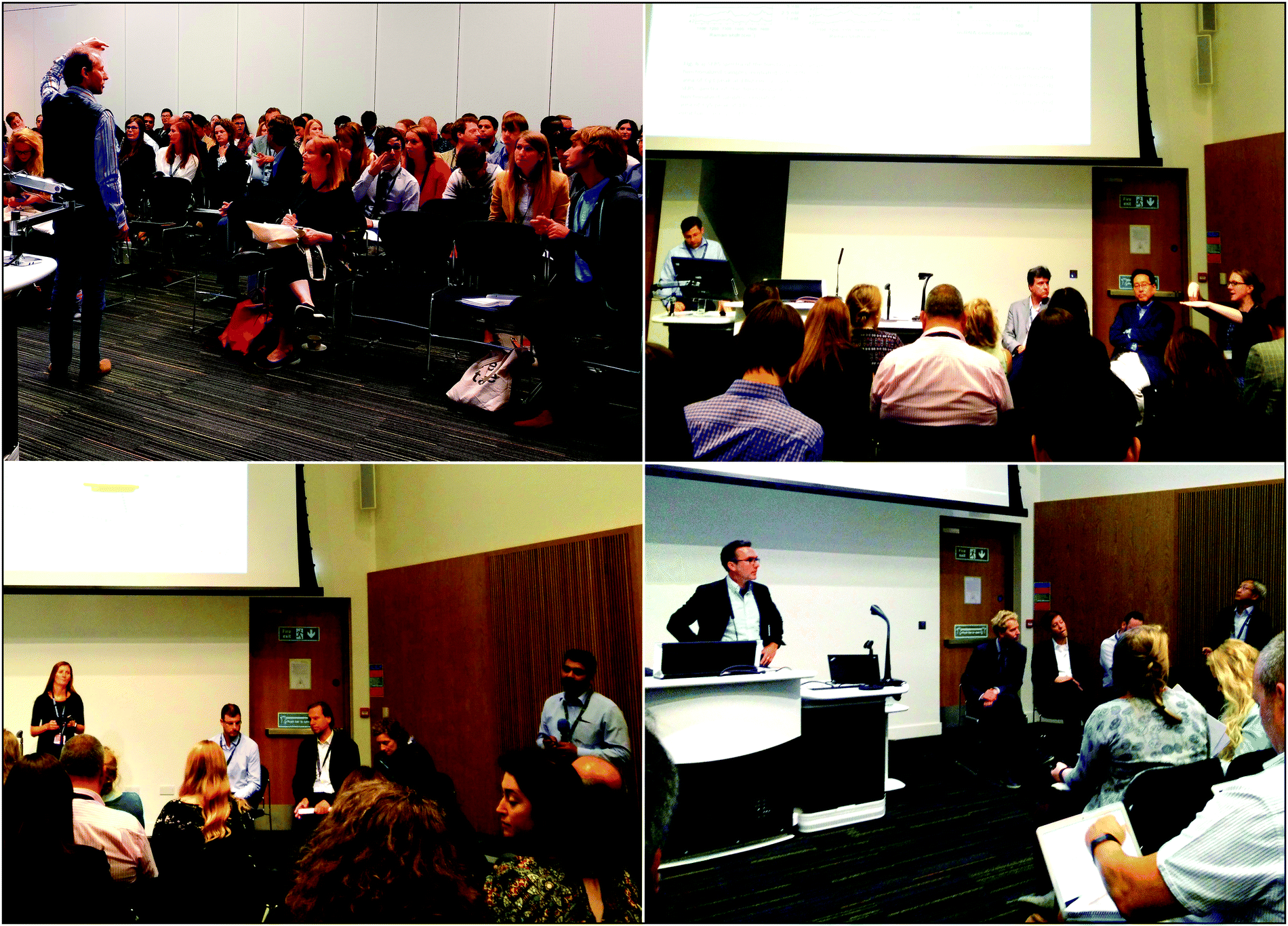 | ||
| Fig. 10 Impressions of discussions in each session of FDSERS17. (a) Prof. J. Baumberg addresses the audience during the first session. (b) Prof. J. Edel chairs a discussion in the second session. Speakers are Prof. F. Giorgis, Prof. K. Murakoshi, and Prof. K. Willets (seated, left to right). (c) Prof. K. Faulds leads a discussion in the third session. Speakers are Prof. J.-F. Masson, Prof. S. Schlücker and Mrs J. Langer (seated, left to right) and Prof. S. Mahajan (standing, on the right) taking part in the discussion. (d) Prof. J. Popp presides a discussion in the fourth session. Speakers are Dr B. de Nijs, Prof. P. Vikesland, Prof. L. Hardwick (seated, left to right) and Prof. Z.-Q. Tian (standing, on the right). | ||
Also in this session, Prof. D. Graham pinpointed important future directions of the field as the “design and use of alternatives to plasmonic materials for enhancement and a move to quantitative SERS for meaningful applications where other techniques such as fluorescence fail, e.g. bioanalysis.”
Socials and poster sessions
The program of the meeting allowed for sufficient time to socialize and interact, for example during the tea times and lunches between sessions (Fig. 11a). The poster sessions at the end of each day were a particularly interesting opportunity to engage in scientific discussions and were busily attended (Fig. 11b). All of the posters had been briefly introduced during the sessions in the form of lightning presentations, in order to kick-start the conversations. “The poster sessions were really vibrant and I have new collaborations already starting based on discussions” remarked Prof. D. Graham.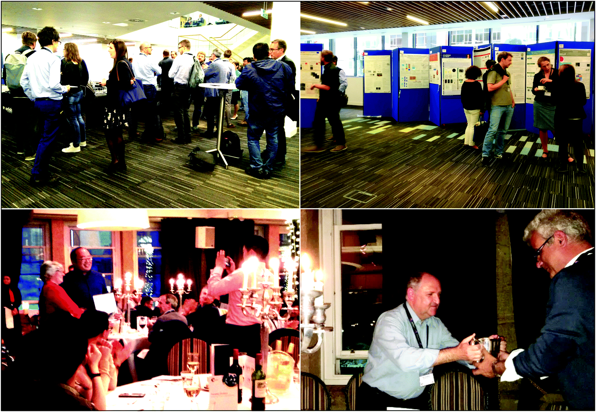 | ||
| Fig. 11 Socials and Poster Sessions. (a) Lively discussions happening during the coffee break. (b) The first poster session is about to begin. (c) Mr W. Lum is presented with the first Poster Prize by Prof. E Campbell during the Conference Dinner. (d) The Loving Cup changes hands during the Ceremony at the end of the Conference Dinner. | ||
Another excellent opportunity to network was incorporated into the programme in the form of the Conference Dinner. The delegates enjoyed a delicious three-course meal on Thursday night at the Supper Club, close to George Square. Prof. E. Campbell gave a formidable speech on her experiences as the president of the Faraday Society and in particular during the current Faraday Discussion. She also conferred the poster prizes, with Mr W. Lum (University of Cincinnati, USA) winning first prize (Fig. 11c) and the runners up being Mr N. Bontempi (University of Brescia, Italy) and Mrs R. Kidd (University of Southampton, UK). As is tradition, the dinner was closed with the Loving Cup ceremony (Fig. 11d). This silver cup, which dates back to 1728 and was crafted by lady silversmith Heslie Fawdery, is used to commemorate G. S. Marlow (Secretary and Editor, 1928–1947) and Angela & Tony Fish (Angela organised the Faraday Discussions 1968–1995). The ceremony involves taking a sip and passing the cup along via an intricate series of bows, which led to the occasional head bump.
Concluding remarks lecture
The concluding remarks were presented by Prof. M. Porter (University of Utah, USA). He recapitulated the meeting by giving a brief overview of all of the topics covered during the Discussion. He stressed the fact that more work on theory needs to be done, and pointed out that most of the presented knowledge comes from empirical work. Moreover, he underlined the presence of a fragmentation of knowledge, inviting the audience to think about whether “we are focusing too much on the physics and forgetting about the chemistry”. The view is shared by Prof. D. Graham who identifies “a challenge for theory and modelling to be more predictive in leading the experimentalists’ design of experiments”.The 2017 Faraday Discussion on SERS has been a vibrant and stimulating meeting. Its success was obvious during the discussion sessions, through the abundance of questions and remarks (more than 60 per session!), which often forced the session chairs to conclude the discussions prematurely due to time restrictions.
Acknowledgements
We acknowledge financial support from EP/G060649/1, EP/L027151/1, EP/G037221/1, EP/N020669/1, EPSRC NanoDTC, and ERC grant LINASS 320503. M. K. was supported by funding from the European Research Council (ERC) under the European Union's Horizon 2020 research and innovation programme (MSCA-IF-2015-EF SPARCLEs 7020005). H. F. was supported by the EPSRC and MRC grant number EP/L016559/1 and the School of Chemistry, University of Edinburgh. F. L. was supported by funding from the Natural Science and Engineering Research Council (NSERC) of Canada.Footnote |
| † All authors contributed equally. |
| This journal is © The Royal Society of Chemistry 2017 |