 Open Access Article
Creative Commons Attribution 3.0 Unported Licence
Evaluation of DFO-HOPO as an octadentate chelator for zirconium-89†
L.
Allott‡
 a,
C.
Da Pieve‡
a,
J.
Meyers
a,
C.
Da Pieve‡
a,
J.
Meyers
 b,
T.
Spinks
a,
D. M.
Ciobota
a,
G.
Kramer-Marek
 a and
G.
Smith
 *a
*a
aDivision of Radiotherapy and Imaging, The Institute of Cancer Research, 123 Old Brompton Road, London, UK. E-mail: graham.smith@icr.ac.uk
bCancer Research UK Cancer Therapeutics Unit, Division of Cancer Therapeutics, The Institute of Cancer Research, 123 Old Brompton Road, London, UK
First published on 12th July 2017
Abstract
The future of 89Zr-based immuno-PET is reliant upon the development of new chelators with improved stability compared to the currently used deferoxamine (DFO). Herein, we report the evaluation of the octadentate molecule DFO-HOPO (3) as a suitable chelator for 89Zr and a more stable alternative to DFO. The molecule showed good potential for the future development of a DFO-HOPO-based bifunctional chelator (BFC) for the radiolabelling of biomolecules with 89Zr. This work broadens the selection of available chelators for 89Zr in search of improved successors to DFO for clinical 89Zr-immuno-PET.
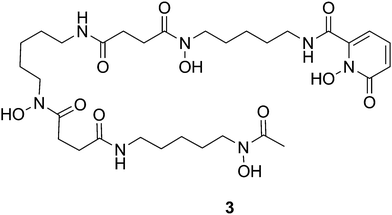
An increasing interest in zirconium-89 (89Zr) for preclinical and clinical immuno-positron emission tomography (immuno-PET) is due to its favourable decay characteristics (t1/2 = 78.4 h, β+ = 22.8%, Eβ+max = 901 keV) for the radiolabelling of antibodies which have long biological half-lives.1–4 Currently, deferoxamine (DFO) is the chelator most commonly used to radiolabel biomolecules with 89Zr.5 DFT modelling showed that the coordination sphere in the Zr–DFO complex consists of the Zr4+ cation, six donor atoms belonging to the DFO molecule, and two other coordination sites being occupied by water molecules.6,7 As a result of this incomplete coordination of 89Zr by the hexadentate DFO molecule, the 89Zr–DFO complex undergoes a certain degree of demetallation in vivo with the released 89Zr taken up by the bone.8 This is of concern because bone uptake of free 89Zr4+ is undesirable owing to the high radiation dose to bone marrow; furthermore this background uptake can confound image acquisition of bone malignancies such as bone metastases. To solve the instability, different strategies have been investigated. Alternative hexadentate macrocycles (i.e. Fusarinine C) and hydroxypyridinone-based compounds (i.e. CP256) have been produced and tested but showed either no improvement or reduced stability in vivo when compared to DFO.9,10 Additionally, a variety of either linear or macrocyclic octadentate chelators have been developed with structures hinged around hydroxamic acid or hydroxypyridinone moieties which resulted in 89Zr-complexes displaying either increased or decreased stability compared to DFO.11–19 White et al. described the use of a DFO-1-hydroxy-2-pyridone ligand (DFO-HOPO) as an effective sequestering agent for the treatment of plutonium(IV) poisoning (Fig. 1).20 The authors showed that the addition of one 1,2-HOPO molecule to DFO produced a low toxicity octadentate chelator which yielded very stable complexes with Pu(IV) at physiological pH. Herein, we report an updated synthesis of DFO-HOPO (3) which was then evaluated as an octahedral ligand for 89Zr. The stability of the radiocomplex was tested in vitro and in vivo and compared to 89Zr–DFO in order to confirm 3 as a viable alternative to the chelators for 89Zr already described in the literature.
 | ||
| Fig. 1 Structure of DFO-HOFO (3) containing three hydroxamic acid and one hydroxypyridone moiety for coordinating 89Zr4+. | ||
The synthesis of DFO-HOPO (3) was adapted from a literature procedure.20 In brief, commercially available DFO was reacted with hydroxamic acid chloride (2) and the product (3) was isolated by semi-preparative RP-HPLC (Scheme S1, ESI†). No protection of the N-hydroxyl group of 1 was necessary. To determine and characterise the coordination capabilities of the chelator, the non-radioactive natZr complex of DFO-HOPO (natZr-3) was prepared in macroscopic scale by mixing the chelator with ZrCl4 at room temperature. Showing the value of 784.247 m/z, the high resolution mass spectrometry (HRMS) analysis of natZr-3 confirmed the expected complex mass to indicate a metal-to-ligand binding ratio of 1![[thin space (1/6-em)]](https://www.rsc.org/images/entities/char_2009.gif) :
:![[thin space (1/6-em)]](https://www.rsc.org/images/entities/char_2009.gif) 1. Examination by RP-HPLC showed the elution of natZr-3 as a single peak at 7
1. Examination by RP-HPLC showed the elution of natZr-3 as a single peak at 7![[thin space (1/6-em)]](https://www.rsc.org/images/entities/char_2009.gif) :
:![[thin space (1/6-em)]](https://www.rsc.org/images/entities/char_2009.gif) 20 (min
20 (min![[thin space (1/6-em)]](https://www.rsc.org/images/entities/char_2009.gif) :
:![[thin space (1/6-em)]](https://www.rsc.org/images/entities/char_2009.gif) s), ca. 33 seconds before HOPO-DFO (3). The coordination of the metal ion by the chelator was further confirmed by infrared spectroscopy (IR) analysis which showed a red-shift in the main carbonyl stretching band from ca. 1620 to ca. 1600 cm−1. Additional characterisation of the complex by ultraviolet-visible spectroscopy (UV-Vis) showed no detectable difference between the absorption spectra of natZr-3 and 3. Moreover, NMR analysis of the natZr complex could not be performed due to its poor solubility in any solvent, which is expected to be resolved upon bioconjugation.
s), ca. 33 seconds before HOPO-DFO (3). The coordination of the metal ion by the chelator was further confirmed by infrared spectroscopy (IR) analysis which showed a red-shift in the main carbonyl stretching band from ca. 1620 to ca. 1600 cm−1. Additional characterisation of the complex by ultraviolet-visible spectroscopy (UV-Vis) showed no detectable difference between the absorption spectra of natZr-3 and 3. Moreover, NMR analysis of the natZr complex could not be performed due to its poor solubility in any solvent, which is expected to be resolved upon bioconjugation.
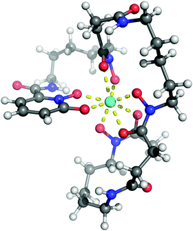
To verify the steric and electronic ability of 3 to form a Zr(IV) octadentate chelate, density functional theory (DFT) calculations were carried out. The optimised geometry (based on the lower energy conformation) shows the metal centre coordinated to eight oxygen atoms of the chelator (Fig. 2). The Zr–O bond distances were in the range of 2.14–2.36 Å, in agreement with values reported in the literature for similar complexes.11,12
 | ||
| Fig. 2 The DFT optimised structure of 89Zr-3. (Atom colour: white = hydrogen; grey = carbon; blue = nitrogen; red = oxygen; cyan = zirconium.) | ||
The preparation of 89Zr-3 was performed as previously described in the literature for 89Zr–DFO.21 Incubating the chelator with a neutralised 89Zr solution at room temperature for 1 h (pH 7) guaranteed a quantitative (>99%) radiolabelling up to a specific activity of 20 MBq nmol−1 even at low concentration of the chelator (3–8 μM). A comparable radiolabelling efficiency was obtained for 89Zr–DFO. All reactions were monitored by radioactive instant thin layer chromatography (radio-ITLC). A variety of mobile and stationary phases were tested to find the optimum analytical conditions for both 89Zr-3 and 89Zr–DFO, which was used as a comparison. The elution profiles of both the radioactive complexes were affected by the type of stationary phase employed, and only the positively charged 89Zr–DFO (consequence of the hexadentate chelation of 89Zr) was influenced also by the mobile phase pH, when SG-ITLC strips were used. The results suggest that, differently from 89Zr–DFO, 89Zr-3 is present in solution as a neutral complex, achievable through the octadentate chelation of 89Zr. This finding further advocates the involvement of the 1,2-HOPO moiety of 3 in the coordination of the metal centre. Enabling the elution of 89Zr-3 and the 89Zr–DFO as well defined and separated bands (Rf of 0.6 and 0.1 respectively on SG-ITLC strips), ammonium acetate (0.1 M, pH 7) was used as mobile phase for the ITLC analysis. Interestingly, the radio-ITLC of 89Zr-3 revealed the presence of two well-separated spots (Rf of ca. 0.6 and 0.1); the relative intensity of the spots was dependent on the specific activity of the product (i.e. concentration of the chelator) and on time. By lowering the specific activities of the product, with a consequent increase of the concentration of 3, a decrease of the band having Rf = 0.1 was observed. After 24 hours at ambient temperature, only the band having Rf = 0.6 was detected. To probe the influence of temperature on the formation of the two products, the radiolabelling reaction was performed at 80 °C. Although the quantity of product eluting with an Rf = 0.1 was reduced, the increased temperature did not prevent it from forming. These observations suggest that the two bands represent two different forms of the 89Zr-3 complex; an initial transitional kinetic product which converted into a final thermodynamically stable product. Examination of the chromatographic data of 89Zr-3 could help explain the phenomenon; the transitional product was detected at the origin of the radio-ITLC strip (Rf = 0.1 at pH 7) suggesting it was charged (similarly to hexacoordinated 89Zr–DFO), possibly as the result of incomplete coordination of the radiometal. With an Rf = 0.6 (at pH 7), the thermodynamically stable final product was most likely neutral, a condition which would be achieved by the complete chelation of 89Zr by octadentate 3. Moreover, radio-HPLC analysis of 89Zr-3 after 24 hours showed only one product (corresponding to the band with Rf = 0.6 on radio-ITLC) having an elution profile very similar to that of natZr-3 suggesting a similar identity as an octadentate complex. Importantly, no 89Zr was released during the transition.
The stability of 89Zr-3 was initially assessed by a simple radio-ITLC analysis using an acidic buffer (pH 2) as mobile phase. Differently from 89Zr–DFO (14.4 ± 4.65% radioactivity not associated with DFO), 89Zr-3 showed no demetallation as a result of the enhanced coordination of the metal centre by the octadentate ligand. To mimic what might happen in vivo, a challenge assay assessed the stability of 89Zr-3 to transchelation in the presence of a large excess of either EDTA or DFO (pH 7). In both challenges, 89Zr-3 showed no transchelation with >99% intact complex after 7 days (Table 1). By comparison, 89Zr–DFO demonstrated transchelation toward EDTA with 65.5% of intact complex after 7 days (Table 1). Moreover, a complete transmetallation of 89Zr–DFO towards 3 was achieved in a matter of hours. Further experiments aiming to test the inertness of 89Zr-3 were performed in mouse serum. With >99% intact complex after incubation at 37 °C for 7 days, 89Zr-3 showed a higher stability compared to 89Zr–DFO (90.6% intact complex) (Table 1).
| Complex | Competitor | Fraction of intact complex (% ±SD) | ||||||
|---|---|---|---|---|---|---|---|---|
| 0 min | 1 h | 3 h | 1 d | 3 d | 7 d | |||
| A | 89Zr-3 | EDTA | >99 | >99 | >99 | >99 | >99 | >99 |
| DFO | >99 | >99 | >99 | >99 | >99 | >99 | ||
| 89Zr–DFO | EDTA | >99 | >99 | >99 | 75.63 ± 3.07 | 63.1 ± 0.75 | 65.5 ± 4.42 | |
| 3 | >99 | 35.8 ± 9.8 | 4.1 ± 2.35 | 0 | 0 | 0 | ||
| B | 89Zr-3 | Mouse serum | >99 | — | >99 | >99 | >99 | >99 |
| 89Zr–DFO | Mouse serum | >99 | — | >99 | 97.7 ± 0.53 | 94.7 ± 0.78 | 90.6 ± 1.75 | |
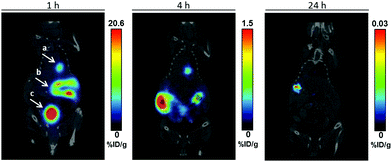
PET imaging and comparative biodistribution studies were performed in healthy mice for 89Zr-3 and 89Zr–DFO. At 1 h p.i. of 89Zr-3, the radioactivity was observed mainly in the bladder and intestine; some activity was also visible in the gall bladder. At 4 and 24 h p.i., most of the residual radioactivity was in the gut. These observations indicate a rapid renal clearance together with slower hepatobiliary excretion. The hydrophilicity of the complexes is an important physiochemical property which regulates their distribution, metabolism, and elimination in vivo. The log![[thin space (1/6-em)]](https://www.rsc.org/images/entities/char_2009.gif) D7.4 of neutral complex 89Zr-3 was found to be −0.87 ± 0.03 which indicates a less hydrophilic character than the positively charged 89Zr–DFO (−3.0 ± 0.01) and can explain the clearance pathway.9 After 24 h, the radioactivity level was minimal therefore no additional imaging studies at longer time points were carried out. Importantly, no uptake of 89Zr in the bone was observed at any time point (Fig. 3).
D7.4 of neutral complex 89Zr-3 was found to be −0.87 ± 0.03 which indicates a less hydrophilic character than the positively charged 89Zr–DFO (−3.0 ± 0.01) and can explain the clearance pathway.9 After 24 h, the radioactivity level was minimal therefore no additional imaging studies at longer time points were carried out. Importantly, no uptake of 89Zr in the bone was observed at any time point (Fig. 3).
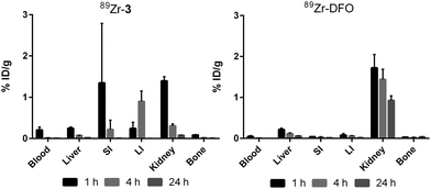
Corroborating the PET images, the biodistribution studies clearly showed the participation of both the renal and hepatobiliary systems in the clearance of 89Zr-3 (Fig. 4). Most of the radioactivity had already cleared through the kidneys at 1 h p.i. (1.39 ± 0.1% ID per g), while at 4 h p.i. the residual activity was localised in the gut (mostly small intestine with 0.898 ± 0.252% ID per g). Differently from 89Zr–DFO (0.93 ± 0.11% ID per g still present in the kidneys), 89Zr-3 was almost completely cleared from the body at 24 h p.i. Although the values are quite low, 89Zr–DFO showed ca. 10-fold higher activity accumulation in the bone than 89Zr-3 at 24 h p.i. (0.037 ± 0.002 and 0.004 ± 0.001 for 89Zr–DFO and 89Zr-3 respectively). This phenomenon could be correlated to either the higher level of radioactivity still present in the animals injected with 89Zr–DFO or to an improved in vivo stability of 89Zr-3 compared to 89Zr–DFO.
 | ||
| Fig. 4 Biodistribution data for 89Zr-3 and 89Zr–DFO at 1, 4 and 24 h p.i. in selected organs. SI = small intestine; LI = large intestine. All experiments were performed in triplicate. | ||
In summary, the 89Zr-3 complex exhibited improved stability compared to 89Zr–DFO in both challenge assays and in serum; the capability and favourability of 3 to form a stable chelate was clearly demonstrated by the complete transchelation of 89Zr from 89Zr–DFO in ca. 3 h. The in vivo studies showed that 89Zr-3 cleared the body via the renal and hepatobiliary systems. However, once conjugated to a biomolecule the pharmacokinetics of the final radioconjugate will depend mainly on the biomolecule itself. Importantly, the straightforward synthesis of 3 from the commercially available DFO is amenable to allow the synthesis of a bifunctional chelator which is currently underway in our laboratory. This could be achieved by using a similar strategy described by Patra et al. for the synthesis of DFO*, where a molecule (or a variety of molecules) containing both the bidentate moiety and a reactive functionality for bioconjugation is attached to the free amine of DFO.11 The promising DFO-HOPO molecule is a valuable addition to the selection of available chelators for 89Zr in search of successful successors of DFO for clinical immuno-PET applications based on important characteristics such as synthesis, chelate stability and in vivo pharmacokinetics.
We thank Tom Burley and Steven Turnock for valuable technical help. This work was supported by the Cancer Research UK – Cancer Imaging Centre (grant ref: C1060/A16464) and Wellcome Trust grant 102361/Z/13/Z. This report is independent research funded by the National Institute for Health Research. The views expressed in this publication are those of the authors and not necessarily those of the NHS, the National Institute for Health Research or the Department of Health.
Notes and references
- M. A. Deri, B. M. Zeglis, L. C. Francesconi and J. S. Lewis, Nucl. Med. Biol., 2013, 40, 3–14 CrossRef CAS PubMed.
- G. Fischer, U. Seibold, R. Schirrmacher, B. Wängler and C. Wängler, Molecules, 2013, 18, 6469–6490 CrossRef CAS PubMed.
- Y. W. S. Jauw, C. W. Menke-vander Houven van Oordt, O. S. Hoekstra, N. H. Hendrikse, D. J. Vugts, J. M. Zijlstra, M. C. Huisman and G. A. van Dongen, Front. Pharmacol., 2016, 7 DOI:10.3389/fphar.2016.00131.
- D. J. Vugts, G. W. M. Visser and G. A. M. S. v. Dongen, Curr. Top. Med. Chem., 2013, 13, 446–457 CrossRef CAS PubMed.
- G. W. Severin, J. W. Engle, R. J. Nickles and T. E. Barnhart, Med. Chem., 2011, 7, 389–394 CrossRef CAS.
- J. P. Holland, V. Divilov, N. H. Bander, P. M. Smith-Jones, S. M. Larson and J. S. Lewis, J. Nucl. Med., 2010, 51, 1293–1300 CrossRef CAS PubMed.
- J. P. Holland and N. Vasdev, Dalton Trans., 2014, 43, 9872–9884 RSC.
- J. P. Holland, V. Divilov, N. H. Bander, P. M. Smith-Jones, S. M. Larson and J. S. Lewis, J. Nucl. Med., 2010, 51, 1293–1300 CrossRef CAS PubMed.
- C. Zhai, D. Summer, C. Rangger, G. M. Franssen, P. Laverman, H. Haas, M. Petrik, R. Haubner and C. Decristoforo, Mol. Pharmaceutics, 2015, 12, 2142–2150 CrossRef CAS PubMed.
- M. T. Ma, L. K. Meszaros, B. M. Paterson, D. J. Berry, M. S. Cooper, Y. Ma, R. C. Hiderd and P. J. Blower, Dalton Trans., 2015, 44, 4884–4900 RSC.
- M. Patra, A. Bauman, C. Mari, C. A. Fischer, O. Blacque, D. Häussinger, G. Gasser and T. L. Mindt, Chem. Commun., 2014, 50, 11523–11525 RSC.
- M. A. Deri, S. Ponnala, B. M. Zeglis, G. Pohl, J. J. Dannenberg, J. S. Lewis and L. C. Francesconi, J. Med. Chem., 2014, 57, 4849–4860 CrossRef CAS PubMed.
- F. Guérard, Y.-S. Lee and M. W. Brechbiel, Chem. – Eur. J., 2014, 20, 5584–5591 CrossRef PubMed.
- D. N. Pandya, S. Pailloux, D. Tatum, D. Magda and T. J. Wadas, Chem. Commun., 2015, 51, 2301–2303 RSC.
- S. E. Rudd, P. Roselt, C. Cullinane, R. J. Hicks and P. S. Donnelly, Chem. Commun., 2016, 52, 11889–11892 RSC.
- J. Rousseau, Z. Zhang, G. M. Dias, C. Zhang, N. Colpo, F. Bénard and K.-S. Lin, Bioorg. Med. Chem. Lett., 2017, 27, 734–738 CrossRef PubMed.
- E. Boros, J. P. Holland, N. Kenton, N. Rotile and P. Caravan, ChemPlusChem, 2016, 81, 274–281 CrossRef CAS PubMed.
- D. J. Vugts, C. Klaver, C. Sewing, A. J. Poot, K. Adamzek, S. Huegli, C. Mari, G. W. Visser, I. E. Valverde, G. Gasser, T. L. Mindt and G. A. van Dongen, Eur. J. Nucl. Med. Mol. Imaging, 2017, 44, 286–295 CrossRef CAS PubMed.
- D. N. Pandya, N. Bhatt, H. Yuan, C. S. Day, B. M. Ehrmann, M. Wright, U. Bierbach and T. J. Wadas, Chem. Sci., 2017, 8, 2309–2314 RSC.
- D. L. White, P. W. Durbin, N. Jeung and K. N. Raymond, J. Med. Chem., 1988, 31, 11–18 CrossRef CAS PubMed.
- M. J. W. D. Vosjan, L. R. Perk, G. W. M. Visser, M. Budde, P. Jurek, G. E. Kiefer and G. A. M. S. v. Dongen, Nat. Protoc., 2010, 5, 739–743 CrossRef CAS PubMed.
Footnotes |
| † Electronic supplementary information (ESI) available: Materials and methods, NMR, HRMS and HPLC data, DFT calculations, radiolabelling and stability studies, in vivo data. See DOI: 10.1039/c7cc03572a |
| ‡ Author contributions: equal contribution for first authorship. |
| This journal is © The Royal Society of Chemistry 2017 |

