
 Open Access Article
 This Open Access Article is licensed under a
 Creative Commons Attribution 3.0 Unported Licence
Retracted Article: Triple-responsive inorganic–organic hybrid microcapsules as a biocompatible smart platform for the delivery of small molecules†
Alexander S.
Timin
 *a,
Albert R.
Muslimov
b,
Kirill V.
Lepik
b,
Natalia N.
Saprykina
c,
Vladislav S.
Sergeev
bd,
Boris V.
Afanasyev
b,
Alexander D.
Vilesov
cd and
Gleb B.
Sukhorukov
*ade
*a,
Albert R.
Muslimov
b,
Kirill V.
Lepik
b,
Natalia N.
Saprykina
c,
Vladislav S.
Sergeev
bd,
Boris V.
Afanasyev
b,
Alexander D.
Vilesov
cd and
Gleb B.
Sukhorukov
*ade
aRASA Center in Tomsk, Tomsk Polytechnic University, Lenin Avenue, 30, 634050, Tomsk, Russian Federation. E-mail: a_timin@mail.ru
bFirst I. P. Pavlov State Medical University of St. Petersburg, Lev Tolstoy str., 6/8, 197022, Saint-Petersburg, Russian Federation
cInstitution of Russian Academy of Sciences Institute of Macromolecular Compounds Russian Academy of Sciences (IMC RAS), Bolshoy Prosp., 31, 199004, Saint-Petersburg, Russian Federation
dRASA Center in St. Petersburg, Peter The Great St. Petersburg Polytechnic University, Polytechnicheskaya, 29, 195251, St. Petersburg, Russian Federation
eSchool of Engineering and Materials Science, Queen Mary University of London, Mile End Road, London E1 4NS, UK. E-mail: g.sukhorukov@qmul.ac.uk
First published on 10th October 2016
Abstract
We designed novel hybrid inorganic/organic capsules with unique physicochemical features enabling multimodal triggering by physical (UV light, ultrasound) and chemical (enzymatic treatment) stimuli. Notably, the UV- and ultrasound response was achieved by a synergetic combination of TiO2 and SiO2 nanostructures which were in situ deposited into the polymer shell of microcapsules during sol–gel synthesis. This results in the formation of a composite hybrid shell with enhanced mechanical stability. Such sol–gel modification reduces the permeability of the capsule shell to allow for small molecule encapsulation. At the same time, these hybrid capsules consist of degradable polypeptides and polysaccharides and can be decomposed in response to enzymatic reaction. Upon employing different modes of treatment (UV-light, ultrasound or enzymatic degradation) we can stimulate different mechanisms of cargo release at desired times. Importantly, such capsules have been shown to be non-cytotoxic and can be internalized into human mesenchymal stem cells (MSCs) and cervical cancer cell lines (HeLa) revealing intracellular degradation. This work demonstrates that our hybrid capsules possess a triple stimuli-responsive effect, which is of capital importance for the future design and application of multimodal responsive platforms to improve externally stimulated release of bioactive compounds and their healthcare performance.
Introduction
The ability to safely and efficiently transfer small and large bioactive molecules into human cells is a fundamental and applied purpose in modern biotechnology and nanomedicine.1,2 Over the past 30 years, a wide variety of different drug delivery systems transferring large-molecular-weight compounds have been developed.3–7 However, most of the drugs being marketed by the pharmaceutical industry have a relatively small molecular weight (below 1000 Da).8,9 Therefore, the design and fabrication of novel carriers with a low permeability, which are capable of loading small drugs and keeping them at a therapeutic concentration for a long period, remains challenge for modern biomedical science and pharmacology.Despite their ability to encapsulate small drugs, such carriers should be capable of controlled release of encapsulated molecules at desired sites and time upon physical or chemical stimuli. At present, various stimuli-responsive drug carriers have been prepared so as to control the release of loaded drugs. The stimuli can be logically divided into three categories: physical (light, electric, magnetic, ultrasound, mechanical, and temperature), chemical (pH, ionic strength, solvent, and electrochemical) and biological (enzymes and receptors).10 Most of them are sensitive to single exposure when only one stimulus is used.11,12 However, a demand for “smart drug delivery systems”, which are capable of enhancing the therapeutic efficiency of the drugs, is significantly increasing and now the therapeutic performance of single-responsive carriers is not satisfactory.13 The improved efficiency of drug carriers can be achieved by the integration of several stimuli-responsive mechanisms in one intelligent carrier.13 Compared to single stimulus-responsive drug carriers, the application of multi-stimuli responsive drug delivery systems has become more interesting because wider and tunable functionalities can be expected.14,15 In particular, multi-functionalized carriers combining two or more stimuli-responsive mechanisms hold greater potential for controlling the drug release process and improving therapeutic performance.16 Notably, multi-stimuli responsive systems possess all the advantages of individual stimulus approaches at the same time, showing unprecedented control over drug delivery to pathological sites for subsequent drug release.16–18 Such unique multiple functionalities attract many research groups to concentrate on the synthesis of dual- or triple-responsive systems with high loading capacities.19,20
One of the most rational approaches for the synthesis of multi-functional platforms is to use inorganic nanoparticles that are intrinsically responsive to different kinds of stimuli. As one type of ultraviolet (UV) light absorbers, TiO2 nanoparticles are a promising choice for modification of drug carriers.21,22 Besides, TiO2 nanoparticles are biocompatible, low-cost, and can be easily obtained via hydrolysis and condensation of sol–gel precursors.23–25 Smart UV responsive carriers could find a wide range of applications in the field of biotechnology, cosmetics and agriculture.26 Despite the advantages of TiO2 nanoparticles, the TiO2 nanostructures are unstable and immediately form aggregates in aqueous solution in the absence of special surfactants. Introducing SiO2 nanostructures could significantly improve the mechanical strength and stability of the formed hybrid materials preventing nanoparticle agglomerations. Besides, the use of SiO2 can enhance the bioactive behavior of the modified materials, due to the possibility of easily internalizing it into cells and SiO2 can be dissolved in biological environments.27 The application of SiO2 in the preparation of titania–silica materials supports the formation of new physicochemical properties due to the combination of high biocompatibility and excellent mechanical strength of SiO2 with the photocatalytic properties of TiO2.28–30 Moreover, the incorporated robust SiO2 nanostructures are supposed to significantly increase their sensitivity to ultrasound.31 Importantly, ultrasound represents a unique and exciting method for achieving spatiotemporal control of the drug release at the desired site.32 Such advanced binary SiO2 and TiO2 nanostructures with versatile physicochemical features extend their applications in the generation of new photo- and ultrasound responsive carriers.33
Polyelectrolyte (PE) microcapsules prepared using the layer-by-layer (LbL) technique have been demonstrated as a unique tool that provides the possibility of in situ deposition of various inorganic nanoparticles resulting in the formation of a composite shell with controllable permeability upon external and internal stimuli.34–38 Such composite capsules have an outstanding encapsulation capacity and they allow us to control drug release at targeted places and time upon internal or external stimuli. Up to now, various types of stimuli-responsive microcapsules have been designed by depositing inorganic nanoparticles onto the polymer shell of microcapsules.39–42 Despite the diversity of composite microcapsules, the stable encapsulation of drugs with a small molecular weight is considerably difficult because of a high permeability of PE capsules to low-molecular-weight compounds.42–44 Fortunately, the LbL method integrated with the sol–gel approach provides an excellent opportunity to fabricate novel hybrid microcapsules with a low permeability and multi-stimuli responsive mechanisms.31,45 For example, H. Gao et al. have recently prepared TiO2-coated microcapsules.46 However, the application of a single TiO2 nanostructure for coating of PE microcapsules leads to the formation of amorphous non-spherical capsules with a high aggregation ability, which limits the implementation of such carriers in common practice. In this study, we use a binary sol–gel system based on orthosilicate and titanium alkoxide for the coating of PE microcapsules in order to overcome this problem. We also focus on biodegradable polyelectrolytes for the preparation of hybrid microcapsules, which are non-toxic and could be spontaneously decomposed after cell internalization.47–49 Such smart designed hybrid capsules decomposing in response to biological stimuli and simultaneously sensitive to physical and chemical (UV light and ultrasound) stimuli are of particular significance for the development of novel stimuli-responsive hybrid platforms that could integrate multiple functionalities such as controllable drug release, improved biocompatibility and biodegradability.
In this work, we have combined the LbL method together with the sol–gel approach to fabricate novel hybrid organic/inorganic microcapsules enabling a triple stimuli-responsiveness upon physical and chemical triggers (UV light, ultrasound and enzymatic treatment). Despite their multi-stimuli sensitivity, such hybrid capsules allow the encapsulation and transportation of small cargo molecules, which was successfully demonstrated on the example of rhodamine B and fluorescein isothiocyanate. The morphology and structural features of the hybrid capsules were investigated using scanning electron microscopy (SEM) and transmission electron microscopy (TEM). These hybrid capsules enable on-demand cargo release via UV-light, ultrasound and enzymatic treatment and represent a leap forward in the search for effective advanced multifunctional platforms. Furthermore, such capsules consisting of biodegradable polymers and inorganic SiO2/TiO2 nanostructures were examined for intracellular degradation in normal human mesenchymal stem cells (MSCs) as an example of healthy cells and the human cervical cancer cell line (HeLa). Capsule degradation and cargo release were monitored using confocal laser scanning microscopy (CLSM).
Experimental section
Materials
Poly-L-arginine hydrochloride (PARG, MW > 70![[thin space (1/6-em)]](https://www.rsc.org/images/entities/char_2009.gif) 000), dextran sulfate (DEXS, MW > 500000), tetraethyl orthosilicate (TEOS, MW = 208.33, 99.9%), titanium(IV) isopropoxide (TIPO, MW = 284.22, 97%), pronase with a packaging pKg of 1 g (10165921001), rhodamine B (Rh-B, MW = 479), fluorescein isothiocyanate (FITC, MW = 389.38), calcium chloride dehydrate, anhydrous sodium carbonate, and ethylenediaminetetraacetic acid trisodium salt (EDTA) were obtained from Sigma-Aldrich and used without further purification. Absolute ethanol (C2H5OH, 98%) was used in the sol–gel synthesis and 0.1 M of HCl was used for template removal. Deionized (DI) water with specific resistivity higher than 18.2 MΩ cm from a three-stage Milli-Q Plus 185 purification system was used.
000), dextran sulfate (DEXS, MW > 500000), tetraethyl orthosilicate (TEOS, MW = 208.33, 99.9%), titanium(IV) isopropoxide (TIPO, MW = 284.22, 97%), pronase with a packaging pKg of 1 g (10165921001), rhodamine B (Rh-B, MW = 479), fluorescein isothiocyanate (FITC, MW = 389.38), calcium chloride dehydrate, anhydrous sodium carbonate, and ethylenediaminetetraacetic acid trisodium salt (EDTA) were obtained from Sigma-Aldrich and used without further purification. Absolute ethanol (C2H5OH, 98%) was used in the sol–gel synthesis and 0.1 M of HCl was used for template removal. Deionized (DI) water with specific resistivity higher than 18.2 MΩ cm from a three-stage Milli-Q Plus 185 purification system was used.
Preparation of SiO2/TiO2-coated capsules using a sol–gel approach
CaCO3 microparticles were used as cores and prepared according to the previously reported method.50 Briefly, 0.615 mL of sodium carbonate (1 M) was added to a 50 mL beaker containing 2.5 mL of DI water, after that 0.615 mL of CaCl2 solution (1 M) was rapidly added under vigorous stirring in order to form porous, spherical CaCO3 particles. Then, the CaCO3 particles were suspended in an aqueous solution of PARG (1 mg mL−1) containing 0.15 M NaCl. The PARG was allowed to adsorb onto CaCO3 particles for 10 min. Next, the dispersion was centrifuged and washed three times with deionized water to remove the non-adsorbed polyelectrolyte. After that, the deposition of DEXS was carried out on the PARG-coated CaCO3 particles using the same manipulation. Such a procedure of PARG/DEXS deposition was repeated several times in order to form a core–shell structure with the following shell architecture: (PARG/DEXS)2 + PARG. It is very important that the last layer should be PARG. Afterwards, the core–shell particles were centrifuged and washed three times in deionized water. In the second step, the sol–gel approach was applied for the SiO2–TiO2 coating. Before the sol–gel synthesis, the core–shell particles were dispersed in 4 mL of ethanol and ultrasonically treated for 3 min in an ultrasonic bath. Then, this suspension was mixed with 17.5 mL of water–ethanol solution (77%) and 100 μL of TEOS was added. The obtained mixture was stirred for 20 min with the addition of 200 μL of ammonia solution (25%). After 20 min of stirring, 33 μL of TIPO was put into the mixture. The molar ratio of TEOS and TIPO was 4:1. The final suspension was stirred for 3 hours at room temperature. Finally, the sample was centrifuged and washed three times with deionized water. The dissolution of the CaCO3 template was accomplished by the exposure of the sample to 0.1 M HCl aqueous solution (2 mL) for 30 min. Finally, hollow SiO2/TiO2-coated capsules were washed with deionized water and re-dispersed in PBS 7.4 for the cell experiments.
For stimuli-responsive cargo release, rhodamine B (Rh-B) was used as a model cargo to facilitate detection and release. The procedure of in situ dye encapsulation was performed in order to prepare Rh-B loading capsules. For this reason, Rh-B was added to 13.5 mL of ethanol to form a solution with a concentration of 200 μg mL−1. Then, this solution was used in the sol–gel synthesis for in situ loading of Rh-B. In addition, the in situ encapsulation of FITC was performed in order to show the possibility of encapsulation of different small cargo molecules. After washing three times with water and removal of CaCO3, the final amount of encapsulated Rh-B was estimated using UV-Vis spectroscopy and equals 8 μg in 6.4 × 10⁶ capsules. The maximum amount of encapsulated FITC was lower than that of Rh-B and equals 5 μg in 6.4 × 10⁶ capsules.
Characterization
The morphology of the SiO2/TiO2-coated capsules was observed on a SUPRA 55VP Scanning Electron Microscope (SEM ZEISS, Germany) at an accelerating voltage of 15 kV. Elemental analysis was conducted using an EDS (Energy Dispersive X-ray Spectrometer) detector. EDX spectra were recorded using an EDS detector. Before SEM analysis, the diluted ethanol suspension of SiO2/TiO2-coated capsules was dropped on a glass slide, air dried, and coated with platinum (Pt) before observation. Furthermore, the morphology and the shell thickness were studied using a JEOL JEM-2100F transmission electron microscope (TEM). Confocal Laser Scanning Microscopy (CLSM) was carried out on a Carl Zeiss, Germany. FT-IR spectra of the samples were recorded on a Nicolet TM 4700 FTIR spectrometer (“Nicolet”, USA). Thermogravimetric analysis (TGA) was performed on a Q500, America, TA Ltd in order to measure the amount of PARG/DEXS in the microcapsules under a nitrogen atmosphere from 24 to 900 °C at a heating rate of 10 °C min−1. UV-Vis spectrometry (Synergy H1 Multi-Mode Reader, BioTek Instruments, Inc., USA) was used to calculate the amount of encapsulated cargo molecules in the obtained hybrid capsules. The zeta-potential measurements were performed on a ZetaSizer Nano-ZS analyzer.Cargo release measurements
To investigate the UV-light, ultrasound and enzymatic stimulated cargo release, the suspension of SiO2/TiO2-coated capsules containing Rh-B was split into several portions. Each portion of SiO2/TiO2-coated capsules was treated with UV-light (ultrasound) irradiation. After UV irradiation (320–400 nm, 50 mW cm−2) or sonication (ultrasonic processor GEX 750, 20 kHz, 50 W), the probe was tested on a CLSM at a desired time. Next, the solution with capsules was centrifuged, the supernatant was collected for the Rh-B quantification and the corresponding fresh solution was refilled to maintain the volume. The amount of Rh-B released was measured at different time points using fluorescence spectroscopy by measuring the fluorescence intensity at 555 nm. Importantly, the suspension with capsules was placed into a cooling bath in order to avoid heating during UV or ultrasound irradiation.To demonstrate the enzymatic degradation of the SiO2/TiO2-coated capsules, a dose of capsules (∼24 × 106 capsules) was put into 1 mL of pronase solution (2 mg mL−1 in PBS 7.4). The capsules were incubated at 37 °C for 24 h and the degradation was monitored using CLSM. After repeating the release experiments three times independently, the mean value was used as the final result.
Cell culture
The human cervical cancer cell line (HeLa) was kindly provided by the colleagues from the Institute of Cytology of Russian Academy of Sciences (RAS). Cells were cultured in Dulbecco's Modified Eagle Medium (DMEM) (Lonza, Switzerland), supplemented with 100 IU mL−1 of penicillin, 0.1 mg mL−1 of streptomycin (Biolot, Russia), and 10% Fetal Bovine Serum (FBS) (Hyclone, USA). After achieving confluence (>80%), the cells were detached using trypsin/EDTA solution (Invitrogen, USA) for 2 min and replated/passaged at a density of 5.0 × 10³ cells per cm2.Mesenchymal stem cells (MSCs) were derived from the bone marrow of healthy donors who have signed informed consent. Cells were isolated using a direct plating procedure. Briefly, 1 mL of whole bone marrow, heparinized, was re-suspended in alpha-MEM (Lonza, Switzerland), supplemented with 100 IU mL−1 of penicillin, 0.1 mg mL−1 of streptomycin (Biolot, Russia), 10% FBS (Hyclone, USA) and 2 mM Ultraglutamine I (Lonza, Switzerland). After achieving confluence (>80%), cells were detached using trypsin/EDTA solution (Invitrogen, USA) for 5 min and replated/passaged at a density of 5.0 × 10³ cells per cm2 up to three passages. Cultures of 2d passage were used for experiments.51
Incubation of MSCs and HeLa cells with SiO2/TiO2-coated microcapsules
The SiO2/TiO2-coated capsules were added to suspensions of cells at a capsule-to-cell ratio of 20:1 and seeded in culturing flasks at a density of 5.0 × 10³ cells per cm2 at 37 °C for 4, 8 and 24 h. At definite time points, the cells were viewed under a CLSM (Carl Zeiss, Germany) with 40× and 63× high numerical-aperture oil immersion objectives. The uptake efficiency was evaluated using flow cytometry (Accuri C6 Cytometer). For this reason, SiO2/TiO2-coated capsules were incubated with HeLa cells and MSCs at 37 °C under a 5% CO2 atmosphere for 3 and 6 h. After incubation, the cells were washed with PBS, detached using trypsin, and centrifuged at 1400 rpm for 5 min. Then, the cells were re-suspended in 0.5 mL of PBS and analyzed using an Accuri C6 Cytometer.
Cell viability assay
The SiO2/TiO2-coated capsules were added and incubated with HeLa cells and MSCs for 24 h and 48 h. Cell viability was defined by differential counting in a hemocytometer using vital staining with trypan blue. Briefly, after 24 or 48 hours of incubation, culture medium containing the detached cells was harvested, and an adherent fraction of cells was detached using trypsin/EDTA solution and mixed with the previously described portion of culture medium, so all the fractions of cells were represented in further analysis. For analysis, 20 μL of cell suspension were mixed with the same amount of 0.4% trypan blue (Vecton, Russia) solution. Stained cells were analyzed ex tempore, and cells free of dye were counted as viable.Results and discussion
Preparation of SiO2/TiO2-coated capsules
Scheme 1 briefly describes the synthetic route of SiO2/TiO2-coated capsules using a combination of the LbL technique and the sol–gel approach. First of all, the LbL method was used to prepare core–shell microparticles consisting of CaCO3 as a core and PARG/DEXS as a shell. According to the LbL method, PARG and DEXS were assembled onto CaCO3 particles to form a core–shell structure with the following shell architecture: CaCO3@(PARG/DEXS)2 + PARG. Zeta potential analysis was used to characterize the surface charge of core–shell microparticles at every step during the LbL process. The exemplary plot of zeta potential (Fig. S1, ESI†) is of the typical zigzag shape which confirms alternating deposition of PARG and DEXS. After deposition of PARG, the surface of the core–shell microparticles became positive (Fig. S1a, ESI†). The positively charged surface is favorable for effective sol–gel coating of negatively charged SiO2/TiO2 nanostructures during the sol–gel method. In the second step, TEOS and TIPO were applied together as precursors in the sol–gel synthesis to form an inorganic SiO2/TiO2 shell on the surface of the CaCO3@PARG/DEXS particles. The molar ratio of TEOS and TIPO was 4:1, which is favorable for the photocatalytic reactivity of the SiO2/TiO2-coated capsules. Many reports have announced that the incorporation of SiO2 into the TiO2 film significantly improves porosity properties such as the surface area and the pore size.52–54 The larger surface area is supposed to enhance the photocatalytic reactivity of SiO2/TiO2 nanostructures.53,54 Also the reduced pore size diameter is advantageous for the encapsulation of small cargo molecules. Importantly, TIPO was put into the reaction mixture only after the addition of TEOS. The hydrolysis rate of TEOS was lower than that of TIPO, therefore, we initially used TEOS to form a silica matrix and then TIPO was added.
 | ||
| Scheme 1 Schematic illustration of the fabrication of SiO2/TiO2-coated capsules (a). The photo-decomposition of the hybrid shell occurs when exposed to UV light due to the photocatalytic properties of induced TiO2 (b). | ||
During the sol–gel synthesis, the hydrolysis reaction of TEOS and TIPO in water/ethanol solution produces reactive Si–OH and Ti–OH species that condense generating three-dimensional composite networks consisting of Si–O–Si, Ti–O–Ti and Si–O–Ti bridges.54,55 After sol–gel coating, the value of zeta potential changed, confirming that the SiO2/TiO2 nanostructures had been deposited as the 6th layer (Fig. S1a, ESI†). In the presence of CaCO3@(PARG/DEXS)2 + PARG core–shell particles, the nucleation and growth of composite SiO2/TiO2 nanostructures will appear directly on the positively charged surface of core–shell particles resulting in the formation of complex hybrid microspheres consisting of inorganic (SiO2/TiO2 and CaCO3) and organic (PARG/DEXS) phases. After removal of CaCO3 by HCl, hollow SiO2/TiO2-coated capsules containing inorganic and organic components were fabricated.
At first, we investigated the influence of SiO2/TiO2 coating on the morphology of novel composite capsules. The SEM micrographs of SiO2/TiO2-coated capsules, as shown in Fig. 1a, demonstrate that after SiO2/TiO2 coating, these capsules are clearly covered by a dense layer of SiO2/TiO2 nanostructures and retain the spherical shape even after template removal. Indeed, comparing the morphology of SiO2/TiO2-coated capsules with that of conventional (PARG/DEXS)3 capsules (Fig. S2a and b, ESI†), it is clearly found that the sol–gel coating has a drastic influence on the morphology of the final capsules: the capsules became robust and maintained a spherical shape even after drying, indicating the enhanced mechanical properties of the hybrid capsules. This is mainly because of the introduction of SiO2/TiO2 components into the polymer shells of the capsules. The EDX spectrum of the SiO2/TiO2-coated capsules revealed the presence of Si, Ti, C, O and N elements that proves the formation of a composite structure consisting of inorganic and organic phases. At the same time, the absence of calcium confirmed the complete removal of CaCO3 by HCl. The amount of PARG and DEXS in the SiO2/TiO2-coated capsules was also estimated using TGA (Fig. S3, ESI†). The strong weight loss (55 wt%) is due to the decomposition of polymers such as PARG and DEXS. The FTIR spectrum of SiO2/TiO2-coated capsules demonstrates the characteristic bands which correspond to the stretching and bending frequencies of SiO2 and TiO2 (Fig. S4, ESI†). The main peak at 1064 cm−1 is assigned to the vibrational band of Si–O–Si. We can also observe a broad band in the range of 3200–3500 cm−1 corresponding to the stretching vibrations of the hydroxyl groups.56 The corresponding Si–OH bending mode is found around 950 cm−1. The broad absorption bands at 400–800 cm−1 are attributed to the vibrations of the Ti–O and O–H bonds.57
 | ||
| Fig. 1 SEM images of SiO2/TiO2-coated capsules (a and c); EDX spectrum of SiO2/TiO2-coated capsules (b) and their corresponding (d–h) elemental mapping images of silicon, carbon, titanium, oxygen and nitrogen, respectively. | ||
We have further studied the morphology and the shell thickness of SiO2/TiO2-coated capsules using transmission electron microscopy (TEM). Before template removal (Fig. 2a–c), the TEM images of the CaCO3@SiO2/TiO2-coated particles clearly demonstrate that they are not hollow. After CaCO3 removal (Fig. 2d–f), the hollow structure of the capsules has been formed with a shell thickness of around 90 nm. In comparison with previously reported investigations concerning SiO2 or TiO2 coating of PE capsules,31,46 we can conclude that the application of CaCO3 as a template during sol–gel synthesis is advantageous because it allows to formate hollow robust capsules with a spherical shape and an ultrathin shell (∼90 nm). Recently, H. Gao et al.31,46 prepared SiO2 and TiO2-coated PE microcapsules using the sol–gel approach. However, hollow capsules (without CaCO3) were used in the sol–gel synthesis leading to the construction of amorphous, non-uniform capsules in a collapsed form, especially, when TiO2 coating was applied. The advantage of using CaCO3 as a template is that there is no need to perform the core dissolution process for template removal, which allows entrapment of cargo molecules inside the capsules in one-go synthesis. W. Parak et al. have successfully prepared degradable and light-addressable silica capsules, which are well dispersed in aqueous solution and did not form agglomerates, using a similar strategy with the application of CaCO3 as a template.27 In addition, the combination of SiO2 and TiO2 using a sol–gel synthesis does allow us to fabricate well-defined spherical capsules avoiding the deformation of capsules as has happened in the case of TiO2 coating of empty capsules.46 Indeed, previous works have reported on the application of SiO2 as a support for titania coating that provides controllable oxide particle growth and formation of defined particles with narrow pore size distributions and high surface areas.58
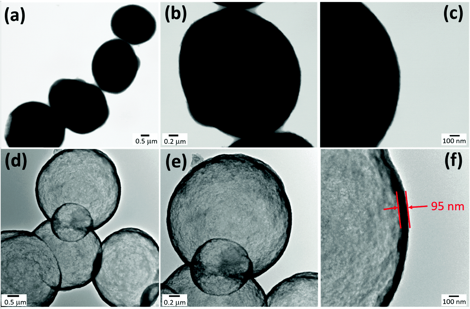 | ||
| Fig. 2 TEM images of CaCO3@SiO2/TiO2-coated particles (a–c) and hollow SiO2/TiO2-coated capsules (the molar ratio of TEOS and TIPO was 4:1) after CaCO3 removal (d–f). | ||
Comparative triggering via UV light, ultrasound and enzymes
The sol–gel approach provides the possibility of in situ growth of an inorganic layer into the polymer shell of core–shell particles resulting in the formation of the composite organic/inorganic capsules after CaCO3 removal. Such modification allows us to control the permeability of the shell and has an influence on the physicochemical properties of the hybrid capsules. In our case, the formation of an inorganic SiO2/TiO2 shell is able to encapsulate small cargo molecules due to the decrease of the permeability of the capsules. W. Parak and G. Sukhorukov in their works have shown that the silica growth on the PE microcapsules can block the possible diffusion of small cargo molecules from capsules.27,31,45 To prove it, we performed the in situ encapsulation of Rh-B and FITC inside the composite SiO2/TiO2-coated capsules. Fig. 3 and Fig. S5 (ESI†) show CLSM images of SiO2/TiO2-coated capsules encapsulating FITC (green fluorescence) and Rh-B (red fluorescence). After successful encapsulation of Rh-B and FITC, the triggered release of the cargo molecules was investigated upon physical and chemical stimuli. For this reason, the SiO2/TiO2-coated capsules containing Rh-B were treated with UV light, sonicated and incubated in pronase solution (PBS 7.4; 2 mg mL−1). Rh-B was chosen instead of FITC because of its longer lifetime.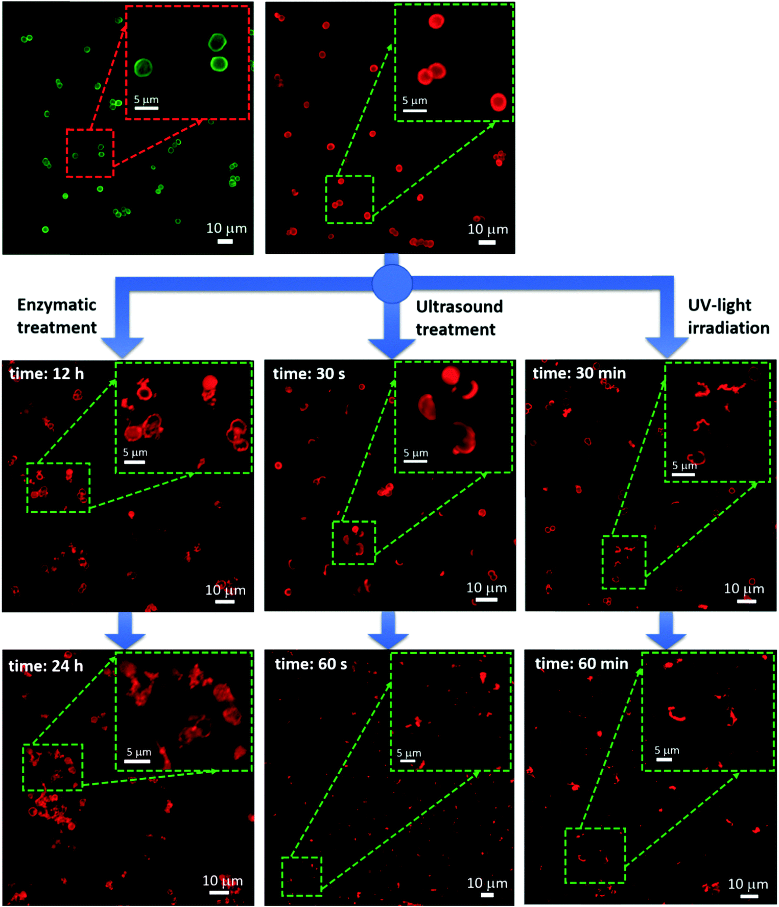 | ||
| Fig. 3 CLSM images of SiO2/TiO2-coated capsules with FITC (green capsules) and Rh-B (red capsules), showing the influence of enzymatic, ultrasound and UV-light on the decomposition of capsules at different periods of time. | ||
It is well known that TiO2 can absorb UV-light.21,22 Therefore, TiO2 has been selected for the modification of capsules as UV-responsive components because TiO2 can decompose the polymer shell consisting of PARG and DEXS via a photocatalytic reaction increasing the permeability of capsules, as demonstrated in Scheme 1b. The UV absorbance spectrum of SiO2/TiO2-coated capsules was measured in order to confirm the absorption in the UV range. As presented in the ESI† (Fig. S6), the SiO2/TiO2-coated capsules showed a wide absorbance peak at 284 nm, while there was no absorbance in case of the (PARG/DEXS)3 capsules. Our results are in good agreement with previously reported work by Wang et al. who fabricated self-assembled multilayer films of titania nanoparticles and polyelectrolytes.59 As shown in Fig. 3 (right) after 30 min of UV irradiation, more than half of the capsules were decomposed and release of Rh-B was observed. More significant changes can be detected after 60 min of UV irradiation; almost all capsules were broken into amorphous parts. The possible reason for capsule decomposition upon UV irradiation can be associated with high photocatalytic activity of the TiO2 in the inorganic layer. Therefore, the polymer shell consisting of PARG and DEXS can be degraded by TiO2 under UV irradiation. As a result, it leads to the destruction of capsules and further leakage of Rh-B (Fig. S7, ESI†). These results are correlated with the reported work of Y. Cong who designed pH and UV dual-responsive microcapsules.19
Another mechanism of capsule breakage is observed when we performed ultrasound irradiation. The ultrasound treatment caused a very rapid destruction of capsules. The incorporated robust layer of SiO2/TiO2 in the polymer shell makes it more responsive to ultrasound.31,45 After 30 s of ultrasound irradiation, almost all capsules were broken into different fragments. Further ultrasound treatment (60 s) leads to complete decomposition of the SiO2/TiO2-coated capsules into small pieces (Fig. 3, middle). The strengthened shell stiffness and low flexibility of the hybrid capsules increased sensitivity to ultrasound and absorption of acoustic energy.57
In order to study the process of enzymatic degradation of SiO2/TiO2-coated capsules, they were put in pronase solution (2 mg mL−1, PBS 7.4) and incubated at 37 °C for 24 h. After 12 h of incubation with pronase, the capsules were deformed and the leakage of Rh-B from the capsules was observed (Fig. 3, bottom left). Further incubation with pronase (24 h) leads to the formation of a mixture of deformed and broken capsules. The results obtained from CLSM clearly indicate that SiO2/TiO2-coated capsules possess the triple-responsive effect when they are triggered by UV (ultrasound) irradiation or enzymatic treatment.
The brief schematic illustration of the decomposition of SiO2/TiO2-coated capsules upon UV (ultrasound) irradiation and enzymatic treatment is shown in Fig. 4a. As has been demonstrated above, UV and ultrasound irradiation caused the breakage of SiO2/TiO2-coated capsules into small fragments. However, enzymatic degradation leads to the deformation of the capsules and cargo leakage. The possible mechanism of enzymatic degradation can be explained as follows: the polymer shell, assembled by PARG and DEXS, is degraded in the presence of a mixture of proteases as fragments of the polymers are still accessible to the enzyme. As a result, it forms hollow channels in the composite shell of SiO2/TiO2-coated capsules resulting in the structural disorder of the capsules, their deformation and a further release of cargo molecules. The encapsulated total amount of Rh-B in the sample of SiO2/TiO2-coated capsules (∼6.4 × 10⁶) was 8 μg. To demonstrate the trigger controlled release of cargo molecules upon UV (ultrasound) irradiation and enzymatic treatment, the Rh-B release time trace of SiO2/TiO2-coated capsules was carried out, as shown in Fig. 4b–d. Fig. 4b shows the release profiles of UV irradiated SiO2/TiO2-coated capsules and non-irradiated capsules as a control. Approximately 40% of the encapsulated Rh-B was released during 30 min of UV irradiation. After 60 min of UV irradiation, the release of Rh-B reached as high as 58% and was yet still slightly increasing while we continued the UV irradiation. In comparison with UV irradiation, the release of Rh-B from SiO2/TiO2-coated capsules treated with ultrasound is much faster; after 60 s of ultrasound treatment, we already observed more than 68% Rh-B release. Finally, the total release amount of Rh-B was around 76% after 100 s of sonication.
 | ||
| Fig. 4 Schematic representations of the different mechanisms of triggered cargo release (a); trigger-induced Rh-B release from SiO2/TiO2-coated capsules under UV irradiation (b), sonication (c) and enzymatic treatment (d). Bars represent mean ± S. D. | ||
It is worth mentioning that ultrasound treatment seems to be more effective than UV irradiation due to the higher amount of released Rh-B within a short duration of sonication. The enzymatic treatment of SiO2/TiO2-coated capsules in the pronase solution also showed a good release of Rh-B from capsules even through the release of Rh-B was much slower compared to UV (ultrasound) irradiation. The release of Rh-B was only 30%, when the time reached 12 h. After 24 h of incubation in pronase solution, SiO2/TiO2-coated capsules revealed 39%. Also, less than 10% of the Rh-B was released during the same period without enzymatic treatment, which indicates good blocking properties of the composite capsules preventing the leakage of Rh-B.
From the obtained results, we can say that the effect of ultrasound was stronger compared to UV- and enzymatic influences, but in our work we just demonstrate the principle of possible combination of these effects, and the desired tuning of responses can be done “a la carte” on particular demand. Thus, upon employing various stimuli (UV, ultrasound irradiation or enzymatic treatment), such smart designed capsules allow us to control the rate of drug release from seconds to hours. This strategy may allow us to achieve different clinical effects using one therapeutic agent. In clinical practice the application of multifunctional carriers may overcome the problems of conventional drug therapy, including the lack of specific targeting, drug degradation, poor distribution in tissues, and low biocompatibility.
As mentioned above, the UV-responsive properties of composite capsules are due to the incorporation of TiO2, which can be a promising strategy for the application of smart UV-responsive carriers in medicine or cosmetics. The different examples of application of UV-responsive carriers in medicine were presented in the field of ophthalmology for photodynamic therapy of macular degeneration and certain types of cancers,60 and the sustained release and suprachoroidal delivery of drugs.61 Nowadays, in dermatology and cosmetics, TiO2 nanoparticles have attracted considerable attention in photo-protection of skin against UV radiation. Despite the effectiveness of TiO2 nanoparticles in blocking the UV light, they might have hazardous effects on the skin suggesting the application of microcapsules based on TiO2. In oncology and internal medicine, UV-responsive capsules provide the possibility of their application in extracorporeal photopheresis,62 supplying the intracellular delivery of a wide variety of biologically active substances such as immunosuppressants, cytotoxic drugs, peptides or nucleic acids with a controllable release with the use of the same equipment and protocols.
Ultrasound-responsive capsules have a great potential for achieving spatiotemporal control of drug release at the desired site without ionizing radiation.31 Ultrasound-responsive micro-carriers may be used as ultrasound contrast agents during the drug delivery.63 Ultrasound-responsive microcapsules can be effectively used in gene therapy for the improved transport of DNA/RNA to the cytosol of the cell.64 In addition, our hybrid microcapsules provide high storage capacity and can induce high local concentration of the delivered therapeutic agents.
Cellular uptake, intracellular cargo release and cytotoxicity study
Although, SiO2/TiO2-coated capsules have shown promising results for triple-responsive cargo release, they should also be biodegradable and non-toxic for further clinical applications. In order to evaluate the biocompatibility and intracellular degradation of SiO2/TiO2-coated capsules, in vitro experiments with human adenocarcinoma HeLa cell lines and primary human mesenchymal stem cells (MSCs), representing the stromal cell of the body, were performed. Due to the fact that MSCs possess inherent tumor-tropic and migratory properties, it allows them to serve as vehicles for the in vivo delivery and treatment of isolated tumors.65 Therefore, the ability of MSCs to internalize hybrid microcapsules is also important in examining the developing cell-microcapsule based approaches.CLSM was applied to visualize the cellular uptake and cargo release in HeLa cells and MSCs. Rh-B was used as a model cargo molecule to visualize intracellular release. At an initial time (t = 0 h), when capsules were added to suspensions of cells at a capsule-to-cell ratio of 20:1, the capsules were stable and no leakage of Rh-B from the capsules was observed (Fig. 5 and 6). However, after 4 h of incubation, as one can see in the CLSM images the capsules have accumulated mainly in the perinuclear compartment of the cells and the Rh-B was spread along the cytosol of the cells. Also it can be clearly seen that most of the capsules exhibited red fluorescence and they maintain their integrity, while some of them became deformed and leaky (at a higher magnification as indicated by the arrows in Fig. 5 and 6). This represents the mechanical deformation due to acting intracellular forces.66 Such results confirmed that capsules began to degrade. After 8 h of incubation, the degraded capsules, internalized by the cells, have lost their spherical shape and deformed. All encapsulated Rh-B was leaked from the capsules into the cytosol of the cells. After a 24 h time point one can see in the CLSM images at a higher magnification that almost all the capsules are degraded and amorphous debris is formed. Notably, in a more detailed magnified view, some of the capsules were cracked into small pieces and these small red pieces from the capsules were still observed (as indicated by the arrows in Fig. 5 and 6 for 24 h of incubation). Also, it is worth mentioning that there was not a significant difference in the degradation rate of capsules internalized by HeLa cells and MSCs. Based on the CLSM images in Fig. S8 and S9 (ESI†), the capsules, which were not internalized by the cells, preserved integrity and stability, i.e. they retained their spherical shape and did not form amorphous fragments.
 | ||
| Fig. 5 CLSM images demonstrating the degradation of SiO2/TiO2-coated capsules and Rh-B release when capsules were incubated with HeLa cells for 24 h. | ||
 | ||
| Fig. 6 CLSM images demonstrating the degradation of SiO2/TiO2-coated capsules and Rh-B release when capsules were incubated with MSCs for 24 h. | ||
To investigate the cellular uptake of SiO2/TiO2-coated capsules with Rh-B into the cells quantitatively, the penetration of SiO2/TiO2-coated capsules into cells was investigated using flow cytometry, after the cells (HeLa and MSCs) were incubated with capsules at different time internals. The longer uptake time of SiO2/TiO2-coated capsules leads to an increase in fluorescence intensity and the ratio of fluorescent-positive cells (Fig. 7). The cellular uptake efficiency of SiO2/TiO2-coated capsules was high for both cell lines (HeLa cells and MSCs). In the case of HeLa cells, more than 85% of cells exhibited red fluorescence signals from Rh-B after 6 h of incubation and more than 95% for MSCs. These results demonstrate that our hybrid capsules are readily internalized by tumorous and normal cell types.
 | ||
| Fig. 7 Flow cytometry histogram profiles of Rh-B within HeLa cells and MSCs incubated with SiO2/TiO2-coated capsules at different time intervals. Dot-plot of forward light scatter cells (FSCs) and side light scatter cells (SSCs) (top); relative cell viabilities of HeLa cells and MSCs incubated with different concentrations of SiO2/TiO2-coated capsules for 24 and 48 h (right). | ||
Next, we examined the effects of SiO2/TiO2-coated capsules (without Rh-B) on the cell viabilities against MSCs and HeLa cells. The cytotoxicity of SiO2/TiO2-coated capsules against MSCs and HeLa cells was evaluated with capsule-to-cell ratios (50:1, 100:1 and 150:1) for 24 h and 48 h, respectively (Fig. 7 right). It is clearly seen that SiO2/TiO2-coated capsules exhibited a low cytotoxicity (>80%) at the concentrations of capsules below 100 per cell for 24 h and 48 h of incubation. However, for a higher concentration of capsules per cell (150:1), the cell viability of SiO2/TiO2-coated capsules decreased below 80% for both cell lines (HeLa cells and MSCs) when the incubation time reached 24 h. The same tendency in the decrease of cell viability of SiO2/TiO2-coated capsules was observed after 48 h of incubation. From the assessment of cell viability we confirm the cellular compatibility of our hybrid capsules, which is highly beneficial for the potential application of these composite hybrid capsules as intracellular delivery vehicles.
In summary, the obtained results clearly demonstrate that SiO2/TiO2-coated capsules have shown a biodegradable ability after internalization by HeLa cells and MSCs within 24 h. Moreover, such composite capsules exhibit a low cytotoxicity, satisfactory biocompatibility profile which plays a main role in the design of a perspective drug delivery system with multi-functional responsive effects for biomedical implementation.
Conclusions
In this work, novel hybrid inorganic/organic capsules with triple-responsiveness were successfully fabricated. The combination of the advantageous structural properties of SiO2 with UV-responsive TiO2 provides an efficient strategy for the fabrication of stable and safe capsules with a low permeability and simultaneous responsiveness to ultrasound and UV light. These capsules are made of polypeptides/polysaccharides and are functionalized by the SiO2/TiO2 shell that supports in the encapsulation of small molecules as illustrated in the example of rhodamine B and at the same time such capsules are subjected to biodegradation upon enzymatic treatment. In vitro experiments have also confirmed that these capsules are not toxic and exhibited intracellular degradation after internalization by HeLa cells and MCSs. These hybrid capsules showed their potential for envisaged drug delivery systems, because varying different modes of treatment (UV light, ultrasound or enzymes) bring multiple leverages to control the release of the bioactive agent at desired times using physical and chemical stimuli. Such a synergetic approach when three stimuli-responsive mechanisms combine in one delivery system might be effectively used to enhance the use of bioactive agents in cosmetics, and biomedical and environmental applications.Acknowledgements
This work was supported by the Russian Foundation of Basic Research grants no. 16-33-50153 mol_nr and no. 16-33-00966 mol_a, and Russian Governmental Program “Nauka”, N: 1.1658.2016, 4002. We also thank Ekaterina Bozhokina who provided us the Human cervical cancer cell line (HeLa).References
- D. Luo and W. Saltzman, Nat. Biotechnol., 2000, 18, 33–37 CrossRef CAS PubMed.
- J. Doerner, S. Febvay and D. Clapham, Nat. Commun., 2012, 3, 990 CrossRef PubMed.
- M. Dzamukova, E. Naumenko, Y. Lvov and R. Fakhrullin, Sci. Rep., 2015, 5, 10560–10571 CrossRef CAS PubMed.
- T. Shutava, P. Pattekari, K. Arapov, V. Torchilin and Y. Lvov, Soft Matter, 2012, 8, 9418–9427 RSC.
- S. De Koker, R. Hoogenboom and B. De Geest, Chem. Soc. Rev., 2012, 41, 2867–2884 RSC.
- A. Timin, E. Balantseva, S. Khashirova, E. Rumyantsev and T. Osadchaya, Colloids Surf., A, 2015, 477, 26–34 CrossRef CAS.
- W. Tong, Y. Zhu, Z. Wang, C. Gao and H. Möhwald, Macromol. Rapid Commun., 2010, 31, 1015–1019 CrossRef CAS PubMed.
- J. Eloy, M. Souza, R. Petrilli, J. Barcellos, R. Lee and J. Marchetti, Colloids Surf., B, 2014, 123, 345–363 CrossRef CAS PubMed.
- A. Kidane and P. Bhatt, Curr. Opin. Chem. Biol., 2005, 9, 347–351 CrossRef CAS PubMed.
- M. Delcea, H. Möhwald and A. G. Skirtach, Adv. Drug Delivery Rev., 2011, 63, 730–747 CrossRef CAS PubMed.
- A. Gulzar, S. Gai, P. Yang, C. Li, M. Ansari and J. Lin, J. Mater. Chem. B, 2015, 3, 8599–8622 RSC.
- S. MacEwan, D. Callahan and A. Chilkoti, Nanomedicine, 2010, 5, 793–806 CrossRef CAS PubMed.
- S. Baek, R. Singh, T. Kim, J. Seo, U. Shin, W. Chrzanowski and H. Kim, ACS Appl. Mater. Interfaces, 2016, 8(14), 8967–8979 CAS.
- J. Wei, X. Ju, X. Zou, R. Xie, W. Wang, Y. Liu and L. Chu, Adv. Funct. Mater., 2014, 24, 3312–3323 CrossRef CAS.
- Y. Yeh, R. Tang, R. Mout, Y. Jeong and V. Rotello, Angew. Chem., Int. Ed., 2014, 53, 5137–5141 CAS.
- V. Zhang, Ch. Ang, M. Li, S. Tan, Q. Qu, Zh. Luo and Y. Zhao, ACS Appl. Mater. Interfaces, 2015, 7, 18179–18187 Search PubMed.
- P. Schattling, F. Jochum and P. Theato, Polym. Chem., 2014, 5, 25–36 RSC.
- D. Xiao, H. Jia, J. Zhang, C. Liu, R. Zhuo and X. Zhang, Small, 2014, 10, 591–598 CrossRef CAS PubMed.
- Y. Cong, K. Chen, Sh. Zhou and L. Wu, J. Mater. Chem. A, 2015, 3, 19093–19099 CAS.
- Z. Xu, Y. Kang, S. Liu and M. Wang, Nanoscale, 2015, 7, 5859–5868 RSC.
- D. G. Shchukin, E. Ustinovich, D. V. Sviridov, Y. M. Lvov and G. B. Sukhorukov, Photochem. Photobiol. Sci., 2003, 2, 975–978 CAS.
- S. A. Ansari and M. H. Cho, Sci. Rep., 2016, 6, 25405 CrossRef CAS PubMed.
- L. Ye, R. Pelton and M. Brook, Langmuir, 2007, 23, 5630–5637 CrossRef CAS PubMed.
- Y. Hu, J. Ge, Y. Sun, T. Zhang and Y. Yin, Nano Lett., 2007, 7, 1832–1836 CrossRef CAS PubMed.
- V. Klimkevicius, T. Graule and R. Makuska, Langmuir, 2015, 31, 2074–2083 CrossRef CAS PubMed.
- R. Raliya, P. Biswas and J. C. Tarafdar, J. Appl. Biotechnol. Rep., 2015, 5, 22–26 CrossRef.
- A. Ott, X. Yu, R. Hartmann, J. Rejman, A. Schütz, M. Ochs and W. Parak, Chem. Mater., 2015, 27(6), 1929–1942 CrossRef CAS.
- M. Manzano and M. Vallet-Regí, J. Mater. Chem., 2010, 20, 5593 RSC.
- J. Vivero-Escoto, I. Slowing, G. Trewyn and V. Lin, Small, 2010, 6, 1952–1967 CrossRef CAS PubMed.
- Z. Li, J. Barnes, A. Bosoy, J. Stoddart and J. Zink, Chem. Soc. Rev., 2012, 41, 2590 RSC.
- H. Gao, D. Wen and G. Sukhorukov, J. Mater. Chem. B, 2015, 3, 1888–1897 RSC.
- J. Paris, M. Cabanas, V. Manzano and M. Vallet-Regi, ACS Nano, 2015, 9, 11023–11033 CrossRef CAS PubMed.
- S. Qourzal, N. Barka, M. Tamimi, A. Assabbane, A. Nounah, A. Ihlal and Y. Ait-Ichou, Mater. Sci. Eng., C, 2009, 29, 1616–1620 CrossRef CAS.
- J. Shao, M. Xuan, T. Si, L. Dai and Q. He, Nanoscale, 2015, 7, 19092–19098 RSC.
- A. Skirtach, C. Dejugnat, D. Braun, A. Susha, A. Rogach, W. Parak, H. Mohwald and G. Sukhorukov, Nano Lett., 2005, 5, 1371–1377 CrossRef CAS PubMed.
- D. Andreeva, D. Gorin, D. Shchukin and G. Sukhorukov, Macromol. Rapid Commun., 2006, 27, 931–936 CrossRef CAS.
- T. Kolesnikova, D. Gorin, P. Fernandes, S. Kessel, G. Khomutov, A. Fery, D. Shchukin and H. Möhwald, Adv. Funct. Mater., 2010, 20, 1189–1195 CrossRef CAS.
- G. Sukhorukov, B. A. Rogach, M. Garstka, S. Springer, W. Parak, A. Munoz-Javier, O. Kreft, A. Skirtach, A. Susha, Y. Ramaye, R. Palankar and M. Winterhalter, Small, 2007, 3, 944–955 CrossRef CAS PubMed.
- J. Huang, C. Luo, W. Li, Y. Li, Y. Zhang, J. Zhou and Q. Jiang, J. Mater. Chem. B, 2015, 3, 4530–4538 RSC.
- C. Zheng, Y. Ding, X. Liu, Y. Wu and L. Ge, Intern. J. Pharm. Practice, 2014, 475, 17–24 CrossRef CAS PubMed.
- K. Katagiri, Y. Imai, K. Koumoto, C. Zheng, Y. Ding, X. Liu, Y. Wu and L. Ge, J. Colloid Interface Sci., 2011, 361, 109–114 CrossRef CAS PubMed.
- K. Katagiri, M. Nakamura and K. Koumoto, ACS Appl. Mater. Interfaces, 2010, 2, 768–773 CAS.
- S. De Koker, L. De Cock, P. Rivera-Gil, W. Parak, R. Auzély Velty, C. Vervaet, J. Remon, J. Grooten and B. De Geest, Adv. Drug Delivery Rev., 2011, 63, 748–761 CrossRef CAS PubMed.
- W. Tong, Y. Zhu, Z. Wang, C. Gao and H. Möhwald, Macromol. Rapid Commun., 2010, 31, 1015–1019 CrossRef CAS PubMed.
- H. Gao, O. Goriacheva, N. Tarakina and G. Sukhorukov, ACS Appl. Mater. Interfaces, 2016, 15, 9651 Search PubMed.
- H. Gao, D. Wen, N. Tarakina, J. Liang, A. Bushby and G. Sukhorukov, Nanoscale, 2016, 8, 5170–5180 RSC.
- I. Marchenko, A. Yashchenok, T. Borodina, T. Bukreeva, M. Konrad, H. Mohwald and A. Skirtach, J. Controlled Release, 2012, 162, 599–605 CrossRef CAS PubMed.
- B. De Geest, R. Vandenbroucke, A. Guenther, G. Sukhorukov, W. Hennik, N. Sanders, J. Demeester and S. De Smedt, Adv. Mater., 2006, 18, 1005–1009 CrossRef CAS.
- V. Vergaro, F. Scarlino, C. Bellomo, R. Rinaldi, D. Vergara, M. Maffia, F. Baldassarre, G. Gianneilli, X. Zhang, Y. M. Lvov and S. Leporatti, Adv. Drug Delivery Rev., 2011, 63, 847–863 CrossRef CAS PubMed.
- G. Sukhorukov, E. Donath, H. Lichtenfeld, E. Knippel, M. Knippel, A. Budde and H. Mohwald, Colloids Surf., 1998, 137, 253–266 CrossRef.
- M. Soleimani and S. Nadri, A protocol for isolation and culture of mesenchymal stem cells from mouse bone marrow, Nat. Protoc., 2009, 4, 102–106 CrossRef CAS PubMed.
- H. Kim, Y. Shul and H. Han, Top. Catal., 2005, 35(3), 287–293 CrossRef CAS.
- K. Gude, V. Gunko and J. Blitz, Colloids Surf., A, 2008, 325, 17–20 CrossRef CAS.
- V. Mangalampalli, P. Sharma, V. Kumari and M. Subrahmanyam, J. Hazard. Mater., 2010, 175, 1101–1105 CrossRef PubMed.
- P. Klankaw, C. Chawengkijwanich, N. Grisdanurak and S. Chiarakorn, Superlattices Microstruct., 2012, 51, 343–352 CrossRef CAS.
- A. Timin, S. Khashirova, E. Rumyantsev and A. Goncharenko, Mater. Sci. and Eng. C, 2016, 64, 20–28 CrossRef CAS PubMed.
- Y. He, J. Peng, W. Chu, Y. Li and D. Ge Tonq, J. Mater. Chem. A, 2014, 2, 1721–1731 CAS.
- R. Grieken, J. Aguado, M. Lopez-Munoz and J. Marugan, J. Photochem. Photobiol., A, 2002, 148, 315–322 CrossRef.
- Z. Wang, T. Sasaki, M. Muramatsu, Y. Ebina, T. Tanaka and M. Watanabe, Chem. Mater., 2003, 15(3), 807–812 CrossRef CAS.
- J. Christie and U. Kompella, Drug Discovery Today, 2008, 13(3–4), 124–134 CrossRef CAS PubMed.
- P. Tyagi, M. Barros, J. Stansbury and U. Kompella, Mol. Pharmaceutics, 2013, 10(8), 2858–2867 CrossRef CAS PubMed.
- D. Tran and R. Salmon, Australas. J. Dermatol., 2011, 52(1), 1–6 CrossRef PubMed.
- H. Ke, J. Wang, Z. Dai, Y. Jin, E. Qu, Z. Xing, C. Guo, X. Yue and J. Liu, Angew. Chem., Int. Ed., 2011, 50, 3017–3021 CrossRef CAS PubMed.
- E. Unger, T. Porter, W. Culp, R. Labell, T. Matsunaga and R. Zutshi, Adv. Drug Delivery Rev., 2004, 56, 1291–1314 CrossRef CAS PubMed.
- Z. Gao, L. Zhang, J. Hu and Y. Sun, Nanomedicine, 2013, 9, 174–184 CAS.
- P. Rivera-Gil, S. Koker, B. Geest and W. Parak, Nano Lett., 2009, 9, 4398–4402 CrossRef CAS PubMed.
Footnote |
| † Electronic supplementary information (ESI) available: Additional figures. See DOI: 10.1039/c6tb02289h |
| This journal is © The Royal Society of Chemistry 2016 |