The substitution of the platinum counter electrode in a plasmonic photoelectrochemical system with near-infrared absorption for solar water splitting

Chih-Jung Chen a, Chih Kai Chena, Tzu-Hsiang Lua, Shu-Fen Hu*b and Ru-Shi Liu*ac
aDepartment of Chemistry, National Taiwan University, Taipei 106, Taiwan. E-mail: rsliu@ntu.edu.tw
bDepartment of Physics, National Taiwan Normal University, Taipei 116, Taiwan. E-mail: sfhu.hu@gmail.com
cDepartment of Mechanical Engineering, Graduate Institute of Manufacturing Technology, National Taipei University of Technology, Taipei 106, Taiwan

Received 2nd October 2016 , Accepted 18th October 2016

First published on 18th October 2016


Abstract

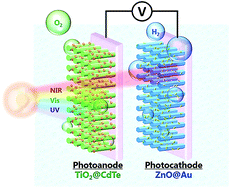
Converting solar energy into a usable chemical fuel has attained great importance in the past decade. The near-infrared and infrared regions contain nearly half of the photons present in the total flux of solar irradiation; however, near-infrared-active and infrared-active materials cannot commonly provide sufficient potential for electron–hole pairs to drive the photoelectrochemical reaction, which limits the development of efficient solar energy conversion devices for future applications. Here, we report on a photoelectrochemical cell that is constructed using ZnO@Au rod nanostructures as the photocathode and TiO2@CdTe quantum dot nanostructures as the photoanode. In this cell, the photoactive materials can utilize a wide range of the solar spectrum (up to the near-infrared). Using a plasmonic photocathode, a maximum efficiency of about 1.4% (at +0.5 V) was exhibited, which was comparable to that attained when using conventional Pt foil as the electrodes. The use of plasmonic materials has several advantages, including easily customizable optical properties and the ability for coupling with plasmon-inducing electromagnetic fields/hot electrons, which can effectively enhance the photocatalytic water splitting reactions. This research aims to provide an alternative photoelectrode to replace conventional Pt electrodes, to improve the conversion efficiency for solar energy.


Introduction

Nowadays, energy consumption per year is approaching 15 TW. To meet this energy requirement, various sustainable energy sources are in demand. Solar energy has been seen as an inexpensive, pollution free and abundant energy source. The solar energy striking the surface of Earth for a given time period vastly exceeds the energy consumption over the same time period. To harvest solar energy efficiently and convert it into a usable energy form has attained great importance in the past few decades.1 As society has become increasingly aware of the adverse effects of human activities on the environment, the development of methods to utilize solar energy effectively has become one of the key scientific challenges of this century.2–5 The photosynthesis reaction supports the existence of life forms on Earth. The core chemical reaction carried out in photosynthesis is the use of solar energy to convert water and carbon dioxide into oxygen gas and carbohydrates, which could act as fuels. Developing an artificial version of the photosynthesis reaction, splitting water into hydrogen and oxygen, is highly desirable due to the attraction of hydrogen as a fuel.6–8 To develop this sustainable energy, the critical factor is converting solar energy into chemical fuels efficiently. Many materials have demonstrated the activity necessary to drive the photoelectrochemical water splitting reaction or half reaction.9–13 Most studies attempted to build the perfect solar cell, in which the absorption of the active materials could cover the entire solar spectrum and thus collect photons from all sunlight. A variety of materials have been synthesized to achieve this goal; numerous organic/inorganic dyes, semiconductors, and semiconductor quantum dots (QDs) have provided the ability to capture UV and/or visible light from solar illumination.14–17 Although there have been many works demonstrating organic/inorganic materials that significantly enhance the solar energy conversion efficiency, these have almost always been performed in the ultraviolet and visible regions of the solar irradiation spectrum, rather than in the near-infrared and infrared regions. Although a few materials, such as up-conversion nanoparticles, can harvest infrared irradiation, the efficiency still has room to improve.18 This is because near-infrared-active and infrared-active materials commonly absorb sub-band gap irradiation, which cannot provide sufficient potential to the electron–hole pairs in order to drive the chemical reactions in electrolytes. For photoelectrochemical water splitting, the minimum energy requirements and the thermodynamic/overpotential loss must be at least 1.8 eV, which corresponds to the onset of light absorption at a wavelength of 688 nm.19,20 Consequently, this effect will limit the efficient utilization of solar energy to meet the requirements of solar photovoltaic devices for future applications. The near-infrared and infrared regions account for more than half of the total solar radiation, making it difficult to achieve a significant enhancement in energy conversion based on the design of current technologies.

Recently, surface plasmon resonance (SPR) was widely applied in studies on solar energy conversion to fuels, and organic photodegradation, which focused on the improvement of the photoanode catalytic efficiency.21–26 In the presented work, we report on a photoelectrochemical cell that is constructed using ZnO@Au rod nanostructures as the photocathode and TiO2@CdTe QD nanostructures as the photoanode. In this cell, the photoactive materials can utilize a wide range of the solar spectrum (up to the near-infrared). Through using a plasmonic photocathode, a maximum efficiency of about 1.4% (at +0.5 V) was exhibited, which was comparable with that obtained when using conventional Pt foil as electrodes.

Experimental

Synthesis of ZnO seeds

ZnO seeds were fabricated through following a previous work.27 Fluorine-doped tin oxide (F:SnO2; FTO) conducting glass, functioning as the substrate, was sequentially ultrasonicated in ethanol and acetone for 10 min. Zinc acetate solution [Zn(CH3CO2)2; 0.1 M] was prepared through dissolution in 100 mL of absolute ethanol. The cleaned-FTO glass was immersed in the zinc acetate solution for 10 s, and subsequently blown to dryness using a stream of argon. This step was repeated 8 times for a uniform coating of zinc acetate on FTO glass, with a sufficient thickness. The zinc acetate coated-FTO substrates were then heated to 350 °C in air for 30 min to yield a layer of ZnO seeds on an FTO substrate.

Synthesis of ZnO nanowires

ZnO nanowires were prepared via a modified growth method.28,29 Zinc nitrate [Zn(NO3)2; 0.06 M] and hexamethylenetetramine (HMT; 0.06 M) were dissolved in 100 mL of DI water, and this solution was transferred to a Teflon vessel. The ZnO seed coated-FTO substrates were placed face down and fixed horizontally in the solution. The Teflon vessel was then sealed in an autoclave and heated to 110 °C to initiate nanowire growth. After a hydrothermal reaction for 24 h, the as-prepared ZnO nanowires was thoroughly washed with DI water, and then blown to dryness under a stream of argon. The samples were subsequently annealed at 450 °C for 30 min in air for the improvement of crystallization and attachment.

Synthesis of TiO2 nanowires

Concentrated hydrochloric acid (HCl; 30 mL) was mixed with 30 mL of DI water, and then stirred for 5 min. Titanium butoxide (C16H36O4Ti; 1 mL) was added to the mixed solution, which was stirred for 5 min. This reagent was subsequently transferred to a Teflon vessel with cleaned-FTO substrates facing down. A hydrothermal reaction was then carried out at 150 °C for 6 h. After the synthesis, the TiO2 nanowires were thoroughly washed with DI water, and then blown to dryness under a stream of argon.

Synthesis of CdTe quantum dots

CdTe quantum dots were synthesized according to our previous works.30,31 Sodium borohydride (NaBH4; 0.08 g) was dissolved in 3.0 mL of DI water to reduce Te powder (0.127 g) for 4 h, to generate 1 M sodium hydrogen telluride (NaHTe) solution. Mercaptopropionic acid (MPA; 38 mM) and cadmium chlorite (CdCl2; 16 mM) were mixed to prepare a solution (74.8 mL) with a tuned pH of 10.8 under a nitrogen gas atmosphere. The NaHTe solution (1.5 mL) was then added to the mixed solution to give a final mixture with a Cd2+/MPA/HTe molar ratio of 1[thin space (1/6-em)]:[thin space (1/6-em)]2.4[thin space (1/6-em)]:[thin space (1/6-em)]0.5. This solution was subsequently heated to 90 °C under reflux for 3 h. After the reaction, CdTe QDs were collected using centrifugation in absolute ethanol, which removed free MPA ligands and unreacted precursor ions to purify the CdTe QDs.

Synthesis of Au nanoparticles

Our previous studies were adapted to prepare Au nanoparticles.31,32 50 mL of aqueous hydrogen tetrachloroaurate(III) hydrate (HAuCl4; 0.4 mM) was heated to 90 °C, then 5 mL of trisodium citrate dihydrate (C6H5Na3O7·2H2O; 1 wt%) was added into the solution, which was keep stirring for 15 min. Then the solution was stirred and cooled to room temperature to obtain an Au nanoparticle suspension.

Synthesis of Au nanorods

Au nanorods were synthesized using a modified seed-mediated growth method.33 A cetyltrimethylammonium bromide [(C16H33)N(CH3)3Br; CTAB] solution was prepared through mixing CTAB (0.36 g) and 10 mL of distilled water, and then heating to 45 °C and stirring to dissolve the CTAB completely. 0.6 mL of 0.1 M sodium borohydride (NaBH4) was added into the solution after 0.25 mL of 10 mM HAuCl4 aqueous solution was added to the CTAB solution kept at 27 °C, which obtains gold nanoparticles as seeds for the following experiment. 4 mL of aqueous HAuCl4 (10 mM) was added into 100 mL of 3.6 wt% CTAB aqueous solution, and then 0.6 mL of 1.7 wt% aqueous silver nitrate (AgNO3) was added into the solution. After that, 0.6 mL of 17.6 wt% aqueous ascorbic acid (C6H8O6) was added into the solution. 0.4 mL of Au seeds was added into the solution after the color of the solution turned from transparent brown to transparent, and this was kept at 27 °C overnight to obtain an Au nanorod suspension.

Surface modification of Au nanorods

1 mL of Au nanorods was added into 10 mL of 0.1 M MPA solution with a pH of 9.0. The solution was ultrasonically agitated for 10 min to let the CTAB be substituted by MPA, to acquire MPA surface modified-Au nanorods.

Preparation of the photoelectrode

The pre-synthesis TiO2 and ZnO nanowire substrates were placed nanowire side up into the CdTe QD and Au solutions, respectively. From our previous studies, a monolayer layer modification of CdTe QDs on ZnO, and the optimized hot electron injection of ZnO@Au nanoparticles were achieved through chemical bath deposition for 24 h.30,32 Therefore, the duration of chemical bath deposition for preparing these photoelectrode materials was kept constant at 24 h. After 24 h chemical bath deposition, the photoelectrodes were taken out and washed with DI water to remove residual QD or Au solutions. The photoelectrodes were pasted with copper wire using silver paste, and dried overnight. The residual space in the substrate was covered using resin to attain TiO2@CdTe QD, ZnO@Au nanoparticle and ZnO@Au nanorod photoelectrodes.

Electrochemical characterization of the water-splitting photoelectrodes

The electrochemical characterization was performed using two electrode methods. A TiO2@CdTe QD photoelectrode was used as the working electrode. The counter electrodes used were a platinum plate, ZnO@Pt nanoparticle electrode, ZnO@Au nanoparticle electrode and ZnO@Au nanorod electrode, separately. All photoelectrochemical (PEC) performances were observed in 0.5 M Na2SO4 (pH = 6.8) solution, which served as a supporting electrolyte. The distance between the photoanode and photocathode was about 5 cm. The PEC measurements for analyzing the photoresponse to the water splitting reaction were taken separately using TiO2@QDs and ZnO@Au rods as the working and counter electrodes, as shown in Fig. 1a. This PEC cell was characterized under simulated solar illumination, utilizing a xenon lamp (PE300BF) equipped with an AM 1.5 filter. The TiO2@QD photoanode was excited using incident ultraviolet and visible light, and the penetrating near-infrared irradiation was absorbed by the ZnO@Au rod photocathode. Another PEC configuration was used for evaluating the hot electron transfer of the ZnO@Au rods (Fig. 7). This set up was obtained by respectively using ZnO@Au rods and Pt foil as the working and counter electrodes under chopped solar simulation, equipped with a monochromater to split the light which corresponded to the SPR absorption of the Au rods. All PEC data was recorded using a potentiostat and the PEC cell was maintained at 25 °C under the simulated solar radiation (100 mW cm−2). Hydrogen and oxygen generation was conducted using a two electrode system with a +0.5 V bias upon simulated solar illumination at 100 mW cm−2 in a self-made cell. The TiO2@CdTe QD photoelectrode was used as the working electrode and the ZnO@Au nanorod photoelectrode was used as the counter electrode. The amount of H2 and O2 accumulated in the glass system was measured using a China Chromatography 2000 gas chromatograph that was equipped with a flame ionization detector (FID).
image file: c6ra24563c-f1.tif
Fig. 1 (a) The schematic structure of a ZnO nanowire@plasmonic//TiO2 nanowire@quantum dot photoelectrochemical cell. (b) The absorption spectra of ZnO, CdTe QDs and Au nanorods.

Characterization of the materials

Ultraviolet-Visible (UV-Vis) absorption spectra were measured using a Thermo EVOLUTION 220 spectrometer. The morphology of the electrode materials were characterized using a JEOL JEM-2100F high-resolution transmission electron microscope (HRTEM), and a JEOL JSM-6700F field-emission SEM (FE-SEM).

Results and discussion

Here, we evaluate a photoelectrochemical device for solar water splitting that is compatible with current state-of-the-art technologies and compatible with other photovoltaic concepts. We integrate three types of materials to collect the ultraviolet (UV), visible (Vis), and near-infrared (NIR) regions of solar energy in a single photoelectrochemical cell, in which TiO2 and ZnO nanowires, CdTe quantum dots (QDs), and gold nanorods (Au rods) are employed as active materials for ultraviolet, visible, and near-infrared irradiation, respectively. This near-infrared active photoelectrochemical cell, which uses plasmonic nanocomposites for solar water splitting, is depicted in Fig. 1a. The figure shows the photoanode, which was composed of TiO2 nanowires configured into an array with a monolayer of quantum dots (TiO2@CdTe QDs). The photoanode exhibited good stability during the photoelectrochemical reaction30 (a structural characterization is shown in Fig. 2). The one-dimensional ordered architecture offers longer electron diffusion lengths and shorter electron transport times than conventional randomly oriented nanoparticle films.2,34,35 Interestingly, ZnO with additional plasmonic (gold nanorod) features, termed a ZnO@Au nanorod photoelectrode, was elaborately used as the photocathode. The ultraviolet and visible light from solar illumination is captured by the photoanode (TiO2@CdTe QDs) and generates photoelectron/hole pairs, through which water is oxidized to release oxygen. Simultaneously, the near-infrared region of solar illumination passes through the photoanode, which is not capable of absorbing NIR, leading to NIR illumination upon the plasmonic nanocomposite photocathode, where localized plasmonic effects were induced on the gold nanostructures. It should be noted that the use of plasmonic metal nanostructures as NIR-active materials, as opposed to using other NIR-active materials such as up-conversion materials, has additional benefits. Aside from localized surface plasmon resonance (LSPR) from metallic nanostructures, which can deliver a significant amount of control over the optical field,36 plasmonic materials are considerably stable and allow for the avoidance of undesired photo-decomposition/corrosion, as compared to semiconductor materials. Therefore, LSPR simultaneously offers significant contributions to photoreactions, such as hot-electron transfer (HET), a localized electromagnetic field (LEMF), and a relatively large optical cross-section.37–39 In addition to its tunable plasmon resonance wavelengths from visible to near-infrared frequencies, gold is thermodynamically stable compared to silver under the tough conditions, such as pH and potential, that are needed for water splitting.40 Unfortunately, no work has been performed on plasmonic materials as bifunctional materials for photocathodes and NIR harvesting in photoelectrochemical devices. Fig. 1b shows the absorption spectra of ZnO nanowires, CdTe quantum dots, and gold nanorods with various LSPR wavelengths, which reveal that a combination of these photoactive materials can cover a wide range of the solar spectrum. Here we synthesized Au rods with different aspect ratios for tuning various different surface plasmon resonance wavelengths. Besides, the longer Au rods captured incident light with longer wavelengths. For convenience, we abbreviated Au rods with different aspect ratios as “SPR-T”, in which T represents the resonance wavelength of the Au rods. This system enables photoelectric conversion for a wide range of wavelengths, from ultraviolet to near-infrared light, which allows the use of light with wavelengths from 300 nm to 800 nm. TEM images of Au rods with various resonance wavelengths are shown in Fig. 3.
image file: c6ra24563c-f2.tif
Fig. 2 (a and b) SEM images of the TiO2 nanorods. The dense TiO2 nanorod array was grown on a conductive substrate (F:SnO2, FTO) via a hydrothermal reaction. The SEM images elucidate that the diameter of the TiO2 was around 200 nm and the length of the TiO2 was around 7 μm. (c) TEM image of the TiO2@CdTe QDs, with the diameter of the TiO2 being consistent with the results from the SEM images. (d) HRTEM image of TiO2@CdTe QDs, showing that CdTe QDs are successfully attached to the surface of the TiO2 nanowires.

image file: c6ra24563c-f3.tif
Fig. 3 TEM images of gold nanorods ((a) SPR-667; (b) SPR-760; and (c) SPR-800), showing that these rods have a uniform size and shape. TEM images of ZnO nanowires decorated with an ensemble of Au rods ((d) SPR-667; (e) SPR-760; (f) SPR-800), showing that these rods are homogeneously attached to the surface of ZnO.

In a typical procedure, ZnO nanowire arrays were first synthesized over the surface of an FTO glass substrate using a hydrothermal method. Scanning electron microscopy (SEM) images reveal the growth of dense and vertically aligned ZnO nanowires on the substrate, and the typical length of the nanowires was approximately 8.5 μm (Fig. 4a). The utilization of single crystalline nanowires with a high aspect ratio will contribute to a high loading of plasmonic materials and effective carrier collection. Transmission electron microscopy (TEM) images of the ZnO nanowires demonstrate that the wires have a uniform diameter (Fig. 4b). Images of the gold nanorods that act as plasmonic active materials (NIR region) and the corresponding electron diffraction patterns are shown in Fig. 4c (SPR-703) and Fig. 4e (SPR-722). The gold nanorods were produced in high yields, and the structures exhibited relatively uniform diameters and lengths. TEM images of Au rod modified-ZnO nanowires indicate that Au rods with a uniform diameter are homogeneously distributed on the surface of ZnO [Fig. 4d (SPR-703) and Fig. 4f (SPR-722)]. The selected area electron diffraction (SAED) pattern (Fig. 4g) reveals that a two component crystalline nature was present in the Au rod decorated-ZnO nanowires. The spot patterns show the [2[1 with combining macron][1 with combining macron]0] zone axis of the ZnO nanowires with a single crystalline wurtzite structure (indicated using the blue hexagon). The ring patterns were indexed as polycrystalline Au rods, which corresponded to a typical face-centered-cubic (fcc) structure. The corresponding elemental mapping of Zn, O, and Au demonstrates that Zn and O are present along the entire length of the nanorods, while Au is found in the areas that correspond to the positions of the gold nanorods in the TEM image (Fig. 4h).


image file: c6ra24563c-f4.tif
Fig. 4 Structural characterization of Au nanostructure-ZnO photoelectrodes. (a) SEM image of an array of ZnO nanowires. (b) TEM image of ZnO nanowires. (c and e) TEM images of gold nanorods ((c) SPR-703 and (e) SPR-722) and the corresponding electron diffraction patterns. (d and f) TEM images of ZnO nanowires decorated with an ensemble of Au rods ((d) SPR-703 and (f) SPR-722). (g) The selected area electron diffraction pattern shows the two crystalline components present in the photoelectrode material. (h) The corresponding elemental mapping of Zn, O, and Au for the photoelectrode material.

To measure the photoelectrochemical performances, we performed a set of linear-sweep voltammogram measurements for the ZnO@Au rod (various SPR wavelengths)//TiO2@QD and Pt foil//TiO2@QD cells under solar simulator illumination in a two electrode system (Fig. 5a). For the ZnO@Au rod//TiO2@QD PEC cell, the electrons in the valence bands of TiO2 and CdTe were excited to the conduction bands using incident UV and Vis light, and the photo-induced holes oxidized water for oxygen evolution. The photo-generated electrons were conducted from the working electrode to the photocathode through the outer circuit under the applied bias, and reduced the protons to produce hydrogen gas. The penetrating near-IR simultaneously excited SPR absorption in the Au rods on the photocathode. Plasmon-induced hot electrons subsequently transferred from Au to ZnO and underwent the hydrogen evolution reaction. Interestingly, Fig. 5a reveals that Au rod-decorated ZnO photocathodes produced a comparable photocurrent density (approximately 1.9 mA cm−2 at 0.5 V) to the value for a conventional PEC (Pt foil//TiO2@QD) when a ZnO@Au rod (SPR-722) plasmonic photoelectrode was substituted for the conventional Pt foil. As a proof of concept, a series of control experiments were performed to reveal the plasmonic effects. A decreasing photocurrent was detected when the pure ZnO photoelectrode replaced the Pt foil. The decrease in the photocurrent might be from the low driving force and poor hydrogen evolution catalytic activity of ZnO. We supposed that the Au rod modified ZnO slightly enhancing the photocurrent could be attributed to the generation of plasmonic effects upon the Au nanorods in the plasmonic photocathode in the NIR region. At present, the Pt noble metal is regarded as the most outstanding catalyst for the hydrogen evolution reaction. Moreover, a previous study from Michael Grätzel's group indicated that the improved onset potential was contributed to by surface chemistry and catalysis.41 However, a larger photoresponse from the ZnO@Au NP photoelectrode was generated compared to the ZnO@Pt NP photoelectrode under identical loading conditions (Fig. 6). Besides, no obvious difference in the turn-on voltage was observed between the ZnO@Au NP and ZnO@Pt NP photoelectrodes (Fig. 5a). These results reveal that the increased photocurrent in the ZnO@Au NPs could be dominantly attributed to SPR enhancement, including LEMF and HET effects, instead of possible reasons such as high surface area and catalytic effects from metallic nanoparticles. The LEMF effect induced vacancies in the conduction band of ZnO to receive photo-generated electrons from the photoanode.32 Moreover, hot electrons were simultaneously injected into the conduction band of ZnO to further increase its efficiency. To quantitatively address the photoactivity of the ZnO@Au rod//QDs@ZnO PEC cells, the ZnO@Au rod (SPR-722) electrode as a photocathode exhibited a maximum photoconversion efficiency of approximately 1.4%, which was comparable with that of conventional Pt foil as an electrode (Fig. 5a). However, the sample Au (SPR-760) shows much better performance than Au (SPR-667), and Au (SPR-667) shows no improvement compared to pure ZnO. We believe LEMF and HET were related to the photocurrent generation. The HET generated electrons could be enhanced through the LEMF effect under certain conditions, which might cause the photocurrent to show evident enhancement.


image file: c6ra24563c-f5.tif
Fig. 5 Photoelectrochemical properties of the plasmon-nanostructural photoelectrodes. (a) Linear-sweep voltammograms from the ZnO@Au rod (various SPR wavelengths)//TiO2@QD, ZnO@Au NP//TiO2@QD, ZnO@Pt NP//TiO2@QD, and Pt foil//TiO2@QD cells. (b) Amperometric It curves for the ZnO@Au rod (various SPR wavelengths) and ZnO@Au NP photoelectrodes. (c) The photocurrent from plasmon inducing hot electrons was plotted as a function of the resonance energy of the illumination light for the ZnO@Au rod photoelectrode. (d) The photocurrent was measured as a function of the incident power of the illumination light for the ZnO@Au rod (SPR-722) photoelectrode.

image file: c6ra24563c-f6.tif
Fig. 6 (a) TEM of the ZnO@Au NP photoelectrode, showing that gold particles with a uniform diameter are homogeneously attached to the surface of ZnO. (b) TEM of the ZnO@Pt NP photoelectrode, showing that platinum particles with a uniform diameter are homogeneously attached to the surface of ZnO.

To further understand the enhancement from plasmonics, several competing processes for damping the plasmon resonance should be considered to clarify the mechanism of the localized plasmonic-inducing enhancement.

The first important property of plasmon damping is non-radiative decay, which involves either intraband transitions within the conduction band or interband transitions between the d band and the conduction band, generating hot electrons.40,42 Another important property of optical antennas is their propensity for energetic generation or “hot” electron–hole pairs through plasmon decay.40 This HET process was elucidated through characterization in solid-state devices,43 is an additional contribution to plasmon damping, broadening the intrinsic linewidth, and is typically considered deleterious to antenna performance.

To explore HET and plasmon damping, modified PEC measurements was conducted according to our previous work. A Pt plate and ZnO@Au rods respectively served as the counter and working electrodes, as shown in Fig. 7. We irradiated the ZnO@Au rod photoelectrode at the plasmon resonance wavelength with chopping conditions to produce plasmonic effects. The monochromatic near-IR irradiation excited the Au rods, and hot electrons overcame the Schottky barrier to inject into the conduction band of ZnO. Under the applied bias, these electrons were further transferred to the Pt counter electrode, contributing to a detectable photocurrent. Fig. 5b shows chronoamperometry curves for the ZnO@Au rod photoelectrodes under illumination from the corresponding plasmonic absorption inducing wavelength with on/off cycles, showing that HET from Au rods under plasmonic irradiation was clearly evident. A maximum hot electron yield from the ZnO@Au rod (SPR-722) photoelectrode was achieved; the hot electron generation from ZnO@Au rod (SPR-722) was about twice that of the ZnO@Au NP photoelectrode. Nevertheless, ZnO@Au rods with a low resonance energy (<1.7 eV) produced a lower detectable current (Fig. 5c), suggesting that the hot electron yield from Au rods with a higher aspect ratio and lower resonance energy greatly decreased. Both experiment and theory have indicated that the scattering cross-section becomes larger than the absorption cross-section when particle size increases.44 This size dependence indicates that HET is most effective at small sizes, and that scattering inhibiting the generation of hot electrons becomes more probable for a larger aspect ratio in Au rods. In addition to the competing absorption/scattering cross-section, the Schottky barrier height for the injection of hot electrons has to be considered as well. To determine the Schottky barrier height between the Au rod and ZnO nanowire, Fowler theory can be employed, since photoelectrons with sufficient energy to overcome the barrier can be described through

image file: c6ra24563c-t1.tif
where CF is the Fowler emission coefficient.43 For most metals, n = 2, and ϕ is the Schottky barrier energy. From the fitting of Fowler's law with the parameter of n = 2, the value of the Schottky barrier ϕ is 0.8 eV, which is determined through the difference between the electron affinity for ZnO and the work function of Au (Fig. 8). Because the work function of Au is 5.1 eV and the electron affinity for ZnO in the (1010) facet is approximately 4.6 eV, the Schottky barrier should be 0.5 eV, which is slightly smaller than the present observation.45 This phenomenon may be owing to the surface state of the Au rods, which commonly alters the work function of Au, especially for nanostructures.46 Specifically, the corresponding photocurrent, as a function of the wavelength of irradiation, shows a divergence from Fowler's law for corresponding Au nanostructures where surface plasmon resonance occurs. This finding suggests that there is a significant contribution from hot electron injection for Au-localized surface plasmon resonance. It is worth noting that Au nanorods exhibit a stronger increase in photocurrent that corresponds to the longitudinal band of the nanorods than from the transverse band. This means that nanorods can generate more hot electrons and contribute to the photoresponse. In general, the photocurrent commonly follows Fowler theory and depends only on the energy-dependent photoemission probability in the absence of electromagnetic effects.43,47 However, the photoresponse to the intensity of incident irradiation exhibits an interesting phenomenon. Incident power variation at a single wavelength (722 nm) results in a linear response from the photocurrent for low-intensity illumination (below 15 mW cm−2), which suggests that the enhancement in photocurrent is dominated by near-infrared-induced plasmonic effects upon Au nanorods over this range of incident light intensities (Fig. 5d). If the photocurrent is merely dominated by the conversion of incident photons to hot electrons, the measured photocurrent should be in good agreement with Fowler's law and demonstrate a linear photoresponse as a function of incident power. At high-intensity illumination (above 20 mW cm−2), the photocurrent saturates, showing a departure from the linear relationship and indicating a significant contribution from other effects.


image file: c6ra24563c-f7.tif
Fig. 7 Measurement configuration for hot electron flow in a 0.5 M aqueous solution of Na2SO4 and the band diagram for plasmonic-inducing hot electrons over an Au–ZnO Schottky barrier. For hot electron generation and plasmon damping, we irradiated the material at a single wavelength to produce plasmonic effects on the ZnO@Au rod composite and the Pt sheet that was used as a counter electrode. The hot electrons that were generated from surface plasmon resonance were injected into the ZnO over the Schottky barrier, contributing to a detectable photocurrent at the desired wavelength. Only NIR radiation was employed to generate the HET signal, which prohibited the excitation of ZnO and allowed us to measure the HET contribution.

image file: c6ra24563c-f8.tif
Fig. 8 Plot of photocurrent as a function of wavelength and the fit to Fowler's law, showing that the photocurrent is dominantly attributed to hot electron flow and an additional contribution from hot electron injection from Au plasmonic resonance, amplified by localized surface plasmon resonance.

Gas evolution using the ZnO@Au rod (SPR-722) photoelectrode for overall water splitting is measured under simulated solar irradiation, and shown in Fig. 9a. The hydrogen and oxygen gas evolution rates for the ZnO@Au rod photoelectrode were about 11.5 and 5.2 μmol h−1. Besides, the Faraday efficiencies for hydrogen and oxygen gas reached 70% and 61%, respectively. Compared to platinum foil without plasmon effects, the ZnO@Au rod photocathode exhibited significant improvement in gas production and faradaic efficiency, suggesting the plasmonic induced enhancement of the photocatalytic evolution of hydrogen and oxygen under solar irradiation. Notably, all measurements were obtained in a neutral medium rather than under acidic/basic conditions. This plasmonic nanocomposite photocathode is not limited to splitting water. It may be substituted for Pt as a photocathode and serve as a potential photoelectrode in various solar photovoltaic devices. Besides, previous studies also show that gas evolution was detectable from plasmon-enhanced solar water splitting under incident illumination, including near infrared excitation.48,49


image file: c6ra24563c-f9.tif
Fig. 9 Gas generation and a schematic band diagram, and the plasmonic-induced effects upon the surface of the ZnO nanowires. (a) Time course of H2 and O2 evolution using ZnO@Au rod (SPR-722) photoelectrodes under simulated solar irradiation in Na2SO4 (0.5 M) aqueous solution at 0.5 V. (b) A schematic band diagram for photoelectrochemical water splitting using a ZnO@Au rod//TiO2@QDs nanostructure. (c) A schematic of the plasmonic-induced effects on the ZnO@Au rod photocathode.

Based on the above observations, a model of the band diagram was proposed for the plasmonic-inducing effects, which is proposed in Fig. 9b. As solar illumination irradiated on the TiO2@CdTe QD photoanode, the UV and visible regions of the incident light were respectively captured through TiO2 nanowires and CdTe QDs. The photo-excited electrons from the photoanode were driven through the outer circuit to the conduction band of ZnO in the photocathode. Simultaneously, the Au rods in the photocathode absorbed plasmonic-inducing irradiation (near-infrared) to generate an LEMF and hot electrons/holes. The LEMF induces additional vacancies at the bottom of the conduction band, which accelerates the migration of photo-induced electrons from the bulk region to the surface of the photocathode.32 The formation rates of charge carriers in a semiconductor are proportional to the local intensity of the electric field. The intensities of the spatially non-homogeneous fields caused by plasmonic excitation are strongest in the areas of the semiconductor that are closest to the gold nanostructures. As a result, the LEMF creates a region inside the ZnO nanorod that acts as a “collecting zone” for photo-generated electrons. The photo-generated electrons accumulate near the collecting zone as a result of localized plasmon resonance and react with protons to generate hydrogen through the semiconductor (ZnO)/liquid interface, rather than the metal (Au)/liquid interface (Fig. 9c). The formation of a collecting zone near the semiconductor surface results in electron–hole pairs that are readily separated and reduces the possibility of recombination owing to the influence of the surface potential. Besides, the carriers traveled a shorter length to the surface of the photocathode and the reduced protons in the electrolyte to be involved in the water splitting reaction. On the other hand, the plasmonic-induced hot electrons overcame the Schottky barrier to directly inject into the conduction band of ZnO, and these hot electrons functioned as “active” electrons for performing the photoelectrochemical reaction. It is worth noting that plasmonic enhancement is attributed to the injection of hot electrons from plasmonic materials into the conduction band of the semiconductor (they become “active” electrons), rather than to the trapping of photo-generated electrons from ZnO. Photogenerated electron transfer from the conduction band to the metal is energetically unfavorable, even if this Schottky barrier is small. Moreover, these induced hot holes on the Au nanorods may take electrons from water as well, and lead to the oxidation of water to generate oxygen, resulting in the enhancement of gas evolution.

Conclusions

In summary, we have demonstrated the characteristics of a photoelectrochemical cell that was constructed using a ZnO@Au rod nanostructure as the photocathode and a TiO2@CdTe QD nanostructure as the photoanode. This PEC cell exhibited a photoconversion efficiency above 1.4% (at 0.5 V), which was comparable to that when using Pt foil. The use of plasmonic materials involves several strategies for the future development of solar energy conversion, including customizable optical properties (up to the infrared region), chemical and physical stability, and the coupling of LEMF and HET, which can effectively enhance the probability of a photoelectrochemical reaction.

Acknowledgements

The authors are grateful for the financial support of the Ministry of Science and Technology (Contract No. MOST 103-2112-M-003-009-MY3 and MOST 104-2113-M-002-012-MY3), Academia Sinica (Contract No. AS-103-TP-A06), and National Taiwan University (Contract No. 104R7563-3).

Notes and references

  1. O. Graydon, Nat. Photonics, 2011, 5, 712 CrossRef.
  2. M. Law, L. E. Greene, J. C. Johnson, R. Saykally and P. Yang, Nat. Mater., 2005, 4, 455–459 CrossRef CAS PubMed.
  3. M. A. Green and S. Pillai, Nat. Photonics, 2012, 6, 130–132 CrossRef CAS.
  4. E. H. Sargent, Nat. Photonics, 2012, 6, 133–135 CrossRef CAS.
  5. R. Won, Nat. Photonics, 2010, 4, 411 CrossRef CAS.
  6. T. J. Meyer, Nature, 2008, 451, 778–779 CrossRef CAS PubMed.
  7. N. Anscombe, Nat. Photonics, 2011, 5, 266–267 CrossRef CAS.
  8. Y. Tachibana, L. Vayssieres and J. R. Durrant, Nat. Photonics, 2012, 6, 511–518 CrossRef CAS.
  9. X. Dang, H. Yi, M.-H. Ham, J. Qi, D. S. Yun, R. Ladewski, M. S. Strano, P. T. Hammond and A. M. Belcher, Nat. Nanotechnol., 2011, 6, 377–384 CrossRef CAS PubMed.
  10. Z. Yi, J. Ye, N. Kikugawa, T. Kako, S. Ouyang, H. Stuart-Williams, H. Yang, J. Cao, W. Luo, Z. Li, Y. Liu and R. L. Withers, Nat. Mater., 2010, 9, 559–564 CrossRef CAS PubMed.
  11. X. Chen, S. Shen, L. Guo and S. S. Mao, Chem. Rev., 2010, 110, 6503–6570 CrossRef CAS PubMed.
  12. M. G. Walter, E. L. Warren, J. R. McKone, S. W. Boettcher, Q. Mi, E. A. Santori and N. S. Lewis, Chem. Rev., 2010, 110, 6446–6473 CrossRef CAS PubMed.
  13. A. Kudo and Y. Miseki, Chem. Soc. Rev., 2009, 38, 253–278 RSC.
  14. A. I. Hochbaum and P. Yang, Chem. Rev., 2010, 110, 527–546 CrossRef CAS PubMed.
  15. J. Li and J. Z. Zhang, Coord. Chem. Rev., 2009, 253, 3015–3041 CrossRef CAS.
  16. X. Wang, G. I. Koleilat, J. Tang, H. Liu, I. J. Kramer, R. Debnath, L. Brzozowski, D. A. R. Barkhouse, L. Levina, S. Hoogland and E. H. Sargent, Nat. Photonics, 2011, 5, 480–484 CrossRef CAS.
  17. H. M. Chen, C. K. Chen, R. S. Liu, L. Zhang, J. Zhang and D. P. Wilkinson, Chem. Soc. Rev., 2012, 41, 5654–5671 RSC.
  18. H.-Q. Wang, M. Batentschuk, A. Osvet, L. Pinna and C. J. Brabec, Adv. Mater., 2011, 23, 2675–2680 CrossRef CAS PubMed.
  19. J. R. Bolton, S. J. Strickler and J. S. Connolly, Nature, 1985, 316, 495–500 CrossRef CAS.
  20. J. A. Turner, Science, 1999, 285, 687–689 CrossRef CAS PubMed.
  21. O. K. Ranasingha, C. Wang, P. R. Ohodnicki, J. W. Lekse, J. P. Lewis and C. Matranga, J. Mater. Chem. A, 2015, 3, 15141–15147 CAS.
  22. Z. H. Shah, J. Wang, Y. Ge, C. Wang, W. Mao, S. Zhang and R. Lu, J. Mater. Chem. A, 2015, 3, 3568–3575 CAS.
  23. T. Wang, B. Jin, Z. Jiao, G. Lu, J. Ye and Y. Bi, J. Mater. Chem. A, 2014, 2, 15553–15559 CAS.
  24. L. Liu, P. Li, B. Adisak, S. Ouyang, N. Umezawa, J. Ye, R. Kodiyath, T. Tanabe, G. V. Ramesh, S. Ueda and H. Abe, J. Mater. Chem. A, 2014, 2, 9875–9882 CAS.
  25. Z. Chen, L. Fang, W. Dong, F. Zheng, M. Shen and J. Wang, J. Mater. Chem. A, 2014, 2, 824–832 CAS.
  26. J. Zhou, F. Ren, S. Zhang, W. Wu, X. Xiao, Y. Liu and C. Jiang, J. Mater. Chem. A, 2013, 1, 13128–13138 CAS.
  27. L. E. Greene, M. Law, D. H. Tan, M. Montano, J. Goldberger, G. Somorjai and P. Yang, Nano Lett., 2005, 5, 1231–1236 CrossRef CAS PubMed.
  28. L. E. Greene, M. Law, J. Goldberger, F. Kim, J. C. Johnson, Y. Zhang, R. J. Saykally and P. Yang, Angew. Chem., Int. Ed., 2003, 42, 3031–3034 CrossRef CAS PubMed.
  29. T. Zhang, W. Dong, M. Keeter-Brewer, S. Konar, R. N. Njabon and Z. R. Tian, J. Am. Chem. Soc., 2006, 128, 10960–10968 CrossRef CAS PubMed.
  30. H. M. Chen, C. K. Chen, Y. C. Chang, C. W. Tsai, R. S. Liu, S. F. Hu, W. S. Chang and K. H. Chen, Angew. Chem., Int. Ed., 2010, 49, 5966–5969 CrossRef CAS PubMed.
  31. C. K. Chen, H. M. Chen, C. J. Chen and R. S. Liu, Chem. Commun., 2013, 49, 7917–7919 RSC.
  32. H. M. Chen, C. K. Chen, C. J. Chen, L. C. Cheng, P. C. Wu, B. H. Cheng, Y. Z. Ho, M. L. Tseng, Y. Y. Hsu, T. S. Chan, J. F. Lee, R. S. Liu and D. P. Tsai, ACS Nano, 2012, 6, 7362–7372 CrossRef CAS PubMed.
  33. C.-W. Chen, P.-H. Lee, Y.-C. Chan, M. Hsiao, C.-H. Chen, P. C. Wu, P. R. Wu, D. P. Tsai, D. Tu, X. Chen and R.-S. Liu, J. Mater. Chem. B, 2015, 3, 8293–8302 RSC.
  34. Z. Fan, H. Razavi, J.-W. Do, A. Moriwaki, O. Ergen, Y.-L. Chueh, P. W. Leu, J. C. Ho, T. Takahashi, L. A. Reichertz, S. Neale, K. Yu, M. Wu, J. W. Ager and A. Javey, Nat. Mater., 2009, 8, 648–653 CrossRef CAS PubMed.
  35. O. K. Varghese, M. Paulose and C. A. Grimes, Nat. Nanotechnol., 2009, 4, 592–597 CrossRef CAS PubMed.
  36. H. M. Chen and R. S. Liu, J. Phys. Chem. C, 2011, 115, 3513–3527 CAS.
  37. K. R. Catchpole and A. Polman, Appl. Phys. Lett., 2008, 93, 191113 CrossRef.
  38. Z. Liu, W. Hou, P. Pavaskar, M. Aykol and S. B. Cronin, Nano Lett., 2011, 11, 1111–1116 CrossRef CAS PubMed.
  39. E. Thimsen, F. Le Formal, M. Grätzel and S. C. Warren, Nano Lett., 2011, 11, 35–43 CrossRef CAS PubMed.
  40. S. C. Warren and E. Thimsen, Energy Environ. Sci., 2012, 5, 5133–5146 CAS.
  41. K. Sivula, F. Le Formal and M. Grätzel, ChemSusChem, 2011, 4, 432–449 CrossRef CAS PubMed.
  42. S. A. Maier, Localized Surface Plasmons, in Plasmonics: Fundamentals and Applications, Springer, New York, 2006 Search PubMed.
  43. M. W. Knight, H. Sobhani, P. Nordlander and N. J. Halas, Science, 2011, 332, 702–704 CrossRef CAS PubMed.
  44. A. Tcherniak, J. W. Ha, S. Dominguez-Medina, L. S. Slaughter and S. Link, Nano Lett., 2010, 10, 1398–1404 CrossRef CAS PubMed.
  45. K. Jacobi, G. Zwicker and A. Gutmann, Surf. Sci., 1984, 141, 109–125 CrossRef CAS.
  46. B. Bröker, R.-P. Blum, J. Frisch, A. Vollmer, O. T. Hofmann, R. Rieger, K. Müllen, J. P. Rabe, E. Zojer and N. Koch, Appl. Phys. Lett., 2008, 93, 243303 CrossRef.
  47. Y. K. Lee, C. H. Jung, J. Park, H. Seo, G. A. Somorjai and J. Y. Park, Nano Lett., 2011, 11, 4251–4255 CrossRef CAS PubMed.
  48. C. Klett, J.-P. Mirica, J.-F. Hergott, F. Lepetit and J.-P. Renault, Chem. Phys. Lett., 2013, 558, 31–35 CrossRef CAS.
  49. Z. Wang, C. Yang, T. Lin, H. Yin, P. Chen, D. Wan, F. Xu, F. Huang, J. Lin, X. Xie and M. Jiang, Adv. Funct. Mater., 2013, 23, 5444–5450 CrossRef CAS.

Footnote

These authors equally contributed to this work.

This journal is © The Royal Society of Chemistry 2016
Click here to see how this site uses Cookies. View our privacy policy here.