Silicon oxycarbide ceramics as anodes for lithium ion batteries: influence of carbon content on lithium storage capacity

Monika Wilamowska-Zawlocka*a, Paweł Puczkarskib, Zofia Grabowskaa, Jan Kasparc, Magdalena Graczyk-Zajacc, Ralf Riedelc and Gian D. Sorarùd
aFaculty of Chemistry, Gdańsk University of Technology, Narutowicza 11/12, 80-233 Gdańsk, Poland. E-mail: monika.wilamowska@pg.gda.pl
bFaculty of Applied Physics and Mathematics, Gdańsk University of Technology, Narutowicza 11/12, 80-233 Gdańsk, Poland
cFachbereich Material und Geowissenschaften, Technische Universität Darmstadt, Jovanka-Bontschits-Straße 2, 64287 Darmstadt, Germany
dDipartimento di Ingegneria Industriale, Università di Trento, Via Mesiano 77, 38123 Trento, Italy

Received 2nd October 2016 , Accepted 3rd October 2016

First published on 27th October 2016


Abstract

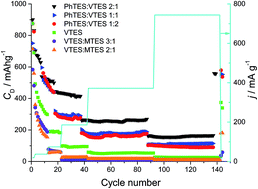
We report here on the synthesis and characterization of silicon oxycarbide (SiOC) in view of its application as a potential anode material for Li-ion batteries. SiOC ceramics are obtained by pyrolysis of various polysiloxanes synthesized by sol–gel methods. The polysiloxanes contain different organic groups attached to silicon, which influence the chemical composition and the microstructure of the final ceramic product. The structure of the SiOC samples is investigated by XRD, micro-Raman spectroscopy, solid state 29Si MAS-NMR and TEM. All investigated samples remain amorphous. However, at the elevated temperature of pyrolysis a phase separation process begins. During this process the carbon clusters become more ordered, which is reflected in the higher intensity and narrowing of the D1 band and decreasing of the D3 band. Moreover, the elevated temperature of pyrolysis promotes consumption of mixed bonds units, SiO3C, SiO2C2, SiOC3, and increases the share of oxygen rich SiO4 and carbon rich SiC4 tetrahedra. Electrochemical studies show a clear dependence between free carbon content and lithium storage capacity. Carbon-rich samples exhibit significantly higher capacities (∼550 mA h g−1 recorded at low current rate after 140 charge–discharge cycles) compared to carbon-poor samples (up to 360 mA h g−1). Moreover, carbon-rich samples exhibit a lower irreversible capacity during their first cycles compared to low carbon samples.


1. Introduction

The aim of research on lithium-ion batteries is to find lightweight and small rechargeable power sources to power not only portable electronics but also bigger applications like electric vehicles. Progress in efficient accumulation of energy is a crucial topic in the field of electrochemistry, because of the increasing energy consumption and demand.1–6 Three different ways of improving the performance of lithium-ion batteries may be observed depending on the components researchers focus on, i.e. electrodes,7–10 electrolytes11,12 and separators.13 Our study is settled in the field of searching for anode materials that can meet recently raised expectations. Negative electrodes should have a low self-discharge, optimal energy density and stability and rapid charging/discharging capability. Polymer Derived Ceramics (PDCs) approach allows designing the electrochemical properties of anodic materials. It provides an opportunity to examine different prepolymer combinations and molar ratios in order to obtain ceramic material of desirable electrochemical properties.14–18 Silicon oxycarbide ceramics (SiOC) is commonly known to display high reversible capacities, stability and slight volume changes during charging/discharging cycles.19–22 Thus, within this work we investigate SiOC obtained through the pyrolysis of several polysiloxanes as an alternative electrode material. To tailor the composition the polysiloxanes were prepared by sol–gel synthesis, in which three preceramic precursors containing different functional groups were used, i.e. methyltriethoxysilane, vinyltriethoxysilane and phenyltriethoxysilane. The mechanism of lithium-ions storage is not still fully understood. A set of electrochemical data was already published in the middle of the 1990s by the group of Dahn.23,24 However, the above publications mostly address the electrochemical performance of the materials with respect to their elemental composition, without considering the mechanism of lithium storage and lithium transport in these materials. As a consequence of increasing interest in this kind of electrode materials, questions related to the lithium storage mechanisms within SiOC based materials have recently been the focus of many research. Raj et al. claim that the mixed bond configuration (tetrahedrally coordinated silicon from SiC4 via SiC3O, SiC2O2 and SiCO3 to SiO4) in SiOC ceramics acts as a major lithiation site.25–29 Other research groups claim that the Li insertion into carbon-rich SiOC compounds occurs in the form of an adsorption and surface storage within the free carbon phase, similar to the storage of Li-ions in disordered carbons. Host sites are considered the edges of graphene sheets, interstitial and defect sites, micro-pores, graphite nano-crystallites and interfacial adsorption at carbon-crystallite interfaces. To the Si–O–C glassy phase, on the contrary, is attributed a minor role in the reversible storage process. The recent investigation of Fukui et al. by means of 7Li MAS NMR (magic angle spinning nuclear magnetic resonance) measurements demonstrates that the free carbon phase within these materials is the major hosting site for Li ions.19,30 However, it is known that the electrochemical properties of final ceramics are strictly dependent on the chemical components of starting polysiloxanes used in synthesis, their molar ratios, crosslinking steps and the temperature of pyrolysis.14,20–22 It has been proven that organic substituents such as phenyl groups increase the final amount of free carbon phase, that affects the capacity. As it has been reported, the relationship between carbon content and lithium storage capacity is not linear, therefore it is important to investigate the structure of materials along with their electrochemical properties.14,31

An important part of this work is to optimize the selection of precursors and their ratios for synthesis in order to improve the electrochemical activity of final SiOC product. Moreover, we focused on the analysis of the influence of different organic functional groups on the structure and the chemical composition of SiOC materials. The structure of the samples were analyzed by various methods such as X-ray diffraction, Raman spectroscopy, 29Si MAS NMR. Furthermore, the electrochemical performance of the obtained anode materials towards lithium ions has been investigated by galvanostatic measurements with potential limitation and cyclic voltammetry.

2. Experimental

2.1. Synthesis of silicon oxycarbides

Silicon oxycarbide samples were prepared by pyrolysis (Ar atmosphere, 1000 °C, selected samples also at 1300 °C) of different polysiloxanes containing various organic functional groups attached directly to silicon (phenyl-, vinyl- and methyl-). As it is known, such functional groups are the source of carbon in the final ceramic material.14,32 Sol–gel method was used for synthesis of polysiloxanes. Three different starting precursors were chosen, namely phenyltriethoxysilane (PhTES), vinyltriethoxysilane (VTES) and methyltriethoxysilane (MTES) and mixed in different ratios in order to adjust type and amount of functional groups present in the final preceramic polymer. Six samples were prepared: VTES, VTES[thin space (1/6-em)]:[thin space (1/6-em)]MTES 3[thin space (1/6-em)]:[thin space (1/6-em)]1, VTES[thin space (1/6-em)]:[thin space (1/6-em)]MTES 2[thin space (1/6-em)]:[thin space (1/6-em)]1, PhTES[thin space (1/6-em)]:[thin space (1/6-em)]VTES 2[thin space (1/6-em)]:[thin space (1/6-em)]1, PhTES[thin space (1/6-em)]:[thin space (1/6-em)]VTES 1[thin space (1/6-em)]:[thin space (1/6-em)]1, PhTES[thin space (1/6-em)]:[thin space (1/6-em)]VTES 1[thin space (1/6-em)]:[thin space (1/6-em)]2 (number means molar ratios between alkoxysilanes used as substrates for sol–gel synthesis). Precursors were mixed together and then ethanol (CH3CH2OH/Si = 2) and acidic water (pH = 4.5, acidified by HCl, H2O/OCH2CH3 = 1) were added to the solution while mixing. The temperature of the synthesis solution was slowly increased to 90 °C and kept for 1 h. Prepared sols, after cooling down to room temperature, were poured to polypropylene test tubes for gelation (4 days at room temp.) and drying (the temperature of drying was increased by 20 °C every three days starting from 40 °C to 100 °C). The dried polysiloxanes were used for pyrolysis (Ar atmosphere, 1000 and 1300 °C).

2.2. Characterization techniques

FTIR spectra were acquired with a FT-IR Nicolet 8700 spectrometer (Thermo Fisher Scientific Inc., USA) operating in ATR mode. Thermogravimetric analysis (TGA) was carried out on a NETZSCH STA 449F1 thermal analyzer (Netzsch Gerätebau GmbH, Germany) under argon gas flow of 70 cm3 min−1 in Al2O3 crucibles. The heating procedure was the same as the pyrolysis program. Chemical composition of the SiOC samples was analyzed using a carbon analyzer (Leco C-200, Leco Corporation, USA) and a nitrogen/oxygen analyzer (Leco TC-436, Leco Corporation, USA). The silicon amount was calculated as a difference to 100 wt%, taking into account that only C, O and Si are present in the samples. Possible small amount of hydrogen in the samples was neglected. X-ray powder diffraction measurements were performed using a flat-sample transmission geometry on a Bruker D8 Advance using Ni-filtered Cu Kα radiation. Raman spectra were recorded using a confocal micro-Raman spectrometer (InVia, Renishaw) with an argon ion laser (514 nm) within the wavenumber range of 100–3200 cm−1. MAS-NMR measurements were performed on a Bruker Avance III 700 MHz spectrometer operating at a proton frequency of 700.24 MHz. 29Si NMR spectra were recorded with following parameters: single pulse sequence, 29Si frequency: 139.11 MHz, π/8 pulse length: 2.5 μs, recycle delay: 100 s (sufficient time to get fully relaxed spectra), 1k scans, external secondary reference: DSS. 3.2 mm zirconia rotors filled with samples were spun at 8 kHz under air flow. Electrochemical cells were tested using Atlas-Sollich 0961 (Atlas-Sollich, Poland). Galvanostatic charging and discharging were performed between 3.0 and 0.005 V at different current rates from 18.6 mA g−1 to 744 mA g−1.

2.3. Preparation of the electrodes

SiOC ceramic samples were milled in a ball mill (Mixer Mill MM200, Retsch, Germany). Zirconia pot and zirconia balls (10 mm and 5 mm) were used. Median particle size (D50) after milling was equal to about 10 μm and D90 was below 40 μm. The active material was mixed with the binder (polyvinylidene fluoride PVdF, Solef, Germany) and the conducting additive (Carbon Black Super P®, Timcal Ltd., Switzerland), with the amounts of 85 wt%, 10 wt% and 5 wt% respectively. The slurry for coating was made by adding NMP (N-methyl-2-pyrrolidone, BASF, Germany) as a solvent (approx. 1 g of NMP for 0.6 g of powder mixture). The slurry was homogenized in the ball mill (Mixer Mill MM200, Retsch, Germany). The electrode layers were tape casted by doctor blade technique (the wet layer was approx. 100 μm thick). Surface treated copper foil was used as substrate for coating (10 μm, copper SE-Cu58 Schlenk Metallfolien GmbH & Co. KG, Germany). The electrode films were dried at 80 °C for 24 h, then the circular electrodes (diameter of 6 mm) were cut and weighted. The active material loading was in a range of 1.2 and 2 mg cm−2. Before assembling the cells, the electrodes were dried under vacuum at 80 °C for 24 h in a Buchi oven and transferred to the glove box (<0.5 ppm O2, <0.5 ppm H2O) without contact with air. The electrochemical measurements were performed in two-electrodes cell configuration (Swagelok®) with SiOC layers as a working electrode and lithium foil (99.9% purity, 0.75 mm thickness, Alfa Aesar, Germany) as a counter/reference electrode. Solution of 1 M LiPF6 in ethylene carbonate (EC)/dimethyl carbonate (DMC) (Sigma-Aldrich) served as an electrolyte and a quartz filter paper MN GF-2 (Macherey-Nagel GmbH & Co. KG, Germany) was used as a separator.

3. Results and discussion

The progress of the hydrolysis, condensation and the drying processes were monitored by FTIR spectroscopy. Fig. 1 shows the spectra of the sample prepared from pure vinyltriethoxysilane (VTES) starting from the precursor through sol–gel synthesis to the final ceramic material. The spectra of the pure alkoxysilane and the sol (synthesis solution after 0.5 h at 90 °C) look similar. The only difference appears in the region characteristic for Si–O–C stretching vibrations. The bands in this region are slightly broader in the sol spectrum, indicating the appearance of Si–O–Si bonds. Moreover, in the spectrum of sol sample new bands appear: (i) small band at ∼880 cm−1 which may be attributed to Si–OH bending vibrations, (ii) broad band at 1620–1660 cm−1 responsible for H2O bending vibration and (iii) broad band at 3100–3600 cm−1 indicating the presence of water and alcohol (O–H stretching vibrations). The spectrum of the dried gel exhibits a broad band between 940 and 1200 cm−1. This region is attributed to stretching vibration of Si–O–Si and Si–O–C bonds. However, significant broadening of bands in 1000–1100 cm−1 region may suggest presence of long chains siloxanes.33,34 Moreover, there are no visible bands at 2975, 2930 and 2885 cm−1 responsible for stretching vibration coming from ethoxy groups, what proves complete hydrolysis and full condensation process. The spectrum of the dried gel does not show bands coming from water or alcohol molecules, while the spectrum of ceramic material shows only broad bands coming from Si–O–Si vibrations. The band at 760 cm−1 (δ(Si–C)), visible in the gel spectrum, disappears in the ceramic sample spectrum, which suggest that most of the carbon from vinyl groups attached to silicon became a free carbon phase after pyrolysis. FTIR spectra of other SiOC samples follows the same scheme as shown in Fig. 1 for VTES sample.
image file: c6ra24539k-f1.tif
Fig. 1 FTIR spectra of VTES sample: precursor, sol (synthesis solution after 0.5 h at 90 °C), dried gel and ceramic sample after pyrolysis at 1000 °C.

TGA measurement revealed that during pyrolysis polysiloxane gels lose around 16–17.5% of mass. The final chemical composition of studied materials is presented in Table 1. The sample PhTES[thin space (1/6-em)]:[thin space (1/6-em)]VTES 2[thin space (1/6-em)]:[thin space (1/6-em)]1 containing the highest amount of phenyl groups contains also the highest amount of carbon in the final ceramic. Moreover, most of the carbon coming from phenyl groups turned into the free carbon as it can be noticed by the empirical stoichiometry of this sample (SiO1.84C0.08 + 2.92Cfree). Replacing phenyl groups with vinyl ones leads to a significant decrease of the amount of carbon in the ceramic material. The sample prepared from pure vinyltriethoxysilane VTES contains only 19.5% of free carbon. On the other hand, replacing vinyl groups with methyl groups in the polysiloxane results in further reduction of free carbon content, and at the same time, causes an increase of carbon atoms bound to silicon. The results of elemental analysis clearly shows that the carbon content and its character can be precisely adjusted by varying functional groups in the preceramic polymers. All investigated samples, pyrolyzed up to 1300 °C, are amorphous. There is only a small difference between the XRD patterns of the samples pyrolyzed at 1000 and 1300 °C (Fig. 2). Higher pyrolysis temperature gives rise to a broad signal at 22° which corresponds to amorphous SiO2 (ref. 35) and may be also attributed to the amorphous phase of SiOC.20 The more pronounced reflex at 22° may suggest a phase separation process at elevated temperature, which is typical behavior for silicon oxycarbide ceramics. However, there is no sign of the presence of silicon carbide (reflexes at 36°, 61° and 72°), which used to segregate already at 1200 °C from other silicon oxycarbide materials.20,35 This result proves the thermal stability of the prepared SiOC samples against crystallization. The small reflex at 43°, attributed to the presence of disordered carbon in the ceramic material,36,37 becomes more pronounced in the diffractograms of the samples pyrolyzed at 1300 °C. The presented XRD patterns show changes of the structure of the SiOC samples upon increasing temperature of pyrolysis.

Table 1 Results of elemental analysis of the investigated SiOC materials pyrolyzed at 1000 °C
Sample C, wt% O, wt% Sia, wt% Cfree, wt% SiOC stoichiometry SiO2(1−x)Cx + yCfree
a Calculated as a difference to 100 wt%.
VTES 22.9 36.8 40.3 19.5 SiO1.60C0.20 + 1.13Cfree
VTES[thin space (1/6-em)]:[thin space (1/6-em)]MTES 3[thin space (1/6-em)]:[thin space (1/6-em)]1 23.7 32.0 44.3 16.7 SiO1.26C0.37 + 0.88Cfree
VTES[thin space (1/6-em)]:[thin space (1/6-em)]MTES 2[thin space (1/6-em)]:[thin space (1/6-em)]1 23.2 33.3 43.5 17.1 SiO1.34C0.33 + 0.91Cfree
PhTES[thin space (1/6-em)]:[thin space (1/6-em)]VTES 2[thin space (1/6-em)]:[thin space (1/6-em)]1 38.5 31.6 30.0 37.5 SiO1.84C0.08 + 2.92Cfree
PhTES[thin space (1/6-em)]:[thin space (1/6-em)]VTES 1[thin space (1/6-em)]:[thin space (1/6-em)]1 38.5 26.2 35.3 33.3 SiO1.30C0.35 + 2.2Cfree
PhTES[thin space (1/6-em)]:[thin space (1/6-em)]VTES 1[thin space (1/6-em)]:[thin space (1/6-em)]2 38.0 27.6 34.4 33.7 SiO1.40C0.30 + 2.28Cfree


image file: c6ra24539k-f2.tif
Fig. 2 XRD pattern of carbon-rich PhTES[thin space (1/6-em)]:[thin space (1/6-em)]VTES 2[thin space (1/6-em)]:[thin space (1/6-em)]1 sample and carbon-poor VTES sample pyrolyzed at 1000 °C and 1300 °C.

Micro-Raman spectroscopy was used to analyze the structure of carbon phase in the SiOC samples (Fig. 3). All the SiOC samples exhibit two overlapping bands at 1330 ± 10 cm−1 and 1595 ± 5 cm−1, which corresponds to D (D1) and G bands, characteristic for carbonaceous materials. The G band represents the in-plane bond-stretching motion of sp2 carbons (E2g symmetry), whereas the D band is related to disorder in graphitic layers (A1g breathing mode) and is not observed in perfect graphite.36,38–40 According to literature reports,36,40–43 a more detailed analysis may be done by fitting the first order Raman spectra of carbonaceous materials with three to five peaks. Typical examples of additional bands are: D4 (I) located at ∼1200 cm−1, D3 (D′′, A) at ∼1500 cm−1 and D2 (D′) at ∼1620 cm−1.36,40–43 In the case of the investigated SiOC samples the best fitting result was obtained by deconvolution of each spectrum with four peaks (examples shown in Fig. 3b). The curve fitting of the spectra was performed for the wavenumber range of 800–2000 cm−1 after background subtraction. D1, D4 and G bands were fitted with Lorentzian line shapes and D3 was fitted with Gaussian line shape. The analyzed spectral parameters and the ratio of absolute intensities of D1 to G bands (ID1/IG) are gathered in Table 2.


image file: c6ra24539k-f3.tif
Fig. 3 Raman spectra of: (a) the SiOC ceramic samples pyrolyzed at 1000 °C; (b) deconvoluted spectra of PhTES[thin space (1/6-em)]:[thin space (1/6-em)]VTES 2[thin space (1/6-em)]:[thin space (1/6-em)]1 sample pyrolyzed at 1000 °C and 1300 °C.
Table 2 Bands positions, bands area and intensity ratio ID1/IG obtained from curve fitting of the Raman spectra of the ceramic samples. Samples pyrolyzed at 1000 °C unless otherwise mentioned
Sample D4 D1 D3 G ID1/IG
cm−1 Area cm−1 Area cm−1 Area cm−1 Area
VTES 1191 7.6 1326 59.6 1508 7.4 1594 25.4 1.03
VTES (1300 °C) 1199 11.6 1340 59.0 1509 3.0 1599 26.4 1.15
VTES[thin space (1/6-em)]:[thin space (1/6-em)]MTES 3[thin space (1/6-em)]:[thin space (1/6-em)]1 1191 2.0 1327 67.8 1509 6.7 1591 23.5 1.31
VTES[thin space (1/6-em)]:[thin space (1/6-em)]MTES 2[thin space (1/6-em)]:[thin space (1/6-em)]1 1190 4.3 1329 65.4 1507 6.8 1596 23.5 1.32
PhTES[thin space (1/6-em)]:[thin space (1/6-em)]VTES 2[thin space (1/6-em)]:[thin space (1/6-em)]1 1199 5.9 1326 61.3 1509 9.3 1593 23.5 1.20
PhTES[thin space (1/6-em)]:[thin space (1/6-em)]VTES 2[thin space (1/6-em)]:[thin space (1/6-em)]1 (1300 °C) 1191 10.6 1342 59.2 1507 3.5 1598 26.8 1.26
PhTES[thin space (1/6-em)]:[thin space (1/6-em)]VTES 1[thin space (1/6-em)]:[thin space (1/6-em)]1 1199 8.1 1324 60.1 1507 7.7 1594 24.1 1.13
PhTES[thin space (1/6-em)]:[thin space (1/6-em)]VTES 1[thin space (1/6-em)]:[thin space (1/6-em)]2 1190 7.9 1336 60.2 1510 6.4 1598 25.5 1.16


In the case of all the SiOC samples the D1 band located at around 1330 ± 10 cm−1 is the main band with the highest area. The second main band is the G band at 1595 ± 5 cm−1. Additionally, the D4 mode can be found at 1191–1199 cm−1 and D3 positioned at ∼1508 cm−1. The D4 band is generally observed as a shoulder of the D1 band for soot and related carbon materials36,43 and it is attributed to the disordered graphitic lattice (C–C and C[double bond, length as m-dash]C stretching vibrations and sp2–sp3 bonds). The D3 band is ascribed to amorphous carbon fractions.36,41,42 As presented in Fig. 3b and Table 2, the D3 band has significant contribution to the spectra of the samples pyrolyzed at 1000 °C. However, its area decreases considerably (2–2.5 times) with increasing temperature of pyrolysis up to 1300 °C. This suggests an ordering of the amorphous carbon phase. Additional changes in the spectra of the samples pyrolyzed at elevated temperature are: (i) the increasing intensity, the narrowing and the shift of the D1 band (from 1326 to 1340 cm−1 for VTES sample and from 1326 to 1342 cm−1 for PhTES[thin space (1/6-em)]:[thin space (1/6-em)]VTES 2[thin space (1/6-em)]:[thin space (1/6-em)]1 sample) and (ii) the increase of ID1/IG ratio (from 1.03 to 1.15 for VTES sample and from 1.20 to 1.26 for PhTES[thin space (1/6-em)]:[thin space (1/6-em)]VTES 2[thin space (1/6-em)]:[thin space (1/6-em)]1 sample). According to Ferrari et al.44 the increase of the ID1/IG ratio suggest an ordering from amorphous carbon to nanocrystalline graphite. The changes observed on Raman spectra confirm the structural evolution of the free carbon phase with the increasing temperature of pyrolysis.

Further investigation of the structure of the prepared SiOC materials was performed by 29Si MAS-NMR spectroscopy. The spectra of the SiOC samples pyrolyzed at 1000 °C are presented in Fig. 4. Spectra of all the samples, except of VTES, exhibit three broad peaks with maxima at chemical shifts of: 108–116 ppm, 76–78 ppm and 40–44 ppm corresponding to the silicon tetrahedra: SiO4, SiO3C, and SiO2C2, respectively.45–47 The spectrum of the VTES sample, synthesized from pure vinyltriethoxysilane, exhibits only one main peak corresponding to SiO4 and one small ascribed to SiO3C. Analyzing the NMR results (Table 3) together with elemental analysis and the empirical stoichiometry (Table 1) one can conclude that methyl groups contribute more to the mixed bonds tetrahedra than vinyl or phenyl groups. An evident change of the structure is observed for the carbon rich sample PhTES[thin space (1/6-em)]:[thin space (1/6-em)]VTES 2[thin space (1/6-em)]:[thin space (1/6-em)]1 pyrolyzed at 1300 °C, compared to the one pyrolyzed at 1000 °C. The peak attributed to the SiO4 unit becomes broader and slightly changes its position toward lower chemical shift values. At the same time, peaks corresponding to the mixed bonds units SiCO3 and SiC2O2 decrease significantly and a small peak at approx. −11 ppm appears, which may be ascribed to the SiC4 tetrahedra.45–47 This result confirm the phase separation process upon an elevated temperature of pyrolysis.


image file: c6ra24539k-f4.tif
Fig. 4 29Si MAS-NMR spectra of the SiOC samples pyrolyzed at 1000 °C; inset: spectra of PhTES[thin space (1/6-em)]:[thin space (1/6-em)]VTES 2[thin space (1/6-em)]:[thin space (1/6-em)]1 sample pyrolyzed at 1000 °C and 1300 °C.

TEM pictures of the PhTES[thin space (1/6-em)]:[thin space (1/6-em)]VTES 2[thin space (1/6-em)]:[thin space (1/6-em)]1 sample pyrolyzed at 1300 °C are shown in Fig. 5a and b. It can be seen that SiOC ceramic sample is amorphous and homogenous. At higher magnification (Fig. 5b) nanocrystalline domains are observed. Based on the identified atomic interlayer distances d(hkl) with d = 2.481 ± 0.08 Å, these nanodomains may be attributed to nanocrystalline silicon carbide (d = 2.510 Å at 2θ = 35.744°)48,49 created during the phase separation process. The presence of SiC was not observed in XRD data, probably due to too small crystals. This in turn, suggests that the phase separation process starts at 1300 °C but is not significant yet.


image file: c6ra24539k-f5.tif
Fig. 5 TEM images of PhTES[thin space (1/6-em)]:[thin space (1/6-em)]VTES 2[thin space (1/6-em)]:[thin space (1/6-em)]1 sample pyrolyzed at 1300 °C.

Electrochemical activity of the prepared ceramics was studied by galvanostatic charging–discharging and cyclic voltammetry. First lithiation–delithiation curves recorded at a low current rate (18.6 mA g−1) are shown in Fig. 6. All materials exhibit significant irreversible capacity and large hysteresis in the first cycle. The large hysteresis, namely the difference in the insertion and extraction potential, is typically observed in the polymer-derived ceramic electrode material.31,50–52 It is attributed to: (i) traces of hydrogen present in the ceramic which trap lithium ions and (ii) presence of the disordered carbon phase,53 which adsorbs lithium at the surface or in nanopores. In both cases, lithium ions are expected to be release at higher potentials. The significant capacity losses observed in the first cycle are most probably related to the presence of defects in the ceramic as well as in the carbon phase. The modelling of lithium insertion into SiOC material (P. Kroll, private communication) reveals that in the first cycle lithium in irreversibly captured in the pores/defects, in particular in the proximity of oxygen.31 The coulombic efficiency of the first cycle is lower for carbon poor samples (49.6–51.1%) than for the samples with a high carbon content (59.6–63.3%). Columbic efficiency η is obtained by simple correlation of first cycle insertion capacity (Cins 1st) and first cycle extraction capacity (Cextr 1st), η = (Cextr 1st/Cins 1st) × 100%. Despite a hysteresis observed in the charge–discharge curves most of the capacity (75–85%) is recovered below 1.5 V. The shape of the curves is similar for all investigated samples. However, small differences can be noticed in the first lithiation curves. The plateau observed at ∼0.4–0.45 V for the PhTES[thin space (1/6-em)]:[thin space (1/6-em)]VTES 2[thin space (1/6-em)]:[thin space (1/6-em)]1 sample with the highest free carbon content during first lithiation is more sloping and at a slightly higher potential than for other materials. On the other hand, for the carbon-poor samples the plateau starts earlier, is long, flat and is situated at a bit lower potential of ∼0.35 V. This observation is in agreement with our previous study, where the lowest potential of the plateau (0.16 V) was recorded for the sample with the lowest carbon content (13.3 wt%).14 The differences between the potentials of the plateaus may suggest different mechanisms of lithium insertion into the SiOC ceramics with different carbon content. This plateau is only observed during the first lithiation (see inset in Fig. 7) and is probably related to irreversible capacity, attributed to lithium bonding to oxygen sites.14,31,54


image file: c6ra24539k-f6.tif
Fig. 6 First lithiation/delithiation cycle of SiOC electrodes.

image file: c6ra24539k-f7.tif
Fig. 7 Cyclic voltammetry curve recorded for carbon-rich sample PhTES[thin space (1/6-em)]:[thin space (1/6-em)]VTES 2[thin space (1/6-em)]:[thin space (1/6-em)]1, inset: 1st and 2nd lithiation/delithiation curves of the PhTES[thin space (1/6-em)]:[thin space (1/6-em)]VTES 2[thin space (1/6-em)]:[thin space (1/6-em)]1 sample. Scan rate 50 μV s−1.

Cyclic voltammetry curves of the first and second cycle recorded for carbon-rich sample PhTES[thin space (1/6-em)]:[thin space (1/6-em)]VTES 2[thin space (1/6-em)]:[thin space (1/6-em)]1 are presented in Fig. 7. The CV curves are in agreement with galvanostatic curves (inset in Fig. 7). The irreversible cathodic peak with maximum at 0.07 V in the first cycle corresponds to the plateau recorded during first lithiation. Second cycle of CV reveals no peak and so the second lithiation galvanostatic curve exhibits no plateau. The second cycle coulombic efficiency (94.5%) is much higher than the first one (61.5%) and increases with further cycles up to 99.8%.

Prolonged galvanostatic cycling was performed in order to investigate long term stability of all investigated SiOC electrodes. Delithiation capacity as a function of cycle number is presented in Fig. 8. Galvanostatic charge and discharge processes were recorded with increasing current density from 18.6 mA g−1 to 744 mA g−1. The sample with the highest content of free carbon phase exhibits the highest capacity values both at low and high current rate. Reversible capacity after 140 charge–discharge cycles measured with low current again (18.6 mA g−1) reaches value of about 550 mA h g−1 for all carbon-rich samples. Capacity values decrease significantly with deceasing carbon content. Samples, which contain less than 20% of free carbon phase exhibit poor electrochemical activity toward lithium ions. The main correlation between the electrochemical performance of the investigated SiOC materials and their carbon content is the following: the more free carbon phase in the sample the higher the lithium storage capacity, especially at high rates. However, such correlation is not always this simple, as presented in our previous study.14 SiOC samples with different amount of free carbon might exhibit similar electrochemical behavior, suggesting a major influence of microstructure on the electrochemical activity towards lithium ions.


image file: c6ra24539k-f8.tif
Fig. 8 Delithiation capacity vs. cycle number recorded at different current rates: (a) investigated SiOC electrodes; (b) PhTES[thin space (1/6-em)]:[thin space (1/6-em)]VTES 2[thin space (1/6-em)]:[thin space (1/6-em)]1 sample pyrolyzed at 1000 °C and 1300 °C.

The electrochemical performance of PhTES[thin space (1/6-em)]:[thin space (1/6-em)]VTES 2[thin space (1/6-em)]:[thin space (1/6-em)]1 sample pyrolyzed at 1000 and 1300 °C is presented in Fig. 8b. At low current rates both samples present similar electrochemical performance. Furthermore, at high current densities the differences in capacity values become pronounced. This result proves that the microstructure of SiOC materials significantly influence their electrochemical properties. Analysis of Raman spectra show diminution of D3 band and increase of the ID1/IG ratio, signifying increase of order of free carbon phase. According to previous investigations of polymer derived SiOC and SiCN ceramics,22,51 the ordering of carbon clusters should lead to much better rate performance, although lower capacitance values recorded at both low and high current rates. Moreover, nanocrystalline SiC visible in TEM micrographs should lead to increase of material conductivity,55 and in consequence facilitate electron transfer and improve rate capability. However, the results of NMR investigation clearly show a considerable phase segregation in the 1300 °C sample, namely the increase of the number of SiO4 units from 57.6% (1000 °C) to 91.5% (1300 °C). Mixed bonds configuration was found to be of crucial importance for better performance of free carbon phase within the ceramic.25 Mixed bonds structure is for the 1300 °C sample replaced by silica and silicon carbide nanodomains. In consequence, we believe that carbon phase becomes less accessible for lithium ions and electrons, due to the presence of insulating silica phase. In comparison with literature, the measured capacity of PhTES[thin space (1/6-em)]:[thin space (1/6-em)]VTES 2[thin space (1/6-em)]:[thin space (1/6-em)]1 (1300 °C) at low current rates (18.6–74.4 mA g−1) is very high compared to other SiOCs pyrolyzed at temperatures higher than 1200 °C.20,22,37

Table 3 29Si MAS-NMR characterization of the SiOC samples. Samples pyrolyzed at 1000 °C unless otherwise mentioned
Sample SiO4 SiO3C SiO2C2 SiOC3 SiC4
δ/ppm % δ/ppm % δ/ppm % δ/ppm % δ/ppm %
PhTES[thin space (1/6-em)]:[thin space (1/6-em)]VTES 2[thin space (1/6-em)]:[thin space (1/6-em)]1 −108.7 57.6 −76.3 25.2 −42.4 13.8 −1.9 3.4
PhTES[thin space (1/6-em)]:[thin space (1/6-em)]VTES 2[thin space (1/6-em)]:[thin space (1/6-em)]1 (1300 °C) −117.1 91.5 −76.3, −55.6 1.4, 1.9 −10.7 5.2
PhTES[thin space (1/6-em)]:[thin space (1/6-em)]VTES 1[thin space (1/6-em)]:[thin space (1/6-em)]1 −108.1 54.0 −76.8 27.5 −41.5 16.0 −0.9 1.5 −16.5 1.0
PhTES[thin space (1/6-em)]:[thin space (1/6-em)]VTES 1[thin space (1/6-em)]:[thin space (1/6-em)]2 −111.8 51.2 −76.2 27.0 −39.2 14.3 −15.4 7.5
VTES −113 92.2 −76.7 7.8
VTES[thin space (1/6-em)]:[thin space (1/6-em)]MTES 3[thin space (1/6-em)]:[thin space (1/6-em)]1 −114.5 56.7 −77.8 31.8 −39.5 10.1 −16.6 1.4
VTES[thin space (1/6-em)]:[thin space (1/6-em)]MTES 2[thin space (1/6-em)]:[thin space (1/6-em)]1 −113.0 53.4 −75.8 33.3 −36.0 13.3


Results obtained from galvanostatic cycling are summarized in Table 4 (and Table 1S in ESI). The results prove a strong influence of free carbon phase on electrochemical properties of silicon oxycarbide ceramics, especially at high current rates. All the carbon-rich sample (PhTES[thin space (1/6-em)]:[thin space (1/6-em)]VTES) contain the same amount of total carbon (38.5–38%). Among the PhTES[thin space (1/6-em)]:[thin space (1/6-em)]VTES mixed samples, PhTES[thin space (1/6-em)]:[thin space (1/6-em)]VTES 2[thin space (1/6-em)]:[thin space (1/6-em)]1 contains the highest amount of free carbon phase (37.5%, compared to 33.3 and 33.7 wt%, cf. Table 1), which explains well its high rate lithium capacity.

Table 4 Comparison of the irreversible Cirrev and reversible CD capacity of the first cycle and average delithiation capacities CD of SiOC electrodes measured at different current rates. Samples pyrolyzed at 1000 °C unless otherwise stated
Sample 1st cycle CD/mA h g−1 Average discharge capacity CD/mA h g−1
Cirrev 18.6/mA g−1 Efficiency/% 74.4/mA g−1 186/mA g−1 372/mA g−1 744/mA g−1 18.6/mA g−1, last (134th) cycles
PhTES[thin space (1/6-em)]:[thin space (1/6-em)]VTES 2[thin space (1/6-em)]:[thin space (1/6-em)]1 562 900 61.5 520 427 260 160 558
PhTES[thin space (1/6-em)]:[thin space (1/6-em)]VTES 2[thin space (1/6-em)]:[thin space (1/6-em)]1 (1300 °C) 484 820 62.8 492 316 184 70 505
PhTES[thin space (1/6-em)]:[thin space (1/6-em)]VTES 1[thin space (1/6-em)]:[thin space (1/6-em)]1 594 876 59.6 487 295 179 110 553
PhTES[thin space (1/6-em)]:[thin space (1/6-em)]VTES 1[thin space (1/6-em)]:[thin space (1/6-em)]2 506 875 63.3 508 279 163 94 558
VTES 763 751 49.6 188 94 53 26 357
VTES[thin space (1/6-em)]:[thin space (1/6-em)]MTES 3[thin space (1/6-em)]:[thin space (1/6-em)]1 580 584 50.1 66 13 9 6 48
VTES[thin space (1/6-em)]:[thin space (1/6-em)]MTES 2[thin space (1/6-em)]:[thin space (1/6-em)]1 683 714 51.1 70 31 18 10 181


To compare electrochemical performance of the investigated samples with other SiOC compounds with similar free carbon content (from 20 to 40%) an overview of various SiOC materials and their electrochemical characteristics is presented in Table 5.

Table 5 Overview of various SiOC compounds and their electrochemical characteristics reported in literature. The SiOC stoichiometry, free carbon content, first cycle reversible (Crev) and irreversible capacity (Cirr), coulombic efficiency (η), current rate and if available the capacity retention upon continuous cycling are given56
SiOC stoichiometry, SiO2(1−x)Cx + yCfree Free C/wt% 1st cycle Rate/mA g−1 Capacity retention Ref.
Crev/mA h g−1 Cirr/mA h g−1 η/%
SiOC0.5 + 2.4C 36.6 560 300 65 14.8 n/a 23
SiO0.61C0.695 + 2.045C 34.7 523 270 66 32.7 n/a 57
SiO1.96C0.02 + 3.44C 40.9 732 381 66 32.7 not stable 58
SiO0.85C0.575 + 1.415C 25.9 794 370 68 100 n/a 27
SiO0.98C0.51 + 1.96C 32.0 605 325 65 18 Cycling stable 59
SiO1.39C0.305 + 0.375C 21.8 322 400 45 18.6 n/a 60
SiO0.6C0.7 + 0.96C 20.0 702 1032 68 100 84% after 1020 cycles 52
SiO1.01C0.495 + 2.435C 36.8 434.3 273.8 61.3 From 37 to 370 58.7% after 60 cycles 61
SiO0.93C0.535 + 1.725C 29.5 501.4 302.7 62.3 From 37 to 370 47.3% after 60 cycles 61
SiO0.87C0.565 + 1.055C 20.6 682.5 495.8 57.9 From 37 to 370 13.5% after 60 cycles 61


Our results as well as data presented in Table 5 clearly show the significance of free carbon phase supporting the hypothesis that free carbon is a major lithium storage host. However, it is hardly possible to quantify the contribution of carbon, with respect to the ceramic, to the electrochemical performance. The goal of the present study is to demonstrate that the electrochemical performance of the SiOC electrode is not linearly dependent on the amount of the free carbon, but it significantly depends also on the microstructure of the material. According to Prof. Kroll54 the presence of a free carbon phase provides low-laying unoccupied states in which electrons can go in, and by consequence significantly decreases the band gap. A strong bonding of Li in the solid SiOC structure is provided, if the interaction of the lithium cation with the host in the form of Li–O bonds outweighs the promotion energy for the electron. Consequently, on the one hand the free carbon phase facilitates lithium bonded to oxygen sites, leading to irreversible lithium uptake; on the other hand, the segregated carbon provides a major part of the reversible lithium storage capacity.54

4. Conclusions

Sol–gel synthesis is an appropriate method to tailor compositions of preceramic polymers and to design the chemical composition and the structure of silicon oxycarbide ceramics. The amount of free carbon phase is a crucial factor for the electrochemical activity towards lithium ions. Moreover, the microstructure plays an important role, in particular in stabilizing the microstructure of free carbon phase. Purely amorphous materials (pyrolyzed at 1000 °C) exhibit better electrochemical performance than materials pyrolyzed at higher temperatures (1300 °C). Moreover, they are very stable at high current rates, what makes them suitable for high power application. The high irreversibility of the first cycle still remains a drawback for possible application of SiOC ceramics.

Acknowledgements

This work is supported by Foundation for Polish Science under grant HOMING PLUS/2012-6/16. JK, MGZ, RR acknowledge the support of German Science Foundation (SPP 1473/JP8).

References

  1. M. Armand and J.-M. Tarascon, Building better batteries, Nature, 2008, 451, 652–657,  DOI:10.1038/451652a .
  2. P. G. Bruce, Energy storage beyond the horizon: rechargeable lithium batteries, Solid State Ionics, 2008, 179, 752–760,  DOI:10.1016/j.ssi.2008.01.095 .
  3. R. Marom, S. F. Amalraj, N. Leifer, D. Jacob and D. Aurbach, A review of advanced and practical lithium battery materials, J. Mater. Chem., 2011, 21, 9938–9954,  10.1039/c0jm04225k .
  4. V. Etacheri, R. Marom, R. Elazari, G. Salitra and D. Aurbach, Challenges in the development of advanced Li-ion batteries: a review, Energy Environ. Sci., 2011, 4, 3243–3262,  10.1039/c1ee01598b .
  5. T. L. Kulova, New electrode materials for lithium-ion batteries (review), Russ. J. Electrochem., 2013, 49, 1–25,  DOI:10.1134/S1023193513010102 .
  6. V. Aravindan, Y.-S. Lee and S. Madhavi, Research progress on negative electrodes for practical Li-ion batteries – beyond carbonaceous anodes, Adv. Energy Mater., 2015, 5, 1402225–1402267,  DOI:10.1002/aenm.201402225 .
  7. P. Poizot, S. Laruelle, S. Grugeon, L. Dupont and J.-M. Tarascon, Nano-sized transition-metal oxides as negative-electrode materials for lithium-ion batteries, Nature, 2000, 407, 496–499,  DOI:10.1038/35035045 .
  8. F. Cheng, J. Liang, Z. Tao and J. Chen, Functional materials for rechargeable batteries, Adv. Mater., 2011, 23, 1695–1715,  DOI:10.1002/adma.201003587 .
  9. M. N. Obrovac and V. L. Chevrier, Alloy negative electrodes for Li-ion batteries, Chem. Rev., 2014, 114, 11444–11502,  DOI:10.1021/cr500207g .
  10. B. Scrosati, J. Hassoun and Y.-K. Sun, Lithium-ion batteries. A look into the future, Energy Environ. Sci., 2011, 4, 3287–3295,  10.1039/c1ee01388b .
  11. M. Marcinek, J. Syzdek, M. Marczewski, M. Piszcz, L. Niedzicki, M. Kalita, A. Plewa-Marczewska, A. Bitner, P. Wieczorek, T. Trzeciak, M. Kasprzyk, P. Łężak, Z. Zukowska, A. Zalewska and W. Wieczorek, Electrolytes for Li-ion transport – review, Solid State Ionics, 2015, 276, 107–126,  DOI:10.1016/j.ssi.2015.02.006 .
  12. J. Kalhoff, G. G. Eshetu, D. Bresser and S. Passerini, Safer electrolytes for lithium-ion batteries: state of the art and perspectives, ChemSusChem, 2015, 8, 2154–2175,  DOI:10.1002/cssc.201500284 .
  13. V. Deimede and C. Elmasides, Separators for lithium-ion batteries–a review on the production processes and recent developments, Energy Technol., 2015, 3, 453–468,  DOI:10.1002/ente.201402215 .
  14. M. Wilamowska, V. S. Pradeep, M. Graczyk-zajac, R. Riedel and G. D. Sorarù, Tailoring of SiOC composition as a way to better performing anodes for Li-ion batteries, Solid State Ionics, 2014, 260, 94–100,  DOI:10.1016/j.ssi.2014.03.021 .
  15. L. M. Reinold, M. Graczyk-Zajac, Y. Gao, G. Mera and R. Riedel, Carbon-rich SiCN ceramics as high capacity/high stability anode material for lithium-ion batteries, J. Power Sources, 2013, 236, 224–229,  DOI:10.1016/j.jpowsour.2013.02.046 .
  16. J. Wu, Y. Li, L. Chen, Z. Zhang, D. Wang and C. Xu, Simple fabrication of micro/nano-porous SiOC foam from polysiloxane, J. Mater. Chem., 2012, 22, 6542–6545,  10.1039/c2jm16840e .
  17. H.-J. Kleebe and Y. D. Blum, SiOC ceramic with high excess free carbon, J. Eur. Ceram. Soc., 2008, 28, 1037–1042,  DOI:10.1016/j.jeurceramsoc.2007.09.024 .
  18. G. Mera, A. Navrotsky, S. Sen, H.-J. Kleebe and R. Riedel, Polymer-derived SiCN and SiOC ceramics – structure and energetics at the nanoscale, J. Mater. Chem. A, 2013, 1, 3826–3836,  10.1039/c2ta00727d .
  19. H. Fukui, H. Ohsuka, T. Hino and K. Kanamura, A Si–O–C composite anode: high capability and proposed mechanism of lithium storage associated with microstructural characteristics, ACS Appl. Mater. Interfaces, 2010, 2, 998–1008,  DOI:10.1021/am100030f .
  20. H. Fukui, K. Eguchi, H. Ohsuka, T. Hino and K. Kanamura, Structures and lithium storage performance of Si–O–C composite materials depending on pyrolysis temperatures, J. Power Sources, 2013, 243, 152–158,  DOI:10.1016/j.jpowsour.2013.05.124 .
  21. M. Graczyk-Zajac, L. Toma, C. Fasel and R. Riedel, Carbon-rich SiOC anodes for lithium-ion batteries: part I. Influence of material UV-pre-treatment on high power properties, Solid State Ionics, 2012, 225, 522–526,  DOI:10.1016/j.ssi.2011.12.007 .
  22. J. Kaspar, M. Graczyk-Zajac and R. Riedel, Lithium insertion into carbon-rich SiOC ceramics: influence of pyrolysis temperature on electrochemical properties, J. Power Sources, 2013, 244, 450–455,  DOI:10.1016/j.jpowsour.2012.11.086 .
  23. A. M. Wilson, J. N. Reimers, E. W. Fuller and J. R. Dahn, Lithium insertion in pyrolyzed siloxane polymers, Solid State Ionics, 1994, 74, 249–254 CrossRef CAS .
  24. A. M. Wilson, G. Zank, K. Eguchi, W. Xing, B. Yates and J. R. Dahn, Polysiloxane Pyrolysis, Chem. Mater., 1997, 4756, 1601–1606.
  25. A. Saha, R. Raj and D. L. Williamson, A model for the nanodomains in polymer-derived SiCO, J. Am. Ceram. Soc., 2006, 89, 2188–2195,  DOI:10.1111/j.1551-2916.2006.00920.x .
  26. P. E. Sanchez-Jimenez and R. Raj, Lithium insertion in polymer-derived silicon oxycarbide ceramics, J. Am. Ceram. Soc., 2010, 93, 1127–1135,  DOI:10.1111/j.1551-2916.2009.03539.x .
  27. D. Ahn and R. Raj, Thermodynamic measurements pertaining to the hysteretic intercalation of lithium in polymer-derived silicon oxycarbide, J. Power Sources, 2010, 195, 3900–3906,  DOI:10.1016/j.jpowsour.2009.12.116 .
  28. J. Shen and R. Raj, Silicon-oxycarbide based thin film anodes for lithium ion batteries, J. Power Sources, 2011, 196, 5945–5950,  DOI:10.1016/j.jpowsour.2011.02.091 .
  29. D. Ahn and R. Raj, Cyclic stability and C-rate performance of amorphous silicon and carbon based anodes for electrochemical storage of lithium, J. Power Sources, 2011, 196, 2179–2186,  DOI:10.1016/j.jpowsour.2010.09.086 .
  30. H. Fukui, Y. Harimoto, M. Akasaka and K. Eguchi, Lithium species in electrochemically lithiated and delithiated silicon oxycarbides, ACS Appl. Mater. Interfaces, 2014, 6, 12827–12836,  DOI:10.1021/am502811f .
  31. M. Graczyk-Zajac, L. Reinold, J. Kaspar, P. Sasikumar, G.-D. Soraru and R. Riedel, New insights into understanding irreversible and reversible lithium storage within SiOC and SiCN ceramics, Nanomaterials, 2015, 5, 233–245,  DOI:10.3390/nano5010233 .
  32. H. Zhang and C. G. Pantano, Synthesis and characterization of silicon oxycarbide glasses, J. Am. Ceram. Soc., 1990, 73, 958–963 CrossRef CAS .
  33. D. R. Anderson, in Analysis of Silicones, ed. A. Lee Smith, Wiley-Interscience, New York, ch. 10, 1974 Search PubMed .
  34. A. L. Smith, Infrared spectra-structure correlations for organosilicon compounds, Spectrochim. Acta, 1960, 16, 87–105,  DOI:10.1016/0371-1951(60)80074-4 .
  35. H. Bréquel, J. Parmentier, S. Walter, R. Badheka, G. Trimmel, S. Masse, J. Latournerie, P. Dempsey, C. Turquat, A. Desmartin-Chomel, L. Le Neindre-Prum, A. Jayasooriya, D. Hourlier, H. J. Kleebe, G. D. Soraru, D. Enzo and F. Babonneau, Systematic structural characterisation of the high temperature behaviour of nearly-stoichiometric silicon oxycarbide glasses, Chem. Mater., 2004, 16, 2585–2598,  DOI:10.1021/cm049847a .
  36. A. Sadezky, H. Muckenhuber, H. Grothe, R. Niessner and U. Pöschl, Raman microspectroscopy of soot and related carbonaceous materials: spectral analysis and structural information, Carbon, 2005, 43, 1731–1742,  DOI:10.1016/j.carbon.2005.02.018 .
  37. J. Kaspar, M. Graczyk-Zajac and R. Riedel, Carbon-rich SiOC anodes for lithium-ion batteries: part II. Role of thermal cross-linking, Solid State Ionics, 2012, 225, 527–531,  DOI:10.1016/j.ssi.2012.01.026 .
  38. F. Tuinstra and L. Koenig, Raman spectrum of graphite, J. Chem. Phys., 1970, 53, 1126–1130,  DOI:10.1063/1.1674108 .
  39. K. Sato, R. Saito, Y. Oyama, J. Jiang, L. G. Cançado, M. A. Pimenta, A. Jorio, G. G. Samsonidze, G. Dresselhaus and M. S. Dresselhaus, D-band Raman intensity of graphitic materials as a function of laser energy and crystallite size, Chem. Phys. Lett., 2006, 427, 117–121,  DOI:10.1016/j.cplett.2006.05.107 .
  40. O. Beyssac, B. Goffé, J. P. Petitet, E. Froigneux, M. Moreau and J. N. Rouzaud, On the characterization of disordered and heterogeneous carbonaceous materials by Raman spectroscopy, Spectrochim. Acta Mol. Biomol. Spectros., 2003, 59, 2267–2276,  DOI:10.1016/S1386-1425(03)00070-2 .
  41. T. Jawhari, A. Roid and J. Casado, Raman spectroscopic characterization of some commercially available carbon black materials, Carbon, 1995, 33, 1561–1565,  DOI:10.1016/0008-6223(95)00117-V .
  42. A. Cuesta, P. Dhamelincourt, J. Laureyns, A. Martínez-Alonso and J. M. D. Tascón, Raman microprobe studies on carbon materials, Carbon, 1994, 32, 1523–1532,  DOI:10.1016/0008-6223(94)90148-1 .
  43. B. Dippel, H. Jander and J. Heintzenberg, NIR FT Raman spectroscopic study of flame soot, Phys. Chem. Chem. Phys., 1999, 1, 4707–4712,  10.1039/a904529e .
  44. A. C. Ferrari and J. Robertson, Interpretation of Raman spectra of disordered and amorphous carbon, Phys. Rev. B: Solid State, 2000, 61, 14095–14107,  DOI:10.1103/PhysRevB.61.14095 .
  45. R. B. Taylor, B. Parbhoo and D. M. Fillmore, in The Analytical Chemistry of Silicones, ed. A. L. Smith, John Wiley and Sons, New York, ch. 12, 1991, http://www.gbv.de/dms/ilmenau/toc/119445506.PDF, accessed September 28, 2015 Search PubMed .
  46. G. T. Burns, R. B. Taylor, Y. Xu, A. Zangvil and G. A. Zank, High-temperature chemistry of the conversion of siloxanes to silicon carbide, Chem. Mater., 1992, 4, 1313–1323,  DOI:10.1021/cm00024a035 .
  47. G. D. Soraru, G. D. Andrea, R. Campostrini, U. Trento, C. De, M. Condensee, U. Pierre and D. Fisica, Structural characterization and high-temperature behavior of silicon oxycarbide glasses prepared from sol–gel precursors containing Si–H bonds, 1995, 87, 379–387.  DOI:10.1111/j.1151-2916.1995.tb08811.x.
  48. N. W. Thibault, Morphological and structural crystallography and optical properties of silicon carbide, Am. Mineral., 1944, 29, 249–278 CAS .
  49. L. G. Ceballos-Mendivil, R. E. Cabanillas-López, J. C. Tánori-Córdova, R. Murrieta-Yescas, P. Zavala-Rivera and J. H. C. González, Synthesis and characterization of silicon carbide in the application of high temperature solar surface receptors, Energy Procedia, 2014, 57, 533–540,  DOI:10.1016/j.egypro.2014.10.207 .
  50. T. Zheng, W. R. McKinnon and J. R. Dahn, Hysteresis during lithium insertion in hydrogen containing carbons, J. Electrochem. Soc., 1996, 143, 2137–2145,  DOI:10.1017/cbo9781107415324.004 .
  51. L. M. Reinold, Y. Yamada, M. Graczyk-Zajac, H. Munakata, K. Kanamura and R. Riedel, The influence of the pyrolysis temperature on the electrochemical behavior of carbon-rich SiCN polymer-derived ceramics as anode materials in lithium-ion batteries, J. Power Sources, 2015, 282, 409–415,  DOI:10.1016/j.jpowsour.2015.02.074 .
  52. L. David, R. Bhandavat, U. Barrera and G. Singh, Silicon oxycarbide glass-graphene composite paper electrode for long-cycle lithium-ion batteries, Nat. Commun., 2016, 7, 10998,  DOI:10.1038/ncomms10998 .
  53. S. Flandrois and B. Simon, Carbon materials for lithium-ion rechargeable batteries, Carbon, 1999, 37, 165–180,  DOI:10.1016/S0008-6223(98)00290-5 .
  54. P. Kroll, Tracing reversible and irreversible Li insertion in SiCO ceramics with modeling and ab initio simulations, MRS Proc., 2011, 1313 DOI:10.1557/opl.2011.707 .
  55. J. Kaspar, M. Graczyk-Zajac, S. Lauterbach, H.-J. Kleebe and R. Riedel, Silicon oxycarbide/nano-silicon composite anodes for Li-ion batteries: considerable influence of nano-crystalline vs. nano-amorphous silicon embedment on the electrochemical properties, J. Power Sources, 2014, 269, 164–172,  DOI:10.1016/j.jpowsour.2014.06.089 .
  56. J. Kaspar, Carbon-rich silicon oxycarbide (SiOC) and silicon oxycarbide/element (SiOC/X, X = Si, Sn) nano-composites as new anode materials for Li-ion battery application, PhD thesis, Technische Universität Darmstadt, 2014 .
  57. H. Fukui, H. Ohsuka, T. Hino and K. Kanamura, Influence of polystyrene/phenyl substituents in precursors on microstructures of Si–O–C composite anodes for lithium-ion batteries, J. Power Sources, 2011, 196, 371–378,  DOI:10.1016/j.jpowsour.2010.06.077 .
  58. H. Fukui, H. Ohsuka, T. Hino and K. Kanamura, Silicon oxycarbides in hard-carbon microstructures and their electrochemical lithium storage, J. Electrochem. Soc., 2013, 160, A1276–A1281,  DOI:10.1149/2.095308jes .
  59. V. S. Pradeep, M. Graczyk-Zajac, R. Riedel and G. D. Soraru, New insights in to the lithium storage mechanism in polymer derived SiOC anode materials, Electrochim. Acta, 2014, 119, 78–85,  DOI:10.1016/j.electacta.2013.12.037 .
  60. X. Liu, M.-C. Zheng, K. Xie and J. Liu, The relationship between the electrochemical performance and the composition of Si–O–C materials prepared from a phenyl-substituted polysiloxane utilizing various processing methods, Electrochim. Acta, 2012, 59, 304–309,  DOI:10.1016/j.electacta.2011.10.073 .
  61. J. Kaspar, M. Graczyk-Zajac, S. Choudhury and R. Riedel, Impact of the electrical conductivity on the lithium capacity of polymer-derived silicon oxycarbide (SiOC) ceramics, Electrochim. Acta, 2016, 216, 196–202 CrossRef CAS .

Footnote

Electronic supplementary information (ESI) available. See DOI: 10.1039/c6ra24539k

This journal is © The Royal Society of Chemistry 2016
Click here to see how this site uses Cookies. View our privacy policy here.