DOI:
10.1039/C6RA24501C
(Paper)
RSC Adv., 2016,
6, 108928-108940
Discovery of novel cinnamylidene-thiazolidinedione derivatives as PTP-1B inhibitors for the management of type 2 diabetes†‡
Received
1st October 2016
, Accepted 26th October 2016
First published on 9th November 2016
Abstract
Protein tyrosine phosphatase-1B (PTP-1B) inhibition is a legitimate approach to combat type 2 diabetes and obesity as it corrects the insulin and leptin signalling cascades. In pursuing this, our goal is to discover compounds bearing small molecular scaffolds, and able to inhibit PTP-1B in a selective manner. In the present work, we have synthesized N3-substituted cinnamylidene-thiazolidinediones (4p–4x), and evaluated in vitro PTP-1B inhibitory activity, and in vivo anti-hyperglycaemic potential in streptozotocin-nicotinamide induced diabetic mice. Among various synthesized compounds, 4w exhibited the most potent in vitro PTP-1B inhibitory activity (IC50 ∼ 6.52 μM) along with excellent in vivo anti-hyperglycaemic activity. Furthermore, molecular docking assisted 3D-QSAR study was performed on the synthesized compounds (4p–4x) for the exploration of the binding mode of interactions, prediction of the binding affinity, and identification of 3D-pharmacophoric features (steric and electrostatic) responsible for inhibitory activity. The docking results were in agreement with the biological activity results i.e. compound 4w showed the highest binding affinity with PTP-1B (MolDock score −123.715), which is comparable with ertiprotafib (MolDock score −125.183). The lead discovered can be used for the further development of cinnamylidene-thiazolidinedione derivatives as antidiabetic agents.
Introduction
Diabetes mellitus (DM) is a metabolic disorder characterized by long-lasting hyperglycaemia.1a,b Type 2 diabetes mellitus (T2DM) is the most common form of diabetes with a share of 90–95% of total cases of diabetes around the world.2 The underlying pathogenesis behind T2DM includes ineffective biological responses to sufficiently produce insulin i.e. insulin resistance.3 Diabetes is associated with typical long-term diabetic complications namely cardiac abnormalities, atherosclerosis, microangiopathy, nephropathy, neuropathy, retinopathy and cataracts.4 The frequency of diabetes is also increasing in parallel with obesity; therefore diabetes is also known as ‘diabesity’ or ‘obesity dependent diabetes mellitus’.1b The prevalence of diabetes worldwide was 285 million in the year 2010 while present data showed that 387 million people worldwide have diabetes in the year 2014, and it is estimated to affect 592 million by the year 2035.3,5 The present threatening data creates urge for pharmacological agents that can be able to manage diabetes and contribute to health scenario and quality of life.
Protein tyrosine phosphatase-1B (PTP-1B) is a cytosolic non trans-membranous prototypic enzyme that belongs to PTPs superfamily,1a,b,3 and expressed in the cells of the liver, adipocytes and muscles.6 Biochemical and cellular studies, chemical and genetic experiments on mouse and human have strongly established this enzyme as a central negative regulator of insulin and leptin signalling cascades.7a–g Mice lacking protein tyrosine phosphatase-1B (PTP-1B) demonstrated enhanced insulin sensitivity with diminished plasma glucose and insulin levels. Furthermore, these mice also exhibit lower adiposity without producing anomalies in growth or fertility or other pathogenetic effects.7a,b,8a,b
PTP-1B inhibition is the legitimate approach to combat type 2 diabetes and obesity as it corrects the insulin and leptin signalling cascades.7c–f,9 A number of PTP-1B inhibitors have been reported over the last decades, although no commercial drugs have been approved till date.7c,10a–e However, ertiprotafib and trodusquemine (Fig. 1) are the only two drug candidates that have entered into clinical trials. Due to side effects and low rate of in vivo efficacy, ertiprotafib was withdrawn in phase II clinical trials.11a–c Trodusquemine is an allosteric, noncompetitive and highly selective inhibitor of PTP-1B currently in phase I clinical trials. Recent study demonstrated that it might also provide a promising way to combat anxiety disorders along with obesity.12a,b
 |
| Fig. 1 PTP-1B inhibitors entered in clinical trials (ertiprotafib and trodusquemine) and marketed TZD derivative (pioglitazone). | |
Development of an orally bioavailable and specific PTP-1B inhibitors bearing small molecular scaffold is not an easy task due to poor cell permeability of the small molecules exhibiting high affinity with PTP-1B due to their hydrophilic nature. A first-in-class PTP-1B inhibitor has yet to be discovered; however, extensive research is under way to develop a potential blockbuster drug. Small molecule PTP-1B inhibitors that not only serve as powerful tools to define the physiological roles of PTP-1B in vivo, but also as excellent lead compounds for the development of new therapeutic agents for the management of T2DM and obesity.10c–e,13
Pioglitazone (Fig. 1), a PPAR-γ agonist, is the only molecule bearing benzyl-2,4-thiazolidinedione (benzyl-2,4-TZD) scaffold that is available in the market to improve glycaemic control by ameliorating insulin resistance and show efficient insulin sensitization both in peripheral tissues and liver in type 2 diabetic patients.14a,b Various researchers around the world reported that 2,4-thiazolidinedione (2,4-TZD) derivatives exhibit inhibitory effects against PTP-1B.15a–i
It has been reported that naturally occurring cinnmaldehyde derivatives possess potent PTP-1B inhibitory activities.16a–c Recently, it has also been reported that arylidene-2,4-TZD derivatives with N-substitution are of particular interest as PTP-1B inhibitors and this structural amendment making them devoid of side effects associated with glitazones.15d,e,17 We have also performed molecular docking assisted 3D-QSAR study to map the spatial fingerprints of arylidine-2,4-TZDs for potent PTP-1B inhibitory activity.3
Taking into consideration above aspects, a series of compounds bearing 2,4-TZD scaffold in conjugation with cinnamaldehyde i.e. N3-substituted cinnamylidene-thiazolidinedione derivatives (4) (Scheme 1) was designed, synthesized and evaluated for their in vitro PTP-1B inhibitory activity as well as in vivo anti hyperglycaemic potential. In addition, a molecular docking assisted 3D-QSAR model was developed on synthesized derivatives (4) for the identification of binding mode of interactions, determination of binding affinity, and exploration of 3D-pharmacophoric (steric and electrostatic) features of cinnamylidene-thiazolidinediones for PTP-1B inhibitory activity.
 |
| Scheme 1 Reagents and conditions: (a) piperidine, ethanol, reflux for 26 h; (b) R-Br, acetone, anhydrous K2CO3, reflux for 26 h; (c) ethyl chloroacetate, anhydrous K2CO3, acetone, reflux for 24 h; (d) glacial acetic acid, hydrochloric acid, reflux for 2 h. | |
Results and discussion
Chemistry
Compounds (4p–x) were synthesized according to the general method depicted in Scheme 1. The condensation of commercially available 2,4-TZD (1) with cinnamaldehyde (2) in the presence of piperidine provided (5Z)-5-[(E)-3-phenylallylidene]-thiazolidine-2,4-dione (3a). The reaction between compound 3a with different alkyl bromide, substituted alkyl bromide and substituted aryl bromide (R1–8) using anhydrous potassium carbonate as base in refluxing acetone, followed by a workup and recrystallization from methanol, provided pure compounds 4p–4w (Scheme 1). Further, the reaction of compound 3a was carried out with ethyl chloroacetate in presence of anhydrous potassium carbonate as base, after removal of solvent the recovered product recrystallized from methanol to yield pure ethyl 2-[(5Z)-5-{(E)-3-phenylallylidene}-thiazolidine-2,4-dione-3-yl]acetate (3b). Acidic hydrolysis of 3b was carried out in refluxing glacial acetic acid, followed by a workup and recrystallization from methanol to yield pure 2-[(5Z)-{(E)-3-phenylallylidene}-thiazolidine-2,4-dione-3-yl]-acetic acid (4x) (Scheme 1).
Biological evaluation
In vitro PTP-1B inhibitory activity. The synthesized compounds (4p–4x) were evaluated in vitro for their ability to inhibit PTP-1B using PTP-1B drug discovery assay kit. Suramin served as reference standard for comparison. Compounds (4p–4x) bearing various substituents (R1–8) at N3 exhibited moderate PTP-1B inhibitory activities (Table 1). Among the series, compound 4w bearing methyl benzoic acid exhibited most potent inhibitory activity with IC50 ∼ 6.52 μM. Increase in chain length from 4p to 4u resulted in compounds with marginal decrease in potency. Compounds bearing bromo group on alkyl chain were found to be more potent as compared to their chloro derivatives. Compound 4x bearing acetic acid substituent at N3 also exhibited good inhibitory activity.
Table 1 In vitro PTP-1B inhibitory activities of N3-substituted cinnamylidene-thiazolidinediones (4p–x)
Compound |
IC50 (μM) |
Compound |
IC50 (μM) |
Data taken from the work of Shrestha et al.18 Data taken from the work of Bhattarai et al.15f |
4p |
30.770 |
4v |
32.006 |
4q |
20.458 |
4w |
6.522 |
4r |
33.271 |
4x |
21.476 |
4s |
32.829 |
Suramin |
11.100 |
4t |
20.916 |
Ertiprotafib |
1.4a |
4u |
33.607 |
Pioglitazone |
220b |
In vivo anti-hyperglycaemic activity. Anti-hyperglycaemic activity of compounds (4p–4x) was carried in STZ-NA induced diabetic mice. Pioglitazone was taken as reference standard. All the compounds as well as reference standard were administered at a fixed dose of 30 mg kg−1 19a,b body weight orally as suspension in carboxy methyl cellulose (CMC). Administration of compounds (4p–x) reduced serum glucose level (SGL) significantly (P < 0.001) in diabetic mice as compared to control group (Fig. 2A).
 |
| Fig. 2 (A) Comparative anti-hyperglycaemic activities of N3-substituted cinnamylidene-thiazolidinediones (4p–x). (B) Anti-hyperglycaemic activities of control, pioglitazone and compound 4w. | |
Compound 4w exhibited activity comparable to reference standard pioglitazone. The in vivo activity data of control, pioglitazone and compound 4w was compared separately and is presented in Fig. 2B. Introducing alkyl substituent (4u) resulted in compound with decreased activity while alkyl chain bearing chloro and bromo substituent resulted in compounds with better anti-hyperglycaemic activity. Compound 4x bearing free carboxylic group also exhibited good anti-hyperglycaemic activity.
Effect on body weight. During the study period of seven days, the mice were weighed and their body weights were recorded. In control mice, a trend of decrease in body weight was observed during seven days treatment period which can be attributed to T2DM. Pioglitazone treatment resulted in increase in the body weight. Effect on body weight after treatment with cinnamylidene-thiazolidinedione derivatives (4p–x) in diabetic mice are presented in Fig. 3. Results indicate that 4w administration has marginally increased in body weight but weight gain was less as compared to reference standard pioglitazone.
 |
| Fig. 3 Graphical presentation of change in body weight after treatment with cinnamylidene-thiazolidinedione derivatives (4p–x) in diabetic mice. | |
Histopathology of pancreas. Histological analysis by Gomori staining of non-diabetic mouse pancreas showed normal histological structure, depicted average sized islets and normal sized β cells (Fig. 4A). Enlarged and inflamed cells of islets, destruction of the β cells, damaged cell membrane, along with necrotic cells were seen in diabetic control animals (Fig. 4B). However, intact cell membrane and normal sized cells were observed in pioglitazone treatment group which indicated a significant protection against diabetes induced histopathological changes (Fig. 4C). Furthermore, the most potent compound 4w also showed similar patterns to that of pioglitazone i.e. intact cell membrane and nucleus were clearly visible (Fig. 4D) indicated similar behaviour in terms of protection against diabetes induced histopathological changes.
 |
| Fig. 4 Histopathological analysis of pancreas of non-diabetic group (A), diabetic control group (B), pioglitazone treated group (C), and treated group with compound 4w (D). | |
Molecular docking assisted 3D-QSAR
In order to shed light on binding affinities as well as binding interactions of cinnamylidene-thiazolidinedione derivatives (4p–x) within the active site of PTP-1B, and generation of 3D-QSAR model for the exploration of pharmacophoric requirements that are mandatory for a chemical entity to be an imminent, potent, selective and bioavailable inhibitor of PTP-1B with reduced toxicity, molecular docking simulations based 3D-QSAR study was performed.
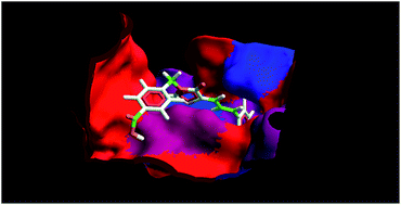
Three distinct cavities (Fig. 5) with different surface area and volume have been mapped within PTP-1B (PDB entry: 2QBS). The volume and surface area of these cavities are depicted in Table 2. The key residues of cavity 1 included Ala 217, Arg 45, Arg 47, Arg 221, Asp 48, Asp 181, Cys 215, Gln 262, Gln 266, Gly 183, Gly 218, Gly 220, Gly 259, Ile 219, Phe 182, Ser 216, Ser 222, Tyr 46 and Val 49 (Fig. 6). The docking simulations in the active site 1 (cavity 1) of PTP-1B with cinnamylidene-thiazolidinedione derivatives (4p–x) were performed by the MVD program, provided excellent result in accordance with the biological activity. The docking scores (MolDock, re-rank and H-bond scores) of compounds (4p–4x) are depicted in Table 3. MolDock, a docking scoring function that takes hydrogen bond directionality into account, combines the differential evolution optimization technique with a cavity prediction algorithm, the use of predicted cavities during the search process allows for a fast and accurate identification of potential binding modes (poses), and a re-ranking score is the indicative of highest ranked poses to further increase docking accuracy.20 It is observed that the most active compound (4w) exhibits highest MolDock score (−123.715) among the series of synthesized compounds, which is comparable with ertiprotafib (−125.183). Compound 4w also exhibits highest re-rank score (−102.381) (Table 3). In particular, the electrostatic interactions and hydrophobicity of the compounds 4w anchored in the cavity 1 of PTP-1B are shown in Fig. 7. The weak hydrogen bond (1) is formed between oxygen of 4-keto present at TZD ring of compound 4w with Asp 48, and the strong hydrogen bond (2) is formed between –OH group of methyl benzoic acid present at N3 of compound 4w with Arg 45 (Fig. 8A). The properties of hydrogen bonds such as H-bond donor/acceptor, energy and length are presented in Table 4. In Fig. 8B the steric interactions of compound 4w with PTP are demonstrated. The phenyl ring of methyl benzoic acid present at N3 of TZD ring in compound 4w showed simultaneous steric interactions with Arg 47 and Tyr 46, and the phenyl ring of phenyl allylidene group present at fifth position of TZD ring in compound 4w showed steric interaction with Cys 215. The root mean square deviation (RMSD) between co-crystallized and re-docked conformation was found 0.99 Å suggesting high docking reliability.
 |
| Fig. 5 Secondary structure of PTP-1B with mapped binding cavities 1, 2 and 3 (green). | |
Table 2 The volume and surface area of cavities (1–3) detected within PTP-1B
Cavity No. |
Volume (Å3) |
Surface area (Å2) |
1 |
53.760 |
165.120 |
2 |
15.360 |
66.560 |
3 |
14.336 |
70.400 |
 |
| Fig. 6 Cavity 1 (green) of PTP-1B with its constituting amino acid residues. | |
Table 3 Docking scores (MolDock, Re-rank and H-bond scores) of compounds (4p–4x) and ertiprotafib with PTP-1B
Compound |
MolDock score (kcal mol−1) |
Re-rank score (kcal mol−1) |
H-Bond score (kcal mol−1) |
4p |
−112.227 |
−93.2859 |
−2.5337 |
4q |
−112.153 |
−92.3753 |
−2.6035 |
4r |
−110.484 |
−94.2814 |
−2.4132 |
4s |
−116.670 |
−93.4941 |
−3.3635 |
4t |
−116.214 |
−91.6772 |
−3.5233 |
4u |
−117.467 |
−95.5568 |
−3.3398 |
4v |
−117.597 |
−96.6064 |
−1.9144 |
4w |
−123.715 |
−97.6551 |
−1.8259 |
4x |
−120.287 |
−102.381 |
−8.7196 |
Ertiprotafib |
−125.183 |
−77.276 |
−4.0603 |
 |
| Fig. 7 Electrostatic interactions (A) (red: electronegative interactions, blue: electropositive interactions) and hydrophobicity (B) (red: hydrophilic interactions, blue: hydrophobic interactions) of compound 4w anchored in the cavity 1 of PTP-1B. | |
 |
| Fig. 8 Hydrogen bond (A) (1 and 2, yellow dotted bonds) and steric (B) (red dotted lines) interactions of compound 4w within the cavity 1 of PTP-1B. | |
Table 4 Properties of hydrogen bonds formed between compound 4w and PTP-1B
H-Bond ID |
H-Bond donor |
Energy (kcal mol−1 Å−1) |
Length (Å) |
1 |
Target (PTP-1B) |
0.332 |
3.492 |
2 |
Ligand (compound 4w) |
1.494 |
3.301 |
3D-QSAR model was generated from a series of nine synthesized N3-substituted cinnamylidene-thiazolidinediones (4p–4x) acting as inhibitors of PTP-1B by using conformational alignment schemes depicted in Fig. 9. As evident from the literature, only 0.5 Å grids spacing has been opted for the present 3D-QSAR investigation. 3D-QSAR calculation for both electrostatic and shape potentials were performed and combined using PLS technique to generate 3D-QSAR model (eqn (1)).3,21 Table 5 demonstrates a statistical summary of the generated 3D-QSAR model. The developed 3D-QSAR model showed excellent cross validated correlation coefficient Q2 (0.828), non-cross validated correlation coefficient r2 (0.866), standard error of estimate S-value (0.089) and high Fisher test (F-test) value (45.405) against PTP-1B. Due to significant statistical measures and robustness of the developed 3D-QSAR model, it is used for the prediction of PTP-1B inhibitory potential of the synthesized compounds (4p–4x) (Table 6). The smaller residual values i.e. moderate difference between actual and predicted values (<1) (Table 6) are the indicator of a good linear correlation of the compounds selected in present 3D-QSAR data set. Further, results from the 3D-QSAR study indicated that the contribution of steric feature is of slight higher significance (51%) as compare to electrostatic feature (49%) for PTP-1B inhibitory potential.
|
pIC50 = 0.0005 + 0.5005 shape + 0.4999 electrostatic
| (1) |
 |
| Fig. 9 Alignment scheme for development of 3D-QSAR model (substituents R1 and R2 used for alignment purpose). | |
Table 5 PLS statistics of 3D-QSAR model
Statistical parameters |
Values |
Q2 |
0.828 |
r2 |
0.866 |
S |
0.089 |
F-Test |
45.405 |
Steric contribution (%) |
51.000 |
Electrostatic contribution (%) |
49.000 |
Table 6 Actual and 3D-QSAR predicted PTP-1B inhibitory activities of compounds 4p–x
Compound |
Actual activity (pIC50) |
Predicted activity (pIC50) |
Residual activity |
4p |
−1.488 |
−1.473 |
−0.015 |
4q |
−1.310 |
−1.476 |
0.165 |
4r |
−1.522 |
−1.456 |
−0.066 |
4s |
−1.516 |
−1.405 |
−0.111 |
4t |
−1.320 |
−1.403 |
0.082 |
4u |
−1.526 |
−1.460 |
−0.066 |
4v |
−1.505 |
−1.504 |
−0.001 |
4w |
−0.814 |
−0.832 |
0.018 |
4x |
−1.332 |
−1.325 |
−0.007 |
The contribution of shape and electrostatic features of the cinnamylidene-thiazolidinedione derivatives required for potent PTP-1B inhibitory activity are displayed in the form of master grid maps (Fig. 10), which also provide a direct visual indication regarding structural features responsible for desired biological activity of molecules under study. The master grid maps also offered an interpretation as to design and optimize novel molecules with superior biological activities. Here, we used most potent compound 4w as reference structure for visualization and interpretation of electrostatic and steric master grid maps (Fig. 10).
 |
| Fig. 10 Electrostatic (A) and steric (B) master grid maps at 0.5 Å grid resolutions. | |
In electrostatic grid maps (Fig. 10A), electrostatic favoured areas have been exemplified by red points (more positive charge increases activity, while more negative charge decreases activity), and un-favoured areas by blue points (more negative charge increases activity, while more positive charge decreases activity).3,21 In the electrostatic master grid map (Fig. 10A), presence of a high density of blue points around the
C
O group of methyl benzoic acid present at N3 position of cinnamylidene-thiazolidinedione scaffold indicates existence of electronegative groups favourable for PTP-1B inhibitory activity. A cluster of red points around the hydrogen atom of –OH group of the same functionality strongly support requirement of electropositive atoms or groups attached with oxygen of –OH group for favourable PTP-1B inhibitory activity. Furthermore, few red points around third and fifth position of phenyl ring of the methyl benzoic acid indicate the presence of electropositive atoms or groups for potent PTP-1B inhibitory activity.
In 3D-QSAR steric master grid map (Fig. 10B), presence of red and blue points indicate sterically favored (more steric bulk increases activity) and un-favored (more steric bulk decreases activity) area, respectively.3,21 Presence of cluster of blue points surrounded by red points around substituent ‘R1’ of cinnamylidene-thiazolidinedione scaffold indicates that ‘R1’ should bear bulky moiety in the core with less branching for favourable steric interaction desired for potent PTP-1B inhibitory activity.
Experimental
Chemistry
All chemicals and solvents were obtained from commercial sources and purified using standard procedures whenever required. Melting points were recorded on Veego-540 melting point apparatus and are uncorrected. The structures of the finally synthesized compounds were confirmed by IR, 1H-NMR, 13C-NMR and mass spectrometry. 1H and 13C nuclear magnetic resonance spectra were obtained using Bruker AC-400F, 400 MHz spectrometer for solutions in deuteriochloroform (CDCl3), deuterated dimethylsulfoxide (DMSO-d6) and were reported in parts per million (ppm), downfield from tetramethylsilane (TMS) as internal standard. The spin multiplicities are indicated by the symbols, s (singlet), d (doublet), t (triplet), q (quartet), m (multiplet) and br s (broad singlet). Infrared (IR) spectra were obtained with Perkin-Elmer 882 Spectrum and RXI, FT-IR model using potassium bromide pellets (cm−1). Mass spectra were obtained with electrospray ionization (ESI), atmospheric pressure chemical ionization (APCI) and matrix assisted laser desorption ionization-time of flight (MALDI-TOF) mass spectrometer. Reactions were monitored and the homogeneity of the products was checked by thin layer chromatography (TLC). For TLC, glass plates coated with silica gel G (E. Merck) were used. The TLC plates were activated at 110 °C for 30 min and visualized by exposure to iodine vapour. Pre-coated aluminium sheets coated with Silica Gel 60 F254, 0.2 mm thickness were used for final monitoring.
Synthesis of (5Z)-5-[(E)-3-phenylallylidene]-thiazolidine-2,4-dione (3a)
A mixture of 2,4-thiazolidinedione (1.17 g, 10.0 mmol) and cinnamaldehyde (1.32 mL, 10.0 mmol) was refluxed in presence of catalytic amount of piperidine (0.25 mL) for 26 h in absolute ethanol (50.0 mL). The solvent was evaporated under reduced pressure to obtain the residue. The residue was poured onto crushed ice with stirring. The separated product was filtered, washed with hydrochloric acid (5%) and dried. It was crystallized from methanol to yield pure 3a.
Yield 79.0%; mp 202–204 °C;22a,b Rf-value: 0.58 (CHCl3
:
MeOH::9
:
1); IR (KBr): νmax/cm−1 3189 (NH), 3026 (CH arom), 1722 (4-CO), 1683 (2-CO) and 1607 (C
C); 1H NMR (DMSO-d6): δ 6.69 (t, 1H, C6H5–CH
CH, J = 11.48 Hz), 7.05 (d, 1H, C6H5–CH
CH; J = 15.28 Hz), 7.33 (t, 3H, –CH arom), 7.42 (d, 1H, C6H5–CH
CH–CH
; J = 11.44 Hz), 7.54 (d, 2H, –CH arom) and 12.21 ppm (br s, NH, exchangeable with D2O), 13C NMR (DMSO-d6): δ 167.61 (–C
O, TZD), 167.03 (–C
O, TZD), 143.12 (CH
CH–CH
), 132.06 (C6H5–CH
CH), 125.42 (C6H5–CH
CH), 122.22 (–C–C
O), 135.67, 129.83, 129.04, 127.74 and 123.28 ppm (C, arom).
General method for the synthesis of (5Z)-3-(2-alkyl/substituted alkyl/substituted aryl)-5-[(E)-3-phenylallylidene]-thiazolidine-2,4-dione (4p–4w)
A mixture of (5Z)-5-[(E)-3-phenylallylidene]-thiazolidine-2,4-dione (3a) (0.46 g, 2.0 mmol), alkyl bromide/substituted alkyl bromide/substituted aryl bromide (R1–8Br) (3.0 mmol) and anhydrous potassium carbonate (1.38 g, 10.0 mmol) in acetone (50.0 mL) was refluxed for 24 h. The solvent was evaporated under reduced pressure to obtain the residue. The residue was stirred in crushed ice and the precipitated product was filtered, washed with water and dried. It was crystallised from methanol to yield compounds 4p–4w.
(5Z)-3-(2-Chloroethyl)-5-[(E)-3-phenylallylidene]-thiazolidine-2,4-dione (4p). Yield 83.5%; mp 165–168 °C; Rf-value: 0.54 (CHCl3
:
MeOH::9
:
1); IR (KBr): νmax/cm−1 3018 (CH arom), 2954 (CH aliph), 1738 (4-CO), 1673 (2-CO), 1605 (C
C) and 737 (C–Cl); 1H NMR (CDCl3): δ 3.48 (t, 2H, –NCH2CH2–), 4.10 (t, 2H, –NCH2CH2–), 6.68 (m, 1H, C6H5–CH
CH–CH–, J = 11.48 Hz), 7.04 (d, 1H, C6H5–CH
CH–CH–, J = 15.24 Hz), 7.36 (m, 3H, CH arom), 7.49 (m, 2H, CH arom) and 7.58 ppm (d, 1H, C6H5–CH
CH–CH
, J = 11.40 Hz); 13C NMR (CDCl3): δ 167.06 (–C
O, TZD), 165.22 (–C
O, TZD), 144.28 (C6H5–CH
CH–CH
), 135.36 (C6H5–CH
CH–CH–), 134.09 (C6H5–CH
CH–CH), 122.22 (–C–C
O), 42.60 (–NCH2CH2–), 25.80 (–NCH2CH2–), 130.16, 129.05, 127.75 and 122.69 ppm (C, arom); mass: 293 (M+) and 295 (M+ + 2).
Synthesis of (5Z)-3-(2-bromoethyl)-5-[(E)-3-phenylallylidene]-thiazolidine-2,4-dione (4q). Yield: 80.3%; mp 175–177 °C; Rf-value: 0.68 (CHCl3
:
MeOH::9
:
1); IR (KBr): νmax/cm−1 3017 (CH arom), 2932 (CH aliph), 1734 (4-CO), 1673 (2-CO), 1605 (C
C) and 736 (C–Br); 1H NMR (CDCl3): δ 3.55 (t, 2H, –NCH2CH2–), 4.10 (t, 2H, –NCH2CH2–), 6.68 (m, 1H, C6H5–CH
CH–CH–, J = 11.64 Hz), 7.04 (d, 1H, C6H5–CH
CH–CH–, J = 15.24 Hz), 7.36 (m, 3H, CH arom), 7.49 (m, 2H, CH arom) and 7.58 ppm (d, 1H, C6H5–CH
CH–CH
, J = 11.44 Hz); 13C NMR (CDCl3): δ 167.06 (–C
O, TZD), 165.22 (–C
O, TZD), 144.28 (C6H5–CH
CH–CH
), 135.36 (C6H5–CH
CH–CH–), 134.08 (C6H5–CH
CH–CH–), 122.23 (–C–C
O), 42.60 (–NCH2–CH2–), 26.98 (–NCH2–CH2–), 130.16, 129.05, 127.75 and 122.70 ppm (C, arom); mass: 336 (M+) and 338 (M+ + 2).
(5Z)-3-Propyl-5-[(E)-3-phenylallylidene]-thiazolidine-2,4-dione (4r). Yield: 82.4%; mp 125–127 °C; Rf-value: 0.68 (CHCl3
:
MeOH::9
:
1); IR (KBr): νmax/cm−1 3021 (CH arom), 2961 (CH aliph), 1736 (4-CO), 1672 (2-CO) and 1605 (C
C); 1H NMR (CDCl3): δ 0.92 (t, 3H, –NCH2CH2CH3), 1.63 (m, 2H, –NCH2CH2CH3), 3.67 (t, 2H, –NCH2CH2CH3), 6.69 (m, 1H, C6H5–CH
CH–CH–, J = 11.44 Hz), 7.02 (d, 1H, C6H5–CH
CH–CH–, J = 15.28 Hz), 7.33 (m, 3H, CH arom), 7.49 (m, 2H, CH arom) and 7.59 ppm (d, 1H, C6H5–CH
CH–CH
, J = 11.64 Hz); 13C NMR (CDCl3): δ 167.39 (–C
O, TZD), 165.82 (–C
O, TZD), 143.56 (C6H5–CH
CH–CH
), 135.50 (C6H5–CH
CH–CH–), 133.17 (C6H5–CH
CH–CH–), 122.93 (–C–C
O), 43.79 (–NCH2CH2CH3), 21.17 (–N–CH2CH2CH3), 11.20 (–NCH2CHCH3), 129.96, 129.01, 127.64 and 123.09 ppm (C, arom); mass: 274 (M+ + 1).
(5Z)-3-(3-Chloropropyl)-5-[(E)-3-phenylallylidene]-thiazolidine-2,4-dione (4s). Yield: 84.0%; mp 144–146 °C; Rf-value: 0.60 (CHCl3
:
MeOH::9
:
1); IR (KBr): νmax/cm−1 3012 (CH arom), 2860 (CH aliph), 1719 (4-CO), 1682 (2-CO), 1605 (C
C) and 735 (C–Cl); 1H NMR (CDCl3): δ 2.13 (m, 2H, –NCH2CH2CH2–), 3.55 (t, 2H, –NCH2CH2CH2–), 3.87 (t, 2H, –NCH2CH2CH2–), 6.69 (t, 1H, C6H5–CH
CH, J = 11.42 Hz), 7.04 (d, 1H, C6H5–CH
CH–, J = 15.24 Hz), 7.26 (t, 3H, CH arom), 7.50 (d, 2H, CH arom) and 7.58 ppm (d, 1H, –CH
CH–CH
, J = 11.60 Hz); 13C NMR (CDCl3): δ 167.28 (–C
O, TZD), 165.62 (–C
O, TZD), 143.99 (C6H5–CH
CH–CH
), 135.42 (C6H5–CH
CH–), 133.65 (C6H5–CH
CH–), 122.78 (–C–C
O), 41.84 (–NCH2CH2CH2–), 39.59 (–NCH2CH2CH2–),30.68 (–NCH2CH2CH2–), 130.07, 129.02, 127.69 and 122.64 ppm (C, arom); mass: 307 (M+) and 309 (M+ + 2).
(5Z)-3-(3-Bromoopropyl)-5-[(E)-3-phenylallylidene]-thiazolidine-2,4-dione (4t). Yield: 79.5%; mp 180–182 °C; Rf-value: 0.68 (CHCl3
:
MeOH::9
:
1); IR (KBr): νmax/cm−1 3016 (CH arom), 2953 (CH aliph), 1736 (4-CO), 1673 (2-CO), 1601 (C
C) and 738 (C–Br); 1H NMR (CDCl3): δ 2.17 (m, 2H, –NCH2CH2CH2–), 3.38 (t, 2H, –NCH2CH2CH2–), 3.86 (t, 2H, –NCH2CH2CH2–), 6.69 (m, 1H, C6H5–CH
CH–CH–, J = 11.44 Hz), 7.04 (d, 1H, C6H5–CH
CH–CH–, J = 15.28 Hz), 7.36 (m, 3H, CH arom), 7.50 (m, 2H, CH arom) and 7.58 ppm (d, 1H, C6H5–CH
CH–CH
, J = 11.64 Hz); 13C NMR (CDCl3): δ 167.27 (–C
O, TZD), 165.59 (–C
O, TZD), 144.01 (C6H5–CH
CH–CH
), 135.38 (C6H5–CH
CH–CH–), 133.67 (C6H5–CH
CH–CH–), 122.58 (–C–C
O), 40.50 (–NCH2CH2CH2–), 30.80 (–NCH2CH2CH2–), 29.69 (–NCH2CH2CH2–), 130.06, 129.01, 127.68 and 122.75 ppm (C, arom); mass: 351 (M+) and 353 (M+ + 2).
(5Z)-3-Butyl-5-[(E)-3-phenylallylidene]-thiazolidine-2,4-dione (4u). Yield: 81.5%; mp 142–144 °C; Rf-value: 0.68 (CHCl3
:
MeOH::9
:
1); IR (KBr): νmax/cm−1 3018 (CH arom), 2929 (CH aliph), 1734 (4-CO), 1672 (2-CO) and 1601 (C
C); 1H NMR (CDCl3): δ 0.92 (t, 3H, –NCH2CH2CH2CH3),1.30 (m, 2H, –NCH2CH2CH2CH3), 1.59 (m, 2H, –NCH2CH2CH2CH3), 3.70 (t, 2H, –NCH2CH2CH2CH3), 6.69 (m, 1H, C6H5–CH
CH–CH–, J = 11.48 Hz), 7.02 (d, 1H, C6H5–CH
CH–CH–, J = 15.28 Hz), 7.33 (m, 3H, CH arom), 7.49 (m, 2H, CH arom) and 7.58 ppm (d, 1H, C6H5–CH
CH–CH
, J = 11.68 Hz); 13C NMR (CDCl3): δ 167.34 (–C
O, TZD), 165.79 (–C
O, TZD), 143.52 (C6H5–CH
CH–CH
), 135.48 (C6H5–CH
CH–CH–), 133.11 (C6H5–CH
CH–CH–), 122.91 (–C–C
O), 41.79 (–NCH2CH2CH2CH3), 29.82 (–NCH2CH2CH2CH3), 19.97 (–NCH2CH2CH2CH3), 13.63 (–NCH2CH2CH2–CH3), 129.93, 128.98, 127.61 and 123.09 ppm (C, arom); mass: 288 (M+ + 1).
(5Z)-3-(4-Bromobutyl)-5-[(E)-3-phenylallylidene]-thiazolidine-2,4-dione (4v). Yield: 82.0%; mp 99–101 °C; Rf-value: 0.68 (CHCl3
:
MeOH::9
:
1); IR (KBr): νmax/cm−1 3021 (CH arom), 2947 (CH aliph), 1737 (4-CO), 1672 (2-CO), 1605 (C
C) and 735 (C–Br); 1H NMR (CDCl3): δ 1.78 (m, 2H, –NCH2CH2CH2CH2–), 1.83 (m, 2H, –NCH2CH2CH2CH2–), 3.41 (t, 2H, –NCH2CH2CH2CH2–), 3.73 (t, 2H, –NCH2CH2CH2CH2–), 6.68 (m, 1H, C6H5–CH
CH–CH–, J = 11.48 Hz), 7.02 (d, 1H, C6H5–CH
CH–CH–, J = 15.24 Hz), 7.26 (m, 3H, CH arom), 7.49 (m, 2H, CH arom) and 7.58 ppm (d, 1H, C6H5–CH
CH–CH
, J = 11.64 Hz); 13C NMR (CDCl3): δ 167.27 (–C
O, TZD), 165.62 (–C
O, TZD), 143.82 (C6H5–CH
CH–CH
), 135.38 (C6H5–CH
CH–CH–), 133.46 (C6H5–CH
CH–CH–), 122.71 (–C–C
O), 40.83 (–NCH2CH2CH2CH2Br), 32.68 (–NCH2CH2CH2CH2–), 29.62 (–NCH2CH2CH2CH2), 26.44 (–NCH2CH2CH2CH2–), 129.99, 128.97, 128.84, 127.64, and 122.77 ppm (C, arom); mass: 365 (M+) and 367 (M+ + 2).
4-[(5Z)-5-{(E)-3-Phenylallylidene}-thiazolidine-2,4-dione-3-ylmethyl]benzoic acid (4w). Yield: 75.4%; mp 216–220 °C; Rf-value: 0.45 (CHCl3
:
MeOH::9
:
1); IR (KBr): νmax/cm−1 3447 (OH), 3035 (CH arom), 2918 (CH aliph), 1739 (CO carboxy), 1686 (4-CO) and 1599 (2-CO); 1H NMR (CDCl3): δ 4.62 (s, 2H, –NCH2–), 6.74 (m, 1H, C6H5–CH
CH–CH–, J = 11.44 Hz), 7.00 (d, 1H, C6H5–CH
CH–CH–, J = 15.24 Hz), 7.32 (m, 3H, CH arom), 7.44 (m, 2H, CH arom), 7.47 (t, 2H, –CH arom), 7.50 ppm (d, 1H, C6H5–CH
CH–CH
, J = 11.68 Hz), 7.55 (d, 2H, –CH arom), 7.94 (d, 2H, –CH arom) and 12.21 ppm (br s, 1H, –COOH, exchangeable with D2O); 13C NMR (CDCl3): δ 167.16 (–COOH), 166.85 (–C
O, TZD), 166.54 (–C
O, TZD), 142.52 (C6H5–CH
CH–CH
), 135.29 (C6H5–CH
CH–CH–), 131.68 (C6H5–CH
CH–CH–), 122.91 (–C–C
O), 32.63 (–NCH2–), 142.25, 130.53, 129.56, 129.10, 128.91, 128.61, 127.37 and 124.92 ppm (C, arom); mass: 388 (M+ + Na).
Ethyl 2-[(5Z)-5-{(E)-3-phenylallylidene}-thiazolidine-2,4-dione-3-yl]acetate (3b)
A mixture of (5Z)-5-[(E)-3-phenylallylidene]-thiazolidine-2,4-dione (3a) (0.92 g, 4.0 mmol), ethyl chloroacetate (0.53 mL, 5.0 mmol) and anhydrous potassium carbonate (2.76 g, 20.0 mmol) in acetone (50.0 mL) was refluxed for 24 h. The solvent was removed under reduced pressure to obtain the residue. The residue was poured onto crushed ice with stirring. The separated product was filtered, washed with water and dried. It was crystallized from methanol to yield pure 3b.
Yield: 85.0%; mp 126–128 °C;23 Rf-value: 0.44 (CHCl3
:
MeOH::9
:
1); IR (KBr): νmax/cm−1 2993 (CH aliph), 1737 (CO ester), 1692 (4-CO), 1608 (2-CO) and 1216 (C–O); 1H NMR (CDCl3): δ 1.27 (t, 3H, –CH2CH3), 4.20 (q, 2H, –COOCH2), 4.43 (s, 2H, –CH2COO–), 6.69 (t, 1H, C6H5–CH
CH, J = 11.46 Hz), 7.04 (d, 1H, C6H5–CH
CH, J = 15.28 Hz), 7.36 (t, 3H, CH arom), 7.50 (d, 2H, CH arom), 7.59 ppm (d, 1H, CH
CH–CH
; J = 11.68 Hz); 13C NMR (CDCl3): δ 166.90 (–CH2COO–), 166.32 (–C
O, TZD), 164.92 (–C
O, TZD), 144.28 (C6H5–CH
CH), 135.38 (CH
CH–CH
), 134.16 (C6H5–CH
CH), 122.36 (–C–C
O), 62.14 (–COOCH2–), 42.08 (–NCH2COO–), 14.11 (–COOCH2CH3), 129.04, 127.75 and 122.69 ppm (C, arom).
2-[(5Z)-{(E)-3-Phenylallylidene}-thiazolidine-2,4-dione-3-yl]-acetic acid (4x). A mixture of 3b (1.0 g, 3.15 mmol), glacial acetic acid (25.0 mL) and hydrochloric acid (12 N, 5.0 mL) was refluxed for 2 h. The solvent was evaporated under reduced pressure to obtain the residue. The residue was poured onto crushed ice with stirring. The product obtained was filtered, dried and crystallized from methanol to yield pure compound 4x.Yield: 72.5%; mp 214–216 °C;24 Rf-value: 0.40 (CHCl3
:
MeOH::9
:
1); IR (KBr): νmax/cm−1 3408 (OH), 2943 (CH aliph), 1727 (CO carboxy), 1691 (4-CO) and 1608 (2-CO); 1H NMR (DMSO-d6): δ 4.31 (s, 2H, –CH2COOH), 6.64 (t, 1H, C6H5–CH
CH–, J = 11.68 Hz), 7.00 (d, 1H, C6H5–CH
CH–, J = 15.24 Hz), 7.29 (t, 3H, CH arom), 7.44 (d, 2H, CH arom), 7.51 (d, 1H, CH
CH–CH
, J = 11.44 Hz) and 8.70 ppm (br s, 1H, –COOH); 13C NMR (DMSO-d6): δ 167.63 (–CH2COOH), 166.28 (–C
O, TZD), 164.32 (–C
O, TZD), 143.64 (C6H5–CH
CH), 134.84, (C6H5–CH
CH–), 133.35 (C6H5–CH
CH), 121.93 (–C–C
O), 41.61 (–NCH2COO–), 129.56, 128.43 and 127.23 ppm (C, arom); mass: 312 (M+ + Na).
Protein tyrosine phoshatase-1B (PTP-1B) assay
The PTP-1B drug discovery assay kit (Enzo Life Sciences) is a colorimetric, non-radioactive assay designed to measure the phosphatase activity of purified PTP-1B. The kit composed of human, recombinant PTP-1B (residues 1–322; MW = 37.4 kDa), expressed in E. coli. The detection of free phosphate released is based on the classic malachite green assay and offers the advantages of convenient, single step detection and excellent sensitivity, without radioactivity. The [pTyr 1146] phosphorylated-peptide, corresponding to the sequence 1142–1153 of the insulin receptor β-subunit domain that must be autophosphorylated to achieve full receptor kinase activation, was used as substrate for the inhibition assays. This ‘activation loop’ is the target for regulators of PTP-1B. All assays were performed at 30 °C in a 96-well microtitre plate.24
Assay buffer (35.0 μL) was added to each well in microtitre plate and warmed to 30 °C. Test sample/inhibitors (10.0 μL) at various concentrations were added to appropriate well. PTP-1B enzyme solution (5.0 μL) was added to each well and enzymatic reaction was initiated by adding warmed substrate (50.0 μL) followed by incubation at 30 °C for 30 min. After incubating wells for desired duration, reaction was terminated by addition of BIOMOL RED™ reagent (BML-KI468) (25.0 μL). Mixing was carried out thoroughly by repeated pipetting with care to avoid producing bubbles. Color was allowed to develop at 30 °C for 20 min. The amount of inorganic phosphate released from the peptide was determined by measuring the absorbance at 620 nm on a micro plate-reading spectrophotometer and the IC50 values were calculated by a regression analysis of the linear portion of the inhibition curves.25
Anti-hyperglycaemic activity using STZ-nicotinamide induced diabetic mice model
Animals. Male albino mice of Laca strain bred were used in the present anti-diabetic screening. They were housed (six mice per cage) under standard (25 ± 2 °C, 60–70% humidity) laboratory conditions, maintained on a 12 h natural day–night cycle, with free access to standard food and water. Animals were acclimatized to laboratory conditions before the experiment. All the experimental protocols regarding animal studies were approved by the ‘Institutional Animal Ethical Committee’ (IAEC) and conducted according to the ‘Committee for the Purpose of Control and Supervision of Experiments on Animals’ (CPCSEA) guidelines on the use and care of experimental animals.
Drugs and reagents. STZ (Sigma Chemical Co., USA), NA (Hi-Media), Tween-80 (Research-Lab, India), D-glucose (S. D. Fine-Chem. Ltd., India), and glucose oxidase peroxidase diagnostic enzyme kit (Span Diagnostic Chemicals, India) were used in anti-hyperglycemic activity. STZ was dissolved in citrate buffer (pH 4.5) and NA in normal physiological saline.
Induction of diabetes and determination of serum glucose. T2DM was induced in overnight fasted mice by a single intraperitoneal (i.p.) injection of STZ (45 mg kg−1 body weight) 15 min after the i.p. administration of 110 mg kg−1 body weight of NA. Animals were fed with glucose solution (5%) for 12 h to avoid hypoglycaemia. The collected blood samples were placed in eppendorffs tube (1.5 mL). The serum was separated by centrifugation maintained at 4 °C and run at speed of 7000 rpm for 15 min. Serum (10.0 μL) and working reagent (GOD/POD) (1.0 mL) were mixed and incubated for 15 min at 37 °C.26 The absorbance of sample (As) and standard (Astd) provided by manufacturer were measured against blank at 505 nm. Glucose was estimated by using the formula:
Glucose (mg dL−1) = As/Astd × 100 |
whereas As = sample reading; Astd = standard reading.The elevated glucose level in serum determined at 72 h confirmed hyperglycaemia. Animals with blood glucose concentration more than 250 mg dL−1 were selected for the study.27a,b
Acute study. In acute study, animals were fasted overnight and the fasting serum glucose (SG), 0 h, levels were calculated. All the compounds were administered at a fixed dose of 30 mg kg−1 body weight orally (homogenized suspension in 0.5% carboxy methyl cellulose (CMC) and permissible amounts of Tween 80). Animals of vehicle treated group were given an equal amount of 0.5% CMC and those of control group were kept as such. Blood samples were removed from all animals at 2, 4, 6 and 24 h by retro orbital puncture method and percentage reduction in SG was calculated with respect to control group. A 10% reduction in serum glucose level versus control group was considered as a positive screening result.25
Sub-acute study. In sub-acute study, animals were fasted overnight and the fasting SG, 0 day, levels were calculated. Now the compounds were administered for a fixed dose of 30 mg kg−1 orally homogenized suspension in 0.5% CMC and permissible amounts of Tween 80 for 7 days at a fixed time. After 7th day, blood samples were removed from all animals and percentage change in SG was calculated. The data obtained were analyzed by one-way ANOVA followed by Dunnett test. The results were expressed as mean ± standard error of mean (SEM) for each group, p < 0.001 was considered as statistically significant.
Effect on body weight in diabetic mice. During the study period of 7 days, the mice were weighed and their body weights were recorded. From this data, mean change in body weight and standard error of the mean (SEM) were calculated.
Histopathology of mice pancreas. The isolated pancreas were trimmed into small pieces and preserved in 10.0% formalin for 24 h. Specimens were cut in section of 3–5 μm in thickness and stained by hematoxyline–eosin stain. The specimen was mounted by disterene phthalate xylene (DPX). The photomicrographs of each tissue section were observed using cell imaging software for life science microscopy (Olympus soft imaging solution GmbH, Munster, Germany). Pancreatic tissue was processed for Gomori staining for morphology of pancreatic β cells.28
3D-QSAR study
Data set and biological activity. The synthesized cinnamylidene-thiazolidinedione derivatives (4p–x) having inhibitory activity against PTP-1B were taken to develop 3D-QSAR based pharmacophoric model. All the molecules were kept into training set. The negative logarithm of IC50 (designated as pIC50, values in μM) against PTP-1B was taken to arrange the data in ascending and linear manner (Table 6).
Molecular modelling, docking and alignment. For the present study, Molegro Virtual Docker (MVD 2013.6.0),29a VLife MDS 3.5,29b SOMFA 2.0.029c and VEGA ZZ29d programs were employed to develop 3D-QSAR model. The chemical structures of all the data set ligands along with ertiprotafib were sketched, subjected to energy minimization using molecular mechanics (MM2) and adopted for re-optimization using the Hamiltonian approximations Austin model 1 (AM1) method available in the MOPAC module of Chem3D Ultra 8.0 until the root mean square (RMS) gradient attains a value smaller than 0.001 kcal mol−1 Å−1 in both the optimization techniques.1a,3,30 Geometrical optimizations included the solvent effect i.e. implicit solvent environment.3 The final active conformation search was performed by docking all the compounds with PTP-1B (PDB entry: 2QBS) using MVD. The 2QBS is a well-established crystal structure of PTP-1B given by W. Xu by using X-ray diffraction method at 2.10 Å resolutions.31 Structural evaluations as well as structural validation1a,3,32a,b and the structural description1b of the target protein (PTP-1B) including its recognition sites have been reported in previous studies. While performing molecular docking, both the protein and ligand molecules were imported into the workspace. All the crystallographic water molecules were removed from the protein during import process. Further, protein and ligands were subjected to molecules preparation. The option ‘detect cavities’ in the preparation window was used to identify cavities (active sites) within the enzyme PTP-1B. During this computational procedure, the maximum numbers of cavities were fixed to 5, grid resolution 0.80 Å and probe size 1.2 Å. The binding radius, grid resolution and maximum iterations parameters were set to 15 Å, 0.3 Å and 1500, respectively; while the other parameters were set as default. The docking algorithm was set to simplex evolution population size 50, RMSD thresholds 1.00 Å for cluster similar poses, RMSD threshold 1.00 Å for ignore similar poses (for multiple runs only) and 10 independent runs were conducted, each of these runs was returning to a single final solution (pose). Only negative lowest-energy representative cluster was returned from each of them after completion of docking and the similar poses were removed keeping the best scoring one. The clusters were ranked through compare the conformation of the lowest binding energy in each cluster. The lowest binding free energy poses were selected for the analysis of the docking results including various intermolecular interactions with PTP-1B, and for generation of 3D-QSAR model.1a,3,30 The lowest binding free energy poses were aligned using substituent based alignment techniques shown in Fig. 9. Among the synthesized cinnamylidene-thiazolidinedione derivatives, compound 4w showed most potent PTP-1B inhibitory potential. Therefore, it was selected as standard reference compound for alignment purpose. All the aligned structures were converted in .cssr file format using VEGA ZZ software. SOMFA program readily takes up .cssr file format for developing QSAR models. Superimposition of all the aligned structures onto the reference compound 4w is depicted in Fig. 11.
 |
| Fig. 11 Superimposition of all the molecules on the template structure. | |
Generation of 3D-QSAR model. In present work, 3D-QSAR model was developed using self-organizing molecular field analysis (SOMFA) approach based on generation of grids from the molecules under consideration. It is a validated 3D-QSAR technique developed by Robinson and co-workers.33 Master grid maps generate intrinsic molecular properties, such as the molecular shape and electrostatic potential which is used in developing the final model using suitable statistical analysis approach.3 Further, grid maps obtained from 3D-QSAR will be helpful in identifying the fundamental structural features of the cinnamylidene-thiazolidinedione derivatives that will be helpful in the exploration of pharmacophoric requirements that are mandatory for a chemical entity to be an imminent, potent and specific inhibitor of PTP-1B with reduced toxicity and improved bioavailability.Initially, structures in .cssr formats were loaded in the 3D-QSAR software along with biological activity (pIC50) against PTP-1B. 3D-QSAR models with a 40 × 40 × 40 Å grid originating at (−20, −20, −20) with a resolution of 0.5 Å were generated around the aligned molecules.3 Only training set molecules were used in developing the 3D-QSAR model. 3D-QSAR model was generated for electrostatic and steric properties against PTP-1B using alignment scheme depicted in Fig. 9. The generated model was used for predicting the electrostatic and steric properties of synthesized cinnamylidene-thiazolidinedione derivatives. Electrostatic and steric properties were combined using partial least square (PLS) technique to develop statistically reliable 3D-QSAR model.
Statistical analysis of generated 3D-QSAR model. PLS methodology was used for the analysis of generated 3D-QSAR model. PLS correlates the electrostatic and steric properties of cinnamylidene-thiazolidinedione derivatives with the inhibitory activities against PTP-1B. PLS algorithm was used in union with leave one out cross validation to develop final 3D-QSAR model. The electrostatic and steric potentials were used independent variables while pIC50 values were used as dependent variables in user defined PLS regression analysis in the V-Life package to derive 3D-QSAR model. Statistical measures obtained from PLS analysis i.e. cross-validated correlation coefficient (Q2), correlation coefficient (r2), standard error of estimate (S-value) and Fischer statistics (F-test) serves as an indicator for a trustworthy QSAR model. Q2 an internal validation indicator and r2 an external indicator takes up values in the range from 1, suggesting a perfect model, to less than 0 where errors of prediction are greater than the error from assigning each compound mean activity of the model. For an acceptable and reliable QSAR model, the value of Q2 and r2 should be greater than 0.5 and 0.6, respectively. For statistically reliable model, S-value should be closer to 0 but should not be greater than 0.5. Further, the model is acceptable if the F-test is above a threshold value. The larger the value of F, greater is the probability that QSAR model is statistically significant.3,34 Interpretation of above said statistical parameters of PLS model is not informative in terms of explaining the behavior. 3D-QSAR generated master grid maps best explain the behavior of the molecule in terms of steric and electrostatic properties by Grid-Visualizer program. These maps are three dimensional and represent the region in space where steric and electrostatic field interactions are responsible for the observed variations in the desired pharmacological activity. Individual compound in data set can be visualized in these grids and variation in activity can be best explained by the master grids.21
Conclusions
In search of PTP-1B inhibitors bearing small molecular scaffold and devoid of side effects associated with glitazones; design, synthesis, evaluation of in vitro PTP-1B inhibitory potential as well as anti-hyperglycaemic potential of N3-substituted cinnamylidene-thiazolidinedione derivatives (4p–x) in STZ-NA induced diabetic mice model was performed. The in vitro assay results show that compound bearing methyl benzoic acid at N3 (4w) exhibits most potent PTP-1B inhibitory activity with IC50 ∼ 6.52 μM and comparable anti-hyperglycaemic potential with insulin sensitizer pioglitazone. In addition, the effect of treatment on body weight of diabetic mice with compounds 4p–x was matched with pioglitazone treatment, and observed that treatment with 4w has marginal increase in body weight but weight gain was less as compared to pioglitazone. The potency of compound 4w was tested by histopathological analysis of mice pancreas; it shows similar pattern of intact cell membrane and clearly visible nucleus as of pioglitazone treated diabetic mice pancreatic tissue. The binding affinities of compounds (4p–x) with PTP-1B in terms of docking scores were determined using molecular docking simulations and the in silico binding affinities were in accordance with biological results i.e. compound 4w showed highest MolDock as well as re-rank scores. The various binding interactions (electrostatic, hydrophobic, H-bond and steric) of 4w within the vicinity of PTP-1B were explored, most importantly it shows steric interaction with Cys215 which is a part of signature motif of PTP-1B and essential for catalysis of PTP-1B.
Although all the synthesized compounds (4p–x) were screened for drug likeness or ADME screening based on “Lipinski's Rule of Five”, which state that for becoming a drug candidate a molecule should have molecular weight < 500, H-bond donor ≤ 5, H-bond acceptor ≤ 10, log
P < 5, rotatable bonds ≤ 10 (Table 7), however further experiments are necessary for evaluation of toxicity and bioavailability parameters of compound 4w. Moreover, the lead generated from 3D-QSAR can be used for further development of potent PTP-1B inhibitors bearing cinnamylidene thiazolidinedione scaffold and devoid of side effects associated with conventional therapies.
Table 7 ADME screening data of compounds 4p–x
Compound |
Mol. wt |
H-Bond acceptor |
H-Bond donor |
log P |
Rotatable bonds |
4p |
293.78 |
2 |
0 |
2.76 |
6 |
4q |
338.23 |
2 |
0 |
2.82 |
6 |
4r |
273.37 |
2 |
0 |
2.86 |
6 |
4s |
307.81 |
2 |
0 |
2.81 |
7 |
4t |
352.26 |
2 |
0 |
2.87 |
7 |
4u |
287.40 |
2 |
0 |
3.26 |
7 |
4v |
366.29 |
2 |
0 |
3.32 |
8 |
4w |
365.42 |
4 |
1 |
3.52 |
7 |
4x |
289.32 |
4 |
1 |
1.59 |
6 |
Acknowledgements
The authors gratefully acknowledge Indian Council of Medical Research (ICMR) for providing the Senior Research Fellowship (SRF). ST pleased to acknowledge University Grants Commission (UGC), New Delhi, Govt. of India for UGC-BSR Research Startup Grant [No. F.20-16(3)/2012(BSR)]. We are also thankful to Dr Rene Thomsen for permission to use MVD software.
Notes and references
-
(a) S. K. Verma, T. Rajpoot, M. K. Gautam, A. K. Jain and S. Thareja, Lett. Drug Des. Discovery, 2016, 13, 295–300 CrossRef CAS;
(b) S. Thareja, S. Aggarwal, T. R. Bhardwaj and M. Kumar, Med. Res. Rev., 2012, 32, 457–517 CrossRef PubMed.
- Diagnosis and Classification of Diabetes Mellitus, http://www.care.diabetesjournals.org/content/diacare/32/Supplement_1/S62.full.pdf, accessed October 2015.
- S. K. Verma and S. Thareja, RSC Adv., 2016, 6, 33857–33867 RSC.
- S. Suzen and E. Buyukbingol, Curr. Med. Chem., 2003, 10, 1329–1352 CrossRef CAS PubMed.
- J. E. Shaw, R. A. Sicree and P. Z. Zimmet, Diabetes Res. Clin. Pract., 2010, 87, 4–14 CrossRef CAS PubMed.
- M. Feldhammer, N. Uetani, D. Miranda-Saavedra and M. L. Tremblay, Crit. Rev. Biochem. Mol. Biol., 2013, 48, 430–445 CrossRef CAS PubMed.
-
(a) L. D. Klaman, O. Boss, O. D. Peroni, J. K. Kim, J. L. Martino, J. M. Zabolotny, N. Moghal, M. Lubkin, Y. B. Kim, A. H. Sharpe, A. Stricker-Krongrad, G. I. Shulman, B. G. Neel and B. B. Kahn, Mol. Cell. Biol., 2000, 20, 5479–5489 CrossRef CAS PubMed;
(b) S. Vakili, S. S. Ebrahimi, A. Sadeghi, S. Gorgani-Firuzjaee, M. Beigy, P. Pasalar and R. Meshkani, Mol. Med. Rep., 2013, 7, 211–216 CAS;
(c) T. O. Johnson, J. Ermolieff and M. R. Jirousek, Nat. Rev. Drug Discovery, 2002, 1, 696–709 CrossRef CAS PubMed;
(d) N. K. Tonks, FEBS Lett., 2003, 546, 140–148 CrossRef CAS PubMed;
(e) J. Montalibet and B. P. Kennedy, Drug Discovery Today: Ther. Strategies, 2005, 2, 129–135 CrossRef CAS;
(f) S. Koren and I. G. Fantus, Best Pract. Res., Clin. Endocrinol. Metab., 2007, 21, 621–640 CrossRef CAS PubMed;
(g) D. E. Moller, Nature, 2001, 414, 821–827 CrossRef CAS PubMed.
-
(a) M. Elchebly, P. Payette, E. Michaliszyn, W. Cromlish, S. Collins, A. L. Loy, D. Normandin, A. Cheng, J. Himms-Hagen, C. Chan, C. Ramachandran, M. J. Gresser, M. L. Tremblay and B. P. Kennedy, Science, 1999, 283, 1544–1548 CrossRef CAS PubMed;
(b) C. M. Rondinone, J. M. Trevillyan, J. Clampit, R. J. Gum, C. Berg, P. Kroeger, L. Frost, B. A. Zinker, R. Reilly, R. Ulrich, M. Butler, B. P. Monia, M. R. Jirousek and J. F. Waring, Diabetes, 2002, 51, 2405–2411 CrossRef CAS PubMed.
- S. Zhang and Z. Y. Zhang, Drug Discovery Today, 2007, 12, 373–381 CrossRef CAS PubMed.
-
(a) A. J. Nichols, R. D. Mashal and B. Balkan, Drug Dev. Res., 2006, 67, 559–566 CrossRef CAS;
(b) S. Lee and Q. Wang, Med. Res. Rev., 2007, 27, 553–573 CrossRef CAS PubMed;
(c) A. J. Barr, Future Med. Chem., 2010, 2, 1563–1576 CrossRef CAS PubMed;
(d) M. E. Sobhia, S. Paul, R. Shinde, M. Potluri, V. Gundam, A. Kaur and T. Haokip, Expert Opin. Ther. Pat., 2012, 22, 125–153 CrossRef CAS PubMed;
(e) R. He, L. F. Zeng, Y. He, S. Zhang and Z. Y. Zhang, FEBS J., 2013, 280, 731–750 CrossRef CAS PubMed.
-
(a) Z. Y. Zhang and S. Y. Lee, Expert Opin. Invest. Drugs, 2003, 12, 223–233 CrossRef CAS PubMed;
(b) D. V. Erbe, S. Wang, Y. L. Zhang, K. Harding, L. Kung, M. Tam, L. Stolz, Y. Xing, S. Furey, A. Qadri, L. D. Klaman and J. F. Tobin, Mol. Pharmacol., 2005, 67, 69–77 CrossRef CAS PubMed;
(c) S. Shrestha, B. R. Bhattarai, H. Cho, J. K. Choi and H. Cho, Bioorg. Med. Chem. Lett., 2007, 17, 2728–2730 CrossRef CAS PubMed.
-
(a) M. Mclane, H. Wolfe, A. Turner and I. Ruiz-White, Keystone Symposia on Molecular and Cellular Biology, Silverthorne, Colorado, 2009 Search PubMed;
(b) Z. Qin, X. Zhou, N. R. Pandey, H. A. Vecchiarelli, C. A. Stewart, X. Zhang, D. C. Lagace, J. M. Brunel, J. C. Beique, A. F. R. Stewart, M. N. Hill and H. H. Chen, Neuron, 2015, 85, 1319–1331 CrossRef CAS PubMed.
- L. F. Iversen, K. B. Moller, A. K. Pedersen, G. H. Peters, A. S. Petersen, H. S. Andersen, S. Branner, S. B. Mortensen and N. P. Moller, J. Biol. Chem., 2002, 277, 19982 CrossRef CAS PubMed.
-
(a) S. Mudaliar and R. R. Henry, Annu. Rev. Med., 2001, 52, 239–257 CrossRef CAS PubMed;
(b) J. Patel, R. J. Anderson and E. B. Rappaport, Diabetes, Obes. Metab., 1999, 1, 165–172 CrossRef CAS.
-
(a) M. S. Malamas, J. Sredy, I. Gunawan, B. Mihan, D. R. Sawicki, L. Seestaller, D. Sullivan and B. R. Flam, J. Med. Chem., 2000, 43, 995–1010 CrossRef CAS PubMed;
(b) R. Maccari, R. Ottana, C. Curinga, M. G. Vigorita, D. Rakowitz, T. Steindl and T. Langer, Bioorg. Med. Chem., 2005, 13, 2809–2823 CrossRef CAS PubMed;
(c) A. P. Combs, E. W. Yue, M. Bower, P. J. Ala, B. Wayland, B. Douty, A. Takvorian, P. Polam, Z. Wasserman, W. Zhu, M. L. Crawley, J. Pruitt, R. Sparks, B. Glass, D. Modi, E. McLaughlin, L. Bostrom, M. Li, L. Galya, K. Blom, M. Hillman, L. Gonneville, B. G. Reid, M. Wei, M. Becker-Pasha, R. Klabe, R. Huber, Y. Li, G. Hollis, T. C. Burn, R. Wynn, P. Liu and B. Metcalf, J. Med. Chem., 2005, 48, 6544–6548 CrossRef CAS PubMed;
(d) R. Maccari, P. Paoli, R. Ottana, M. Jacomelli, R. Ciurleo, G. Manao, T. Steindl, T. Langer, M. G. Vigorita and G. Camici, Bioorg. Med. Chem., 2007, 15, 5137–5149 CrossRef CAS PubMed;
(e) R. Ottana, R. Maccari, R. Ciurleo, P. Paoli, M. Jacomelli, G. Manao, G. Camici, C. Laggner and T. Langer, Bioorg. Med. Chem., 2009, 17, 1928–1937 CrossRef CAS PubMed;
(f) B. R. Bhattarai, B. Kafle, J. S. Hwang, D. Khadka, S. M. Lee, J. S. Kang, S. W. Ham, I. O. Han, H. Park and H. Cho, Bioorg. Med. Chem. Lett., 2009, 19, 6161–6165 CrossRef CAS PubMed;
(g) B. R. Bhattarai, B. Kafle, J. S. Hwang, S. W. Ham, K. H. Lee, H. Park, I. O. Han and H. Cho, Bioorg. Med. Chem. Lett., 2010, 20, 6758–6763 CrossRef CAS PubMed;
(h) Z. Liu, Q. Chai, Y. Y. Li, Q. Shen, L. P. Ma, L. N. Zhang, X. Wang, L. Sheng, J. Y. Li, J. Li and J. K. Shen, Acta Pharmacol. Sin., 2010, 31, 1005–1012 CrossRef CAS PubMed;
(i) Z. Wang, Z. Liu, W. Lee, S. N. Kim, G. Yoon and S. H. Cheon, Bioorg. Med. Chem. Lett., 2014, 24, 3337–3340 CrossRef CAS PubMed.
-
(a) B. S. Lakshmi, S. Sujatha, S. Anand, K. N. Sangeetha, R. B. Narayanan, C. Katiyar, A. Kanaujia, R. Duggar, Y. Singh, K. Srinivas, V. Bansal, S. Sarin, R. Tandon, S. Sharma and S. Singh, J. Diabetes, 2009, 1, 99–106 CrossRef CAS PubMed;
(b) E. J. Moran, S. Sarshr, J. F. Cargill, M. M. Shahbaz, A. Lio and A. M. M. Mjalli, J. Am. Chem. Soc., 1995, 117, 10787–10788 CrossRef CAS;
(c) H. Fu, J. Park and D. Pei, Biochemistry, 2002, 41, 10700–10709 CrossRef CAS PubMed.
- R. Maccari, R. Ottana, R. Ciurleo, P. Paoli, G. Manao, G. Camici, C. Laggner and T. Langer, ChemMedChem, 2009, 4, 957–962 CrossRef CAS PubMed.
- S. Shrestha, B. R. Bhattarai, K. J. Chang, K. H. Lee and H. Cho, Bioorg. Med. Chem. Lett., 2007, 17, 2760–2764 CrossRef CAS PubMed.
-
(a) R. Sakhardande, V. Kulkarni, N. Sachan and M. Nimbalkar, Eur. Pat., WO2010082212A2, 2010;
(b) N. Sachan, S. Thareja, R. Agarwal, S. S. Kadam and V. M. Kulkarni, Int. J. PharmTech Res., 2009, 1, 1625–1631 CAS.
- R. Thomsen and M. H. Christensen, J. Med. Chem., 2006, 49, 3315–3321 CrossRef CAS PubMed.
- S. Thareja, T. Rajpoot and S. K. Verma, Steroids, 2015, 95, 96–103 CrossRef CAS PubMed.
-
(a) Y. Hu, T. Xie, K. Fu, H. Kang, P. Wei and H. Huang, Heterocycles, 2009, 78, 757–761 CrossRef CAS;
(b) D. Hardej, C. R. Ashby, N. S. Khadtare, S. S. Kulkarni, S. Singh and T. Talele, Eur. J. Med. Chem., 2010, 45, 5827–5832 CrossRef CAS PubMed.
- S. M. Rida, H. M. Salama, I. M. Labouta and Y. S. A. Ghany, Pharmazie, 1985, 40, 727–728 CAS.
- E. V. Tkachenko, R. B. Lesik and V. D. Lukyanchuk, Farming Magazine, 2004, 5, 88–93 Search PubMed.
- PTP-1B Tyrosine Phosphatase Drug Discovery Kit Manual, http://www.enzolifesciences.com/fileadmin/enzo/BML/ak822.pdf, accessed October 2014.
- S. L. Badole and S. L. Bodhankar, Eur. J. Pharmacol., 2010, 632, 103–109 CrossRef CAS PubMed.
-
(a) J. C. Huralikuppi, Phytother. Res., 1991, 5, 217–223 CrossRef;
(b) E. U. Etuk, Agric. Biol. J. North Am., 2010, 1, 130–134 CAS.
- G. L. Gomori, Am. J. Clin. Pathol., 1950, 20, 665–666 CAS.
-
(a) Molegro Virtual Docker, version 6.0.0, CLC Bio, 8200 Aarhus N, Denmark, 2012 Search PubMed;
(b) VLife Molecular Design Suite (VLife MDS), version 3.5, VLife Sciences Technologies Pvt. Ltd., Pune, India, 2013 Search PubMed;
(c) D. D. Robinso, SOMFA2, version 2.0.0, Computational Chemistry Research Group, Oxford University, UK, 2006 Search PubMed;
(d) VEGA ZZ, version 3.0.3.18, Drug Design Laboratory, Department of Pharmaceutical Sciences, University of Milan, Milan, Italy, 2014 Search PubMed.
- S. K. Verma, S. K. Sharma and S. Thareja, Lett. Drug Des. Discovery, 2015, 12, 284–291 CrossRef CAS.
- Protein data bank, http://www.rcsb.org/pdb/explore/explore.do?structureId=2QBS, accessed October 2015.
-
(a) D. P. Wilson, Z. K. Wan, W. X. Xu, S. J. Kirincich, B. C. Follows, D. Joseph-McCarthy, K. Foreman, A. Moretto, J. Wu, M. Zhu, E. Binnun, Y. L. Zhang, M. Tam, D. V. Erbe, J. Tobin, X. Xu, L. Leung, A. Shilling, S. Y. Tam, T. S. Mansour and J. Lee, J. Med. Chem., 2007, 50, 4681–4698 CrossRef CAS PubMed;
(b) R. Ottana, R. Maccari, J. Mortier, A. Caselli, S. Amuso, G. Camici, A. Rotondo, G. Wolber and P. Paoli, Eur. J. Med. Chem., 2014, 71, 112–127 CrossRef CAS PubMed.
- D. D. Robinson, P. J. Winn, P. D. Lyne and W. G. Richards, J. Med. Chem., 1999, 42, 573–583 CrossRef CAS PubMed.
- A. Golbraikh and A. Tropsha, J. Mol. Graphics Modell., 2002, 20, 269–276 CrossRef CAS PubMed.
Footnotes |
† The authors declare that in the present study none of the experimentation was performed using the human subjects. |
‡ Electronic supplementary information (ESI) available. See DOI: 10.1039/c6ra24501c |
|
This journal is © The Royal Society of Chemistry 2016 |
Click here to see how this site uses Cookies. View our privacy policy here.