Thiago de Castro Rozadaa,
Rodrigo Meneghetti Pontesa,
Roberto Rittnerb and
Ernani Abicht Basso*a
aDepartamento de Química, Universidade Estadual de Maringá, Av. Colombo, 5790, 87020-900 Maringá, PR, Brazil. E-mail: eabasso@uem.br; Fax: +55 44 30114125; Tel: +55 44 30114125
bInstituto de Química, Universidade Estadual de Campinas, 13083-970 Campinas, SP, Brazil
First published on 21st November 2016
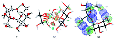
The stereoelectronic effects involved in the conformations shown by D-sucrose were analysed at the M06-2X/6-31++G(d,p) level, both under vacuum and in water with the continuum solvation model IEF-PCM, and employing the natural bond orbital theory (NBO) and non-covalent interactions (NCI). Different groups of conformations for D-sucrose in relation to the glycosidic linkage were evaluated and the results showed that not only were hydrogen bonds important to explain the relative energy observed, but it was also necessary to consider any stabilizing orbital interactions involving the glycosidic linkage. The most stable conformation observed in water had dihedral angles for the glycosidic linkage with values of 110.8° and −44.9° for ϕ and ψ, respectively.
Disaccharides are particularly interesting because they have the same rotational degree of freedom as oligo- and polysaccharides4 (a pair of dihedral angles identified as ϕ and ψ), but have a structure small enough to allow the application of sophisticated quantum mechanical methods. Different approaches have been used to determine the lower energy combinations of these dihedral angles, including classical based methods (molecular dynamics and molecular mechanics),4–8 and ab initio9,10 and density functional theory (DFT)11 methods.
A comprehensive understanding of the stereoelectronic effects involved in the different conformations of disaccharides can bring great contributions to the understanding of how these molecules interact with proteins and can assist in the development of new force fields to describe more complex carbohydrates.
D-sucrose is a disaccharide composed of a glucose and a fructose unit connected in the sequence α-D-Glc-(1 → 2)-β-D-Fru (Fig. 1). In the crystalline structure, D-sucrose presents values of 180° and −55° for ϕ and ψ.12 The conformational behavior in water, however, has been a point of controversy. While some NMR studies indicate that only one rigid conformation (similar to the crystalline structure) is present,13–17 others suggest that the scenario is not so clear and that additional conformations need to be considered in order to get a good correlation between the experimental and theoretical data.18–24 These differences arise from the fact that interpretation of the NMR data depends on the structural model applied, and the structural model, in turn, can be very problematic for a flexible molecule like a disaccharide. To make the task even harder, structural models are highly dependent on the force field and solvation method applied.
Most of the studies on the conformations of D-sucrose reported in the literature were based solely on classical force fields.18,25–27 While this could be admissible at a time when computer resources were not available for a system of this size, at present the application of sophisticated electronic structure methods is indispensable. Refined DFT methods are expected to provide more reliable information about the conformational preference of D-sucrose. Also, electronic structure procedures allow the use of tools that can interpret the conformational behavior, like the natural bond orbital (NBO)28 and non-covalent interaction (NCI)29 methods. A study of these conformations is essential for a posterior evaluation of the interactions of D-sucrose with the aromatic residues of amino acids, since carbohydrates are recognized by proteins through interactions with the amino acid residues that form the proteins. Among these interactions, the C–H–π interaction stands out, where the C–H bonds of the carbohydrate interact with the π bonds of the aromatic residue of some amino acids. In this way, the conformational study of a carbohydrate is important for the comprehension of what regions of the molecule can interact with the amino acid residues for example. Although D-sucrose presents a different type of glycosidic linkage between two anomeric positions, the conclusions shown in this work with respect to the stereoelectronic effects that stabilize the different conformations of the carbohydrate can contribute to the study of other disaccharides with more usual glycosidic linkages (aldoses). Thus, the main goal of this work is to unequivocally elucidate the conformational behavior of D-sucrose, as it is not clear in the literature as evidenced by the different analyses involving NMR data. For this purpose, we employed a conformational search starting from the crystalline structure (using MCMM/OPLS_2005) and submitted the resulting conformer candidates to calculations using the Minessota M06-2X functional.30 The final structures were then studied with a combination of NBO and NCI methods. As a result, a complete documentation of the conformational behavior of D-sucrose is reported hereafter.
31 software package for the electronic structure calculations, the NBO 5.9
32 program for analysis involving the natural bond orbital theory (NBO), the program NCIPlot 3.0
29 to obtain data for the non-covalent interaction (NCI) analysis and the PyMOL33 program to visualize the structures and surfaces.
The different conformations of D-sucrose were optimized with the DFT method M06-2X30 and the 6-31++G(d,p)34 basis set, both under vacuum and in water. The IEF-PCM solvation method was applied for optimization in water using the Bondi radii for the description of the molecular cavities. The hybrid functional M06-2X is adequate for the obtainment of thermodynamic and kinetic data of main group elements and for cases in which non-covalent interactions are important.35 Besides, this method shows an excellent performance for the energy evaluation of carbohydrates36 with a favorable computational cost. Frequency calculations at the same theory level were performed to characterize the optimized structures as stationary points, and also to obtain the zero point energy correction (ZPE)37 and thermal corrections. Single-point energy calculations with the MP2 method38 and 6-311++G(2df,2pd) basis set functions were also performed for selected conformations of the carbohydrate under vacuum and in water (IEF-PCM/Bondi).
The NBO analysis was performed under vacuum at the M06-2X/6-31++G(d,p) level with the structures optimized in water at the same level of theory. The wave functions utilized for the non-covalent interaction analysis (NCI) were obtained at the M06-2X/6-31++G(d,p) level in water (IEF-PCM/Bondi).
| Entry | Conformer | ΔEvac. | ΔEwater |
|---|---|---|---|
| 1 | S1-gg-tg-gg | 0.38 | 0.08 |
| 2 | S1-gt-tg-gg | 0.58 | 0.00 |
| 3 | S1-tg-tg-gg | 0.00 | 1.01 |
| 4 | S1-gt-tg-ggccw | 2.27 | 0.75 |
| 5 | S2-gg-gt-tg | 10.09 | 6.95 |
| 6 | S2-gg-gt-gg | 5.67 | 2.07 |
| 7 | S2-gg-tg-gg | 7.51 | 5.11 |
| 8 | S2-gt-gt-gg | 7.16 | 1.79 |
| 9 | S2-tg-gt-gg | 9.74 | 7.58 |
| 10 | S2-tg-gt-gg | 5.73 | 3.10 |
| 11 | S3-gt-tg-gt | 7.73 | 3.21 |
| 12 | S3-tg-tg-tg | 8.21 | 4.49 |
| 13 | S4-gg-gt-tg | 10.92 | 7.67 |
| 14 | S4-gg-gt-tg | 9.55 | 8.34 |
| 15 | S4-gg-gt-gt | 10.38 | 6.95 |
| 16 | S4-gt-gg-gt | 9.20 | 5.74 |
| 17 | S4-gt-gt-tg | 11.21 | 7.57 |
| 18 | S4-tg-gt-tg | 11.46 | 8.50 |
| 19 | S4-tg-gt-gt | 7.84 | 5.39 |
| 20 | S5-gg-tg-gg | 7.90 | 4.15 |
| 21 | S6-gg-tg-tg | 7.72 | 8.13 |
| 22 | S7-gg-tg-gg | 8.19 | 4.89 |
The S1 group of conformations obtained were based on the dihedral angles ϕ and ψ of the glycosidic linkage of the X-ray structure (108° and −55°, respectively) and they also correlate with the dihedral angles of the most stable conformation found by Case18 (105° and −60° for ϕ and ψ, respectively). The average dihedral angle of 111.5° of ϕ for the conformations of the group S1 in water was only 3.5° larger than the same dihedral angle of the crystalline structure, while on the structure M1 obtained by Case this dihedral angle was 3° smaller than the crystalline structure. For the dihedral angle ψ, the average dihedral angle of the conformations of the group S1 was 5° below the crystalline structure and around 10° less than M1. The dihedral angles ϕ and ψ of the S3 group conformations are also close to the crystalline structure and to the M1 structure. Nevertheless, the average dihedral angle ϕ of 99.3° for the group S3 conformations is about 9° smaller than the crystalline structure, while for the conformations of the group S1 this dihedral angle is larger than the crystalline structure. However, the average dihedral angle ψ of −53.3° for the conformations of the group S3 in water differs by only 0.7° from the dihedral angle in the crystalline structure. The conformations of the group S2 in turn have the dihedral angles ϕ and ψ of the glycosidic structure (94.1° and −164.3°, respectively) close to the structure M3 obtained by Case (85° and −165° for ϕ and ψ, respectively).
Under vacuum, only the conformations of the group S1 have a relevant contribution to the equilibrium (entries 1–4, Table 1). The other groups showed a pronounced reduction in the relative energy compared to the group S1 with an inclusion of the continuous solvation model, with the exception of the group S6. However the group S1 still was the predominant conformation in the equilibrium.
In relation to the dihedral angle ω1 of the group S1 conformations, it was observed that the conformation trans–gauche was the most stable under vacuum, but it became the least stable of the three conformations in water. The orientation of the exocyclic groups can also contribute to the stability of one conformation allowing interactions like hydrogen bonds to form between the hydroxyl groups of the pyranosidic and furanosidic portions of the disaccharide.
Regarding the hydroxyl group orientation, the clockwise arrangement (S1-gt-tg-gg) was more stable than the counterclockwise arrangement (S1-gt-tg-ggccw) both under vacuum and in water. These hydroxyl orientations are an outcome of the destabilizing steric effects and the attractive interactions of hydrogen bonds. The QTAIM and NCI analysis for D-glucose showed that the hydrogen bonds forming five membered rings are weak or non-existent and the hydroxyl arrangements are directed by the lone pair repulsion between the endocyclic oxygen and the oxygen bonded to the anomeric carbon.39
To evaluate the stereoelectronic effects involved in the conformational preference of D-sucrose, the most stable conformations of the groups S1–S3 in water were selected for the NBO and NCI analysis. The different conformations for the group S1 regarding the dihedral angle ω1 were also analysed to investigate the influences on the glycosidic portion of the molecule. The relative energies for these conformations calculated at different theory levels are shown in Table 2 and the optimized structures are presented in Fig. 2.
| Entry | Conformer | M06-2Xa | MP2b | ||
|---|---|---|---|---|---|
| ΔEvac. | ΔEwat. | ΔEvac. | ΔEwat. | ||
| a Optimization with the 6-31++G(d,p) basis set function, ZPE correction and IEF-PCM/Bondi solvation model when in water.b Energy calculation with the 6-311++G(2df,2pd) basis set function and IEF-PCM/Bondi solvation model when in water. | |||||
| 1 | S1-gt-tg-gg | 0.58 | 0.00 | 0.25 | 0.00 |
| 2 | S2-gt-gt-gg | 7.16 | 1.79 | 8.14 | 2.29 |
| 3 | S3-gt-tg-gt | 7.73 | 3.21 | 9.32 | 3.21 |
| 4 | S1-gg-tg-gg | 0.38 | 0.08 | 0.00 | 0.44 |
| 5 | S1-tg-tg-gg | 0.00 | 1.01 | 0.31 | 1.44 |
| 6 | S1-gt-tg-ggccw | 2.27 | 0.75 | 2.45 | 0.66 |
The range of the dihedral angles ϕ observed for the more stable conformations of the groups S1–S3 in water was below 20°. For the most stable conformation of the group S1 this dihedral angle was 110.8°, while for the conformations of the groups S2 and S3 the dihedral angles were close, with values of 93.8 and 95.5°, respectively. However, the dihedral angle ψ showed greater variations between the different groups of conformations. The S1 conformation had a dihedral angle ψ of −44.9°, while for the conformation of the group S2 this dihedral angle was −160.1°. For the conformation of the group S3 it was −61.9°. Based on these dihedral angles and on the structures a–c presented in Fig. 2 it can be seen that the main difference between the conformation S1 and S2 is related to the orientation of the furanosidic part of D-sucrose, while the main difference between S1 and S3 was the observed hydrogen bond interactions.
![]() | ||
| Fig. 4 Plot of the reduced density gradient versus the electron density multiplied by the sign of (λ2)ρ for the conformations of D-sucrose. (a) S1-gt-tg-gg; (b) S2-gt-gt-gg; (c) S3-gt-tg-gt. | ||
It was observed that there were three interaction points which were characterized as hydrogen bonds by the NCI analysis for the conformation of the group S1. The first one is between O5 and the hydroxyl O12–H (1.97 Å), the second one is between O12 and the hydroxyl O6–H (1.82 Å) and the third one is between O2 and the hydroxyl O7–H (1.97 Å). For the conformation of the group S2 only one hydrogen bond interaction was observed, between O2 and the hydroxyl O9–H (1.94 Å), and for the conformation of the group S3 three interactions were observed, one between O1 and the hydroxyl O9–H (2.01 Å), one between O2 and the hydroxyl O7–H (1.93 Å) and one between O6 and the hydroxyl O12–H (1.84 Å).
To investigate the importance of the hyperconjugative interactions for the greater stability of the conformation of the group S1 over the conformations of the groups S2 and S3, energy calculations were performed where electron transfer from a donor orbital to an acceptor orbital was prevented. That is, the bonding orbitals contained the maximum occupancy possible (two electrons), similar to the Lewis description. Then this energy was compared with the energy obtained before the removal of the hyperconjugative interactions and the difference in energy indicates the importance of the hyperconjugative interactions for each conformer. The energy variation obtained in this analysis is presented in Table 3. The conformation S1-gt-tg-gg presents the biggest stabilizing effects due to the hyperconjugative interactions. The preference for this conformation over the other conformations of the group S1 was also correlated with its larger hyperconjugative effect. However, the most stable conformation of the group S1 also presents the biggest destabilizing energy due to repulsion between the occupied orbitals. One reason for this greater destabilizing effect is the greater proximity between the hydroxyl groups provided by the hydrogen bonding interactions.
| Entry | Conformer | ΔEhyperconj. | Steric energy | |
|---|---|---|---|---|
| Vacuum | Water | |||
| 1 | S1-gt-tg-gg | 1005.850 | 991.662 | 835.34 |
| 2 | S2-gt-gt-gg | 997.713 | 981.088 | 827.59 |
| 3 | S3-gt-tg-gt | 996.652 | 981.135 | 832.25 |
| 4 | S1-gg-tg-gg | 993.202 | 980.545 | 829.01 |
| 5 | S1-tg-tg-gg | 994.186 | 981.725 | 832.76 |
| 6 | S1-gt-tg-ggccw | 1011.711 | 995.378 | 835.74 |
A detailed investigation of the effects involved in the conformational preference of D-sucrose could be obtained by the NBO analysis of the orbital interaction.† Interactions were observed like hydrogen bonds where the oxygen lone pairs act as the donor orbital and the σ* of the O–H bond acts as the acceptor orbital, interactions involving the orbitals of the glycosidic bonds C1–O1 and O1–C8, through-space interactions different to hydrogen bonds between the pyranosidic and furanosidic ring and interactions involving the dihedral angle ω1.
The hydrogen bond interactions favor the conformation of the group S1 (37.56 kcal mol−1), followed by the conformations of the groups S3 and S2 (34.04 and 27.29 kcal mol−1, respectively). The conformation of the group S2 has the largest stabilizing hyperconjugative effect involving the orbitals of the bonds C1–O1 and O1–C8 (110.02 kcal mol−1), but with a small difference compared to the conformation of the group S1 (109.50 kcal mol−1). For the conformation of the group S3 this interaction has an energy of 104.90 kcal mol−1. Through-space interactions, other than hydrogen bonds, were only observed for the conformations of the groups S2 and S3 (1.26 and 2.40 kcal mol−1, respectively).
Altogether, a great contribution of the hydrogen bond interactions and hyperconjugative interactions involving the orbitals of the glycosidic linkage to the stabilization of the conformations of the group S1 was observed and that overrides the biggest repulsive steric interactions observed for this group. The conformation of the group S2 presents a stabilization energy only slightly smaller than the conformation of the group S3, but with repulsive interactions that favoured this conformation over the conformation of the group S3. Although the stabilizing energies of the conformations of the groups S2 and S3 are close to each other, they differ in the type of interactions. For the conformation of the group S3 it is the hydrogen bonds and other through-space interactions that stand out over the conformation of the group S2, while for this last conformation it is the interactions involving the glycosidic linkage and the dihedral angle ω1 that stand out in the conformation of the group S3.
Table 4 presents the occupancy of some of the acceptor orbitals for the different conformations of D-sucrose. Higher occupancy values for acceptor orbitals indicate higher electron delocalization to these orbitals.
| Conformer | ||||||
|---|---|---|---|---|---|---|
| S1-gt-tg-gg | 0.02316 | 0.02883 | 0.03103 | 0.00977 | 0.05448 | 0.07749 |
| S2-gt-gt-gg | 0.02641 | 0.01881 | 0.00504 | 0.02864 | 0.05748 | 0.08299 |
| S3-gt-tg-gt | 0.02572 | 0.03049 | 0.00670 | 0.01476 | 0.05926 | 0.07330 |
| S1-gg-tg-gg | 0.02570 | 0.02860 | 0.00726 | 0.01148 | 0.05464 | 0.07873 |
| S1-tg-tg-gg | 0.02649 | 0.02823 | 0.00476 | 0.01127 | 0.05458 | 0.07859 |
| S1-gt-tg-ggccw | 0.00953 | 0.02966 | 0.03091 | 0.00465 | 0.05689 | 0.07655 |
The first four orbitals shown in Table 4 are the acceptor orbitals of the hydrogen bond interactions. The interaction involving the orbital
is relevant for the three groups of conformations. The interactions involving the orbital
are more pronounced for the conformations of the groups S1 and S3, while the interactions involving the orbital
have more relevance for the conformation of the group S2. The occupancy also highlights the importance of the delocalization involving the orbital
to the greater stabilization of conformation S1-gt-tg-gg.
Regarding the dihedral angle ω1 for the conformations of the group S1, it was observed that the conformation S1-gt-tg-gg (with a conformation gauche–trans for the referred dihedral angle) presents the largest energy variation with deletion of the hyperconjugative interactions both under vacuum and in water. This conformation showed the largest destabilizing interactions due to repulsion between the occupied orbitals (835.34 kcal mol−1), followed by conformations S1-tg-tg-gg and S1-gg-tg-gg (832.76 and 829.01 kcal mol−1, respectively).
The higher stability of the conformation gauche–trans is directly related to the interaction involving the lone pairs of oxygen O12 with the orbital
(4.71 and 12.36 kcal mol−1). This interaction is not possible for the conformations gauche–gauche and trans–gauche of the dihedral angle ω1. If the interaction
was not considered, the observed hyperconjugative energy related to the dihedral angle ω1 would be larger for the conformation gauche–gauche (19.30 kcal mol−1), followed by the conformations gauche–trans (18.65 kcal mol−1) and trans–gauche (13.97 kcal mol−1).
To summarize, the dihedral angles of 110.8° and −44.9° for ϕ and ψ, respectively, observed for the most stable conformation of the group S1 allowed better hydrogen bond interactions and good stabilizing orbital interaction involving the glycosidic linkage. For the conformation of the group S2 the dihedral angle of 93.8° for ϕ and −160.1° for ψ led to a good stabilization energy due to the interactions between the orbitals involved in the glycosidic linkage, but with this spatial arrangement only one hydrogen bond was observed. The dihedral angles of 95.5° and −61.9° (entry 11, Table S2†) for ϕ and ψ, respectively, observed for conformation S3 led to three hydrogen bond interactions, which are smaller than the observed dihedral angles for the conformation of the group S1 and the orbital interactions involving the glycosidic linkage were the smallest of the three conformations analysed by NBO.
Footnote |
| † Electronic supplementary information (ESI) available: Dihedral angles, detailed NBO interactions and Cartesian coordinates of the optimized conformations. See DOI: 10.1039/c6ra24413k |
| This journal is © The Royal Society of Chemistry 2016 |