Direct observation of spatial configuration and structural stability of locked Y-shaped DNA structure†
Tapas Paul and
Padmaja P. Mishra*
Chemical Sciences Division, Saha Institute of Nuclear Physics, 1/AF Bidhannagar, Kolkata 700064, India. E-mail: padmaja.mishra@saha.ac.in; Fax: +91-33-2337-4637; Tel: +91-33-2337-5345-49
First published on 17th October 2016
Abstract
Here we introduce a new building block unit (locked Y-DNA) and its structural properties for a self-assembled, bottom-up, three-dimensional supramolecular nanoarchitectural probe. The spatial structural conformation and overall stability of this system have been explored using meticulous nanometre measurement techniques to predict essential structural characteristics as a building block for programmable nanomaterial.
Nucleic acids are the fundamental ‘material of life’ as they store and transmit hereditary information during the central dogma of fundamental life process. In addition to their integral biological functions, they can be used for bottom-up molecular self-assembly into stable structures by spontaneous reorganisation of the molecular components.1,2 The nanoscaled DNA double helix has been rather used as a structural material than as a carrier of biological information in vitro systems. DNA strands can be selectively bent at the molecular level to design structures with several virtual shapes.3 Divergent DNA superstructures have been obtained from the basic building blocks either by crystallization or by self-assembled aggregation through minimizing their free energy.4,5 Numerous structural conformations like tetrahedral, cubes, dodecahedral, buckyballs, etc. have been used to constitute advanced three-dimensional systems to serve as indispensable material in DNA nanotechnology.6–8 One such important building block is Y-shaped DNA (Y-DNA) that self-assembles into dendrimer-like DNA.9,10 This novel formation of the supramolecular nanoarchitectures has been used in developing DNA nanobarcode sensor.11,12 They have also been established to be potentially beneficial in numerous in vivo applications that include drug and gene delivery, imaging, tissue repair, etc., due to their excellent biocompatibility and chemical homogeneity.13–16 The structural properties of Y-DNA play a significant role in the overall supramolecular nanoarchitectures, as the nature of these assemblies depends critically on conformational stability and the structural flexibility of Y-DNA.9,13,17 Hence, investigation of Y-DNA's structural properties is imperative for developing self-assembly techniques and using them as a building unit.
Analogous to the X-shaped Holliday junctions,18,19 Y forks DNAs continuously appear as key intermediate states during triplet repeat expansion or deletion of a DNA replication process.20 The three arms constituting Y-DNA spontaneously orient to adopt preferred stable structures.21,22 Like holliday junctions, Y-DNA transforms its conformation to a structurally rigid and unfavourable T-shaped isomer at higher Mg2+ ion concentration.21 The angles at the junction, as well as separation distances of the individual arms are different, even at room temperature and physiological salt concentrations.23,24 This suggests that DNA structure rather stabilises by adoption of a three-dimensional architecture over a planer structure. Increasing the temperature to near the melting point, it experiences a conformational change due to partial denaturation at the junction and the distance between the arms changes accordingly.23 Steady state and time resolved FRET measurements of Y-DNA have extended information about the structural integrity of the vertices that is crucial for the overall integrity of the nanostructures.24
Here, we contemplate to study the spatial structural conformation and overall stability of self-assembled locked Y-DNA using ensemble and single molecule FRET (smFRET) methods. Locked Y-DNAs form due to face-to-face joining of two Y-DNAs, resulting in a shape that closely resembles to replication bubble. Thus, it has two single stranded arms sandwiched between two double stranded arms. Addition of complementary sequences to the single stranded portions would form locked Y-DNA with having double stranded arms. It is very important to study the overall shape and angle at the junction of this structure to improve understanding of the formation of self-assembled three-dimensional supramolecular nanoarchitecture. The novelty of this work lies in the use of short DNAs with locked arms with added complementary sequences, producing a very rigid system to give the concept of a DNA nanocluster with overall compact structural integrity. The short DNAs used for this purpose have 23 nucleotides, labelled at the middle of the Y face locked DNA with Cy3 (donor) and Cy5 (acceptor) as efficient FRET pair (Fig. 1). This would allow monitoring of nanometre and/or sub-nanometre structural fluctuation at single molecular resolution using the smFRET method.
 | ||
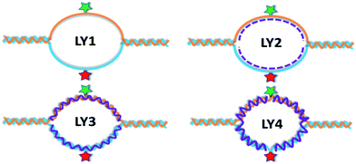
| Fig. 1 Schematic of the four different systems of locked Y-DNA, labelled at the middle of the locked Y arms with Cy3 (green star) and Cy5 (red star). | ||
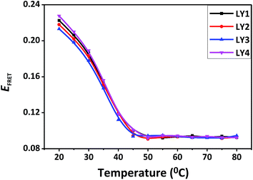
Similarly to Y-DNA, this newly probe locked Y-DNA can also be projected as an important building block for DNA nanoarchitectures. The two sets of DNAs used here are designed in such a way that once the individual strands are annealed, they form a structure where two Y faces are bridge-locked at the centre that is sandwiched between two dsDNA having an equal number of base pairs (appears as if two Ys join together in a face to face manner). In particular, one has 11 nucleotides at each of the locked Y faces, sandwiched between two dsDNA having six base pairs (named as 6-11-6 composition) and the other has 7 nucleotides at each of the locked Y faces, sandwiched between two dsDNA having 8 base pairs (named as 8-7-8 composition). In both of the cases, Cy3 and Cy5 are labelled at the centre of the two Y faces respectively, to be followed up by energy transfer efficiencies. In this particular system, the two bridge Y arms are believed to behave as two flexible single strands (LY1, Fig. 1). We applied mixture of nucleotides (i.e., dNTP) and two different sets of complementary sequences at the bubble region of both these systems separately, in contemplation of monitoring the structural and conformational flexibility by imposing restriction on the single stranded backbones. Upon addition of dNTP, the single stranded arms involve in hydrogen bonding with the free complementary nucleotides (LY2, Fig. 1). Out of the two complementary sets added, one is the complementary of individual single strands (LY3, Fig. 1), and the other is the continuous complementary of two half single strands from the centre and extending towards the two Y arms (LY4, Fig. 1). The former patterns an open structure, while the latter forms a closed structure at the junction. Ensemble FRET measurements of these architectural probes show different FRET efficiency (Een-FRET) having values of 0.23, 0.22, 0.21, and 0.23 for LY1, LY2, LY3, and LY4 systems of 6-11-6 composition, respectively (Fig. 2). A comparatively lower Een-FRET for LY2 than the LY1 system is an indication of decreases in the distance between the two faces. This could be due to the formation of complementary hydrogen bonding with the free nucleotides that leads an increase in molecular crowding and hence the distance. Similarly, for LY3 also the Een-FRET is lower than LY1 and LY2. This again suggests that the duplex formation with the complementary strands at the two single stranded arms effectively increases the distance between them. On the contrary, LY4 shows higher Een-FRET values compared to the other systems. Here the complementary strands rather compact the two single stranded fork arms by forming a closed structure at the junction, resulting in a decrease in the distance. The more rigid 8-7-8 composition system behaves closely similar to the 6-11-6 system as well, as described above (Fig. S1, ESI†). The Een-FRET values were found to decrease consistently with increase in temperature. The Tm calculated from the melting FRET curve (34 ± 2 °C for 6-11-6 and 38 ± 2 °C for 8-7-8 composition) is an indication of their comparable stability. Using Een-FRET, the distances between the locked arms and angles at the junctions were calculated to speculate their probable three-dimensional structures that might provide information for designing DNA nanoarchitecture. Since all the above measurements were carried out in bulk phase that provides ensemble averaging parameters, we performed smFRET measurements to get accurate distance and clear information about structural conformation of the locked Y probe.
 | ||
| Fig. 2 Change of ensemble FRET values with temperature for four different systems; LY1 (black), LY2 (red), LY3 (blue), and LY4 (magenta) respectively. | ||
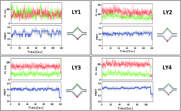
The advantage of measuring the distances of nanometre resolution from single molecule FRET experiments for heterogeneous or dynamic biological processes is to avert averaging over a number of events. smFRET studies provide detailed dynamics and conformational information by exploring every tether molecule,25 and that is onerous to accomplish through ensemble FRET measurements. In this particular study, this technique is truly appropriate to furnish clear information about three-dimensional conformations. The LY1 system of 6-11-6 composition shows the existence of three distinct FRET states, having single molecule FRET efficiency (Esm-FRET) values of 0.46, 0.52, and 0.72 (Fig. 3). This specifies that two single stranded bridged Y arms continuously fluctuate within a distance between 5.6 nm to 4.61 nm. Also, the kinetic rate parameters propose that this particular system stabilises an intermediate state which is closer to the configuration associated with the low FRET state (unpublished results). The structure developed with addition of complementary nucleotides (dNTP) in the LY1 system still has three FRET states with Esm-FRET values 0.54, 0.61, and 0.66. However, as can been seen, the fluctuation distances are less in comparison to that of LY1 system (Fig. 3). Two distinct FRET states with Esm-FRET 0.58 and 0.66 have been observed for LY3 system, though the flexibility at two Y arms is expected to facilitate the conformational changes of the overall system (Fig. 3). Similarly, as the complementary strands form a closed structure at the junction in LY4 system thus making it more rigid, it has two closely spaced but distinct FRET states of 0.67 and 0.72 (Fig. 3).
All the individual FRET states attended by different systems and their corresponding distances are represented in Table 1. Upon addition of dNTP or complementary strands to LY1, the overall system appears to experience conformational change and thus the fluctuation of the two locked Y arms is reduced. Though both of these two arms form a duplex with the added complementary strand in both LY3 and LY4 systems, the distance between the two faces, as well as the rate of fluctuation are higher for LY3 than LY4. This discrepancy could be attributed to the composition at the Y junction as LY3 has a nick at the junction that is relatively flexible, whereas LY4 has a hinge. This corroborates that formation of hydrogen bonding with complementary strands minimise the conformational energy to contract the two double stranded Y arms in the LY4 system; however, effects are exactly in reverse manner for the LY3 system. Very similar trends have also been observed for the 8-7-8 composition as well (Fig. S2 and Table S2, ESI†). As the system is initially more rigid for 8-7-8 composition over 6-11-6 composition, the rate of fluctuations is also reduced upon treating with complementary sequences.
| Different systems | LY1 | LY2 | LY3 | LY4 | ||||||
|---|---|---|---|---|---|---|---|---|---|---|
| FRET efficiency (Esm-FRET) | 0.46 | 0.56 | 0.72 | 0.54 | 0.61 | 0.66 | 0.58 | 0.66 | 0.67 | 0.72 |
| Distance between the two attached fluorophore (nm) | 5.55 | 5.19 | 4.61 | 5.26 | 5.01 | 4.84 | 5.12 | 4.84 | 4.80 | 4.61 |
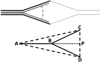
Molecular self-assembly of a particular building block unit can provide assembled nanoarchitecture of different 2D/3D conformational shapes,7,27 depending on the nature of their arrangement. One among the important deciding parameters could be the bending at the junction (i.e. junction angle).23 In our designed probes, the arm constituting one of the legs of ‘Y’ has a fixed sequence, whereas the two locked Y arms have various compositions depending on whether it is LY1, LY2, LY3, or LY4. Angular changes of different systems at the junction can now be speculated considering the length of the corresponding base/nucleotide units and the distance between the two faces calculated from FRET measurements. The dyes in our systems are positioned at the centre of locked Y-DNA and the nucleotides are symmetrically distributed to both sides. The schematic shown in Fig. 4 represents one half of a locked Y-DNA structure, where CD is the donor–acceptor distance obtained from the FRET values; BC impersonates the corresponding nucleotides distance. P is the midpoint of CD, where C and D are the position of the dyes and also the centre of the locked arms. Using the schematic model and considering a contribution of corresponding nucleotide bases along with the calculated FRET distance, the angles at the Y junction have been determined for both of the compositions. As the arm BC = 3.85 nm for LY1 of the 6-11-6 composition (11 × 0.7/2), the angle created at the Y junction (∠CBD) is found to be 92°, while the donor and accepter attained the maximum possible distance of 2.78 nm (CP) in order to attain one of the energetically favourable conformations. In a similar way, the angle ∠CBD calculated to be 74° for another conformation, where the locked arms approached the closest possible distance from each other (i.e. CP = 2.32 nm). This construes that during the conformational fluctuations of the locked arms, the angle at the Y junction experiences changes that depend on the distances achieved due to the movement of the Y arms. That might also induce stretching and/or contraction in the axial chain length of the overall DNA structure, resulting in conformational alternations which have not been explored in this work. In spite of the formation of hydrogen bonding with the free nucleotides in the LY2 system, it is still challenging to explicitly define the backbone behaviour of locked Y arms. However, since the complementary strands form a duplex with both of the locked Y arms in LY3 and LY4, hence the backbones behave scrupulously similar to dsDNA. In this case, BC is expected to be 1.87 nm (11 × 0.34/2). This value is less than the length of CP; which is quite unconventional as BC should either be equal or greater than that of CP. This is a cohesive indication that the complementary strands do not form a perfect duplex at the locked arms. Keeping this in mind, we computed the possible distances that BC would make, with various probable combination of dsDNA and ssDNA nucleotides behaviour (using nucleotide contribution of 0.34 nm for dsDNA and 0.7 nm ssDNA)28,29 (see ESI†). From this, we speculated that a maximum of 7 base pairs behave to be perfectly double stranded and a minimum of 4 base pairs behave to be perfectly single stranded at the two locked Y arms in the LY3 system (Table S3, ESI†). Similarly, LY4, locked Y arms behave to have maximum 8 double stranded base pairs and minimum 3 single stranded base pairs (Table S3, ESI†). Proportionately, the maximum junction angle should be 162° for LY3 and 170° for LY4. However, it is convincing that the angle at the junction of LY3, LY4, and even for LY2 systems should not be greater than that of the maximum possible angle of LY1 (92°). This suggests that a maximum of one nucleotide in the LY3 system and two nucleotides in the LY4 system have perfect dsDNA character. This result also signifies that the nucleotides neither have perfect dsDNA nor perfect ssDNA character. Rather, they develop an unpredictable intermediate character that impose complicacy in calculating the accurate distance. Though the complementary strands minimize the fluctuation due to hydrogen bonding, they are unable to maintain the characteristic behaviour of dsDNA. Similar clarification is also valid for the LY2 system. We have also observed similar experimental outcomes for the comparatively rigid composition (8-7-8), (see ESI, Table S4†).
In summary, we presented the structural viability and conformational details of a newly developed building block unit (locked Y-DNA) of a rational three-dimensional DNA nanoarchitecture using a highly sensitive distance dependent smFRET process. The range within which two bridge strands continuously fluctuate was accurately monitored at nanometre resolution and observed to be significantly reduced upon addition of complementary sequences. Our results provide direct evidence and convincing information of structural integrity and overall configuration of bottom-up fabricated probe locked Y-DNA as a programmable nonmaterial.
Acknowledgements
We would like to thank Manindra Bera for useful scientific discussions. This work is supported by BARD project under the Department of Atomic Energy (DAE, Government of India).Notes and references
- H. Li, J. D. Carter and T. H. LaBean, Mater. Today, 2009, 12, 24–32 CrossRef CAS.
- S. H. Ko, M. Su, C. Zhang, A. E. Ribbe, W. Jiang and C. Mao, Nat. Chem., 2010, 2, 1050–1055 CrossRef CAS PubMed.
- P. W. Rothemund, Nature, 2006, 440, 297–302 CrossRef CAS PubMed.
- L. Jaeger and A. Chworos, Curr. Opin. Struct. Biol., 2006, 16, 531–543 CrossRef CAS PubMed.
- A. V. Pinheiro, D. Han, W. M. Shih and H. Yan, Nat. Nanotechnol., 2011, 6, 763–772 CrossRef CAS PubMed.
- A. A. Greschner, K. E. Bujold and H. F. Sleiman, J. Am. Chem. Soc., 2013, 135, 11283–11288 CrossRef CAS PubMed.
- C. Zhang, Y. He, M. Su, S. H. Ko, T. Ye, Y. Leng, X. Sun, A. E. Ribbe, W. Jiang and C. Mao, Faraday Discuss., 2009, 143, 221–233 RSC.
- C. Lin, Y. Liu and H. Yan, Biochemistry, 2009, 48, 1663–1674 CrossRef CAS PubMed.
- N. C. Seeman, J. Theor. Biol., 1982, 99, 237–247 CrossRef CAS PubMed.
- Y. Li, Y. D. Tseng, S. Y. Kwon, L. d'Espaux, J. S. Bunch, P. L. McEuen and D. Luo, Nat. Mater., 2004, 3, 38–42 CrossRef CAS PubMed.
- N. A. Bell and U. F. Keyser, Nat. Nanotechnol., 2016, 11, 645–651 CrossRef CAS PubMed.
- J. B. Lee, M. J. Campolongo, J. S. Kahn, Y. H. Roh, M. R. Hartman and D. Luo, Nanoscale, 2010, 2, 188–197 RSC.
- J. M. Fréchet, Proc. Natl. Acad. Sci. U. S. A., 2002, 99, 4782–4787 CrossRef PubMed.
- K. O. Freedman, J. Lee, Y. Li, D. Luo, V. B. Skobeleva and P. C. Ke, J. Phys. Chem. B, 2005, 109, 9839–9842 CrossRef CAS PubMed.
- A. Brenlla, R. P. Markiewicz, D. Rueda and L. J. Romano, Nucleic Acids Res., 2014, 42, 2555–2563 CrossRef CAS PubMed.
- C. Am Hong, B. Jang, E. H. Jeong, H. Jeong and H. Lee, Chem. Commun., 2014, 50, 13049–13051 RSC.
- C. C. Lee, J. A. MacKay, J. M. Fréchet and F. C. Szoka, Nat. Biotechnol., 2005, 23, 1517–1526 CrossRef CAS PubMed.
- M. L. Carpenter, G. Lowe and P. R. Cook, Nucleic Acids Res., 1996, 24, 1594–1601 CrossRef CAS PubMed.
- Y. Liu and S. C. West, Nat. Rev. Mol. Cell Biol., 2004, 5, 937–944 CrossRef CAS PubMed.
- D. M. Lilley, Q. Rev. Biophys., 2000, 33, 109–159 CrossRef CAS PubMed.
- Q. Guo, M. Lu, M. Churchill, T. Tullius and N. R. Kallenbach, Biochemistry, 1990, 29, 10927–10934 CrossRef CAS PubMed.
- M. Lu, Q. Guo and N. R. Kallenbach, Biochemistry, 1991, 30, 5815–5820 CrossRef CAS PubMed.
- S. Chatterjee, J. B. Lee, N. V. Valappil, D. Luo and V. M. Menon, Nanoscale, 2012, 4, 1568–1571 RSC.
- J. B. Lee, A. S. Shai, M. J. Campolongo, N. Park and D. Luo, ChemPhysChem, 2010, 11, 2081–2084 CrossRef CAS PubMed.
- A. A. Deniz, S. Mukhopadhyay and E. A. Lemke, J. R. Soc., Interface, 2008, 5, 15–45 CrossRef CAS PubMed.
- S. Buckhout-White, C. M. Spillmann, W. R. Algar, A. Khachatrian, J. S. Melinger, E. R. Goldman, M. G. Ancona and I. L. Medintz, Nat. Commun., 2014, 5, 5615 CrossRef CAS PubMed.
- M. Endo, K. Hidaka, T. Kato, K. Namba and H. Sugiyama, J. Am. Chem. Soc., 2009, 131, 15570–15571 CrossRef CAS PubMed.
- J. Yan and J. F. Marko, Phys. Rev. Lett., 2004, 93, 108108 CrossRef PubMed.
- C. Yuan, E. Rhoades, X. W. Lou and L. A. Archer, Nucleic Acids Res., 2006, 34, 4554–4560 CrossRef CAS PubMed.
Footnote |
| † Electronic supplementary information (ESI) available: Experimental details and all results of 8-7-8 composition. See DOI: 10.1039/c6ra23983h |
| This journal is © The Royal Society of Chemistry 2016 |


