
Enhanced green upconversion photoluminescence from Ho3+/Yb3+ co-doped CaZrO3 phosphor via Mg2+ doping
A. Maurya,
R. S. Yadav*,
R. V. Yadav,
S. B. Rai and
A. Bahadur*
Department of Physics, Institute of Science, Banaras Hindu University, Varanasi 221 005, India. E-mail: amreshbhu@gmail.com; ramsagaryadav@gmail.com; Fax: +91-542-230-7308; Fax: +91-542-2369889; Tel: +91-542-230-7308 Tel: +91-542-2369889
First published on 28th November 2016
Abstract
This paper reports enhanced green upconversion photoluminescence from Ho3+/Yb3+ co-doped CaZrO3 phosphor via Mg2+ doping synthesized through a solid state reaction method. The X-ray diffraction measurements confirm a shift in the peak position due to the presence of Mg2+ in the CaZrO3 phosphor. The scanning electron micrographs reveal an increase in the particle size for doping with Mg2+ ions. The Ho3+/Yb3+ co-doped CaZrO3 phosphor gives an intense monochromatic green upconversion emission centered at 543 nm due to 5F4/5S2 → 5I8 transition along with weak UV, blue, red and NIR emissions on excitation at 976 nm. The emission intensity of Ho3+ ions was optimum for 3 mol% Yb3+. The doping of Mg2+ ions slightly changes the band gap of the CaZrO3 phosphor; thereby enhancing the emission intensity significantly. When Mg2+ ions are doped in the Ho3+/Yb3+ co-doped CaZrO3 phosphor the emission intensity of the green band is enhanced by up to 4 times. This enhancement is due to substitution of Ca2+ by the Mg2+ ions, which decreased the lattice parameters and increased the crystallinity. The lifetime of the 5F4/5S2 level increases with the increase in the concentration of Mg2+ ions. Thus, the Ho3+/Yb3+/Mg2+ co-doped CaZrO3 phosphor could be a suitable candidate for intense monochromatic green light and optical devices.
1. Introduction
Rare earth doped photoluminescent materials have been fascinating to researchers in recent years due to their wide applications in various fields such as lasers, solar cells, temperature sensing, optical storage, bio-thermal treatment, bio-imaging, etc.1–9 Rare earth ions possess a unique property to convert low energy near infrared (NIR) photons into a visible photon with high energy known as upconversion (UC). It is a non-linear optical process and characterized by either successive absorption of two or more low energy photons via intermediate meta-stable levels or cooperative absorption of two photons resulting in a photon of high energy.10 The photon conversion has also attracted the attention of researchers in the field of energy harvesting by cooperative downconversion energy transfer.4 Researchers are still in search of efficient phosphor materials that could enhance the emission intensity for longer performance. The emission intensity of Ho3+/Yb3+ co-doped phosphors has been extensively studied in which Yb3+ transfers its excitation energy to a Ho3+ ion and enhances the emission intensity. Thus, it acts as a sensitizer.5,11–16 Some efforts have been made to enhance the emission intensity of Ho3+/Yb3+ co-doped phosphors using different impurity ions/elements such as Mg2+, Zn2+ and Li+ ions.5,15,17 When these ions are incorporated in the host matrices they modify the crystal field in such a way that a large emission intensity could be obtained. However, further attention is needed to synthesize it in a suitable host material with a suitable combination of activator and impurity ion for longer life and wide applications.In recent years, the impurity elements have proved their suitability to enhance the emission intensity of the phosphors to a great extent.5,15,17–19 They not only modify the local crystal field but also enhance the emission intensity significantly. Among the various impurity elements, Li+ has been studied extensively to improve the optical properties of the activators.17–19 The effect of Li+ on the emission intensity of Ho3+/Yb3+ co-doped Y2O3 phosphor has been studied by Yadav et al.20 They have found that Li+ changes the local crystal field, which tailors the emission intensity appreciably. The emission intensity of Ho3+/Yb3+ co-doped phosphor is also affected by incorporation of Zn2+ ions in the Y2O3 host.15 The addition of Zn2+ in the phosphor enhances the emission intensity significantly. However, the emission intensity of Ho3+/Yb3+ co-doped CaMoO4 phosphor is also enhanced greatly by incorporating Mg2+ ions. It has been observed that Mg2+ with lower ionic radii substitutes Ca2+ from the host and creates shrinkage in the lattice, which gives rise to an enhancement in the emission intensity.5 The effect of Mg2+ on the emission intensity of Ho3+/Yb3+ co-doped phosphor is found rarely. However, the optical properties from Ho3+/Yb3+ in the CaZrO3 host is not reported to our knowledge and needs further attention. The enhancement in the emission intensity from none of the impurity ion in the Ho3+/Yb3+ co-doped CaZrO3 phosphor is also not investigated.
The Mg2+ ion has been used in different combinations of activators and host matrices to enhance the emission intensity.21–26 The effect of Mg2+ has been studied by Wang et al. in non-rare earth activated BaMgAl10−2xO17:xMn4+,xMg2+ phosphor. It has been found that Mg2+ doping not only affects narrow band red emission of the Mn4+ ions but also reduces the concentration quenching caused by Mn4+ dipole–dipole interaction.24 The Mg2+ doped materials show good color purity and thermal stability. Similarly, the effect of alkaline earth metal ions (i.e. Mg2+, Ca2+, Sr2+ and Ba2+) has been studied in non-rare earth activated ZnO nanoparticles by Hameed et al.25 It has been reported that these ions enhance the emission intensity of ZnO nanoparticles originating from defect levels. In the case of rare earth doped compound, the effect of Mg2+ has been studied by Dey et al. in the Ho3+/Yb3+ co-doped CaMoO4 phosphor. They have explained an enhancement in the emission intensity due to shrinkage in the lattice dimension by incorporation of Mg2+ in the host matrix.5 However, it has been also noticed that the Mg2+ doping promotes overlapping between CTS and 4f–5d in Eu3+ ion, which increases the absorption cross section; thereby gives larger emission intensity.26 The Mg2+ doped materials are also useful in biological systems as an efficient fluorescent probe to improve the resolution of bio-imaging.25,27 Thus, the Mg2+ is a potential candidate for enhancing the photoluminescence intensity of different activator doped materials.
The host material also plays an important role to enhance the emission intensity. The host with low phonon frequency reduces the non-radiative relaxation and gives intense emission intensity.28,29 Calcium zirconate (CaZrO3) is a low phonon frequency host (∼505 cm−1) and is widely studied for optical properties. It possesses strong chemical, thermal and structural stability and thus has long lifespan.30–33 The Ho3+ and Yb3+ ions have been used as co-dopants for CaZrO3 in which Yb3+ ions transfer their excitation energy successively and cooperatively in the different levels of Ho3+ ion and enhance the emission intensity appreciably.11–16 The magnesium (Mg2+) ion has been used as an impurity element, which improves the emission intensity of Ho3+/Yb3+ co-doped phosphor. Addition of Mg2+ ion influences the structural as well as the optical properties drastically.
In this paper, the Ho3+/Yb3+ co-doped CaZrO3 phosphor has been synthesized through solid state reaction method. The structural and morphological analyses of the synthesized phosphor were carried out using X-ray diffraction (XRD) and scanning electron microscopy (SEM) techniques. The vibrational features of the synthesized phosphors were studied using Fourier transform infrared (FTIR) measurements. The phosphor samples give very intense and almost monochromatic green emission on excitation with 976 nm radiation from a diode laser via upconversion process (i.e. two photon processes). The emission intensity of the phosphor enhances many times in the presence of Mg2+ ion. The lifetime measurement reveals the factor responsible for enhancing the emission intensity. Thus, the Ho3+/Yb3+/Mg2+ co-doped CaZrO3 phosphor can be a potential candidate for intense monochromatic green light and optical devices.
2. Experimental
2.1 Materials and method
The 0.5 mol% Ho3+, x mol% Yb3+ (i.e. x = 3, 5, 7, 10, 15 and 20) and 0.5 mol% Ho3+, 3 mol% Yb3+, y mol% Mg2+ (i.e. y = 0, 5, 10, 15, 20 and 25) co-doped CaZrO3 phosphor samples were prepared by solid state reaction method.4 The Ho2O3 (99.99%), Yb2O3 (99.99%), CaCO3 (99%), ZrO3 (99.99%) and MgO (99.9%) were used as starting materials. These materials were crushed rigorously with the help of an agate mortar and pestle for 30 minutes using acetone as mixing solution. The homogeneous product thus obtained was kept in an alumina crucible for annealing in a closed programmable furnace maintained at 1300 °C for 5 hours. The product thus obtained is known as phosphor. We have also prepared phosphor sample by adding different concentration of Mg2+ in the phosphor above stoichiometric ratio. The phosphor samples were examined for structural and optical properties using different characterization techniques.2.2 Characterization
The X-ray diffraction (XRD) technique has been used to identify the crystallite phase and effect of doping element using Cu, Kα radiation (λ = 0.15406 nm) from a RINT/DMAX 2200H/PC (Rigaku, Japan) machine with 2° min−1 scan speed at room temperature. The phase of the XRD patterns was analyzed using International Centre for Diffraction Data (ICDD). The microstructure features of the sample were studied using a scanning electron microscopy (SEM) with JEOL-TM Model JSM 5410 unit. The Fourier transform infrared (FTIR) spectra of the phosphors were recorded using Perkin Elmer IR spectrometer (FT-IR/FIR spectrometer Frontier) to see the presence of different molecular species in the host. The UV-vis-NIR (ultraviolet-visible-near infrared) spectra of the phosphor samples were monitored in diffuse reflectance method from a Perkin Elmer UV-vis-NIR spectrometer (Lambda 750). The upconversion emission spectra of different samples were recorded using 976 nm radiation from a diode laser and a iHR320, Horiba Jobin Yvon, spectrometer attached with PMT (photomultiplier tube) was used to record the spectra. The lifetime of Ho3+ ion in the samples for 5F4/5S2 transition in green region were measured by chopping the continuous radiation of 976 nm laser using 150 MHz digital oscilloscope (Model no. HM 1507, Hameg Instruments).3. Results and discussion
3.1 Structural measurements
The XRD pattern gives clear information about the nature of the material whether it is crystalline or non-crystalline. It facilitates the crystallinity, phase and average crystallite size of the phosphor. The XRD patterns of the Ho3+/Yb3+ co-doped CaZrO3 and its Mg2+ co-doped phosphor samples have been recorded in the 20–80° region and they are shown in Fig. 1. The XRD patterns match well with the JCPDS file no. 35-0790 having lattice parameters a = 5.755, b = 8.010, c = 5.592. The indexing of the XRD patterns has been done using the JCPDS file (see Fig. 1(a)). The phase of the crystal is orthorhombic with the space group Pbmn. The XRD pattern of Mg2+ co-doped phosphor shows a single phase phosphor with improved crystallinity.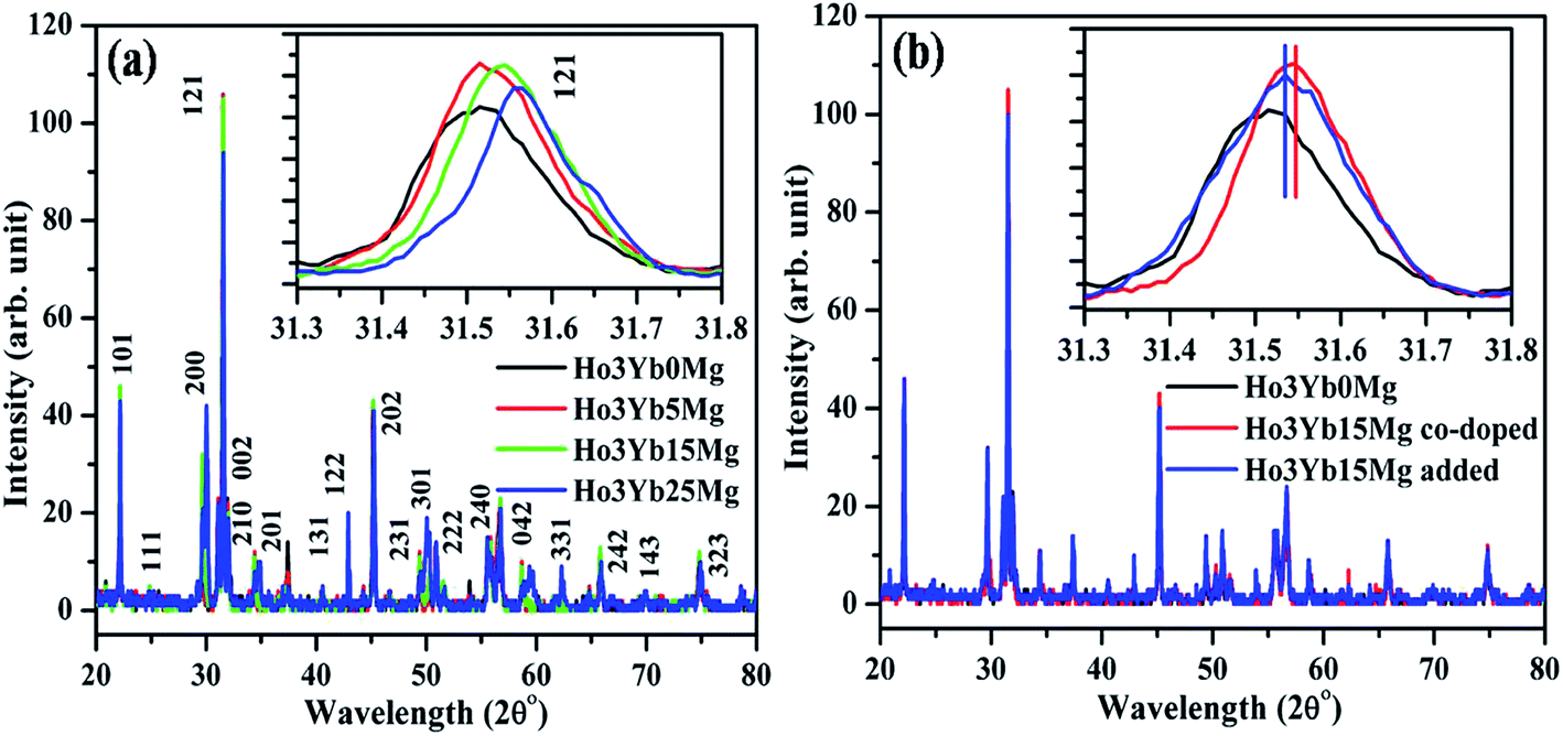 | ||
| Fig. 1 (a) XRD patterns of the Ho3+/Yb3+ co-doped and its Mg2+ co-doped CaZrO3 phosphor samples (annealed at 1300 °C/5 h). The inset figure shows the variation in FWHM and a shift of the most intense XRD peaks in the two cases. (b) XRD patterns of the Ho3+/Yb3+ co-doped; its Mg2+ co-doped and added CaZrO3 phosphors. The insets show the variation in FWHM of the XRD peaks in the three cases. | ||
When Mg2+ is co-doped in the Ho3+/Yb3+ co-doped CaZrO3 phosphor the peaks of the XRD patterns are shifted towards higher angle side. As the concentration of Mg2+ increases the XRD peaks are regularly shifted towards higher angle side. Since Mg2+ has smaller ionic radii (72 pm) than Ca2+ ion (100 pm), therefore, it substitutes Ca2+ from the CaZrO3 host. This causes a shift in the XRD peaks towards higher angle side. As a result, Mg2+ ion creates shrinkage in the lattice parameters and improves the crystallinity.5 It is clear from the figure that all the samples are highly crystalline. On the other hand, when Mg2+ is added (15 mol%) above the stoichiometric ratio the XRD peaks are slightly shifted towards lower angle side.34 It may be due to the fact that some of the Mg2+ ions may occupy at the substitutional sites and remaining the interstitial sites in the CaZrO3 host (see Fig. 1(b)).
The Voigt function is a single line profile method used to analyze the crystallite size and the lattice strain. The crystallite size ‘D’ and the lattice strain ‘e’ have been calculated from the integral breadths of the broadened profile of the XRD peaks. The Cauchy and Gaussian components of the XRD peak are obtained from the ratio of full width at half maximum (FWHM) intensity (2ω) and the integral breadths (β). In a single line analysis, the ‘D’ and ‘e’ are related to the Cauchy (βc) and Gaussian (βG) widths of the XRD peak at Bragg angle θ;
D = kλ/βc![[thin space (1/6-em)]](https://www.rsc.org/images/entities/char_2009.gif) cosθ cosθ
| (i) |
|
e = βG/4tanθ
| (ii) |
The constituent Cauchy and Gaussian components are given as
| βc = (a0 + a1ψ + a2ψ2)β |
| βG = (b0 + b1/2(ψ − 2/π)1/2 + b1ψ + b2ψ2)β |
| a0 = 2.0207, a1 = −0.4803 and a2 = −1.7756 |
| b0 = 0.6420, b1/2 = 1.4187, b1 = −2.2043 and b2 = 1.8706 |
The crystallite size thus calculated are found to be 78, 82, 110 and 264 nm for 0, 5, 15 and 25 mol% concentration of Mg2+ ions, respectively. However, on addition of Mg2+ ions (15 mol%) above stoichiometric ratio the crystallite size is found to be 88 nm, which is comparatively less than the crystallite size obtained in doped condition (110 nm). It reveals that doping the ions (15 mol%) improves crystallinity in the phosphor compared to adding the ions.
The lattice strains have been also calculated for doping the 0, 5, 15 and 25 mol% concentration of Mg2+ and they are found to be 0.51, 0.48, 0.46 and 0.45, respectively.31 However, in the case of Mg2+ addition (15 mol%) the lattice strain is increased to 0.57. This suggests that the crystal strain reduces regularly on doping whereas addition of the ions creates strain in the phosphor. Thus, an ordered crystal structure is obtained for doping the ions.
We have also analyzed the XRD data for Rietveld refinement. The observed XRD profile matches well with the theoretical profile and their intensity counts are also close to each other. The Rietveld refinement is good if the goodness of fit (GOF) factor is achieved ∼1. In our case, we have obtained the GOF value to 0.64 after refining the XRD data.18 The Rietveld refinement of XRD data for the annealed Ho3+/Yb3+/Mg2+ co-doped CaZrO3 phosphor is given in Fig. 2.
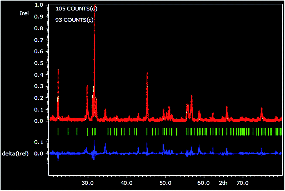 | ||
| Fig. 2 Rietveld refinement of XRD data of the Ho3+/Yb3+/Mg2+ co-doped CaZrO3 phosphor sample (annealed at 1300 °C/5 h). | ||
The SEM micrographs of the Ho3+/Yb3+ co-doped (annealed at 1300 °C/5 h) and its Mg2+ co-doped CaZrO3 phosphor samples are shown in Fig. 3(a) and (b). In both the cases, the shape of the particle is almost spherical and agglomerated with each other in different orientations. The particle size is found to be increased on Mg2+ doping (see Fig. 3(b)).5,19
 | ||
| Fig. 3 SEM micrographs of the Ho3+/Yb3+ co-doped CaZrO3 phosphor samples in absence (a) and presence (b) of Mg2+ ions. | ||
3.2 Optical measurements
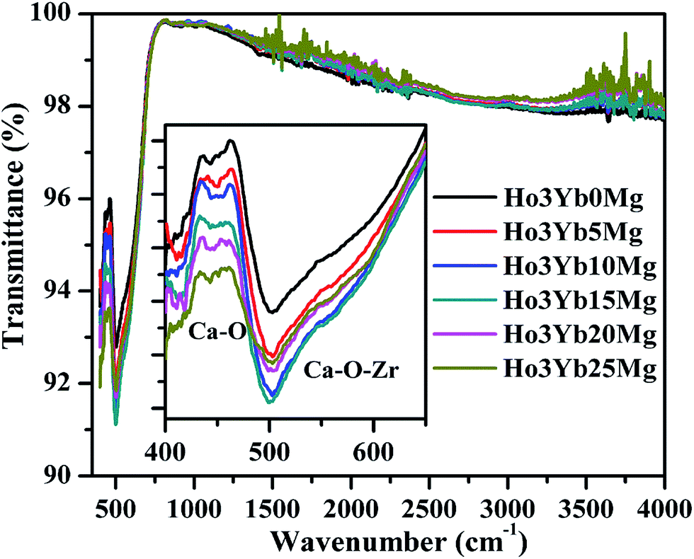 | ||
| Fig. 4 FTIR spectra of the Ho3+/Yb3+ co-doped CaZrO3 phosphor (annealed at 1300 °C/5 h) in absence and presence of Mg2+ ions. | ||
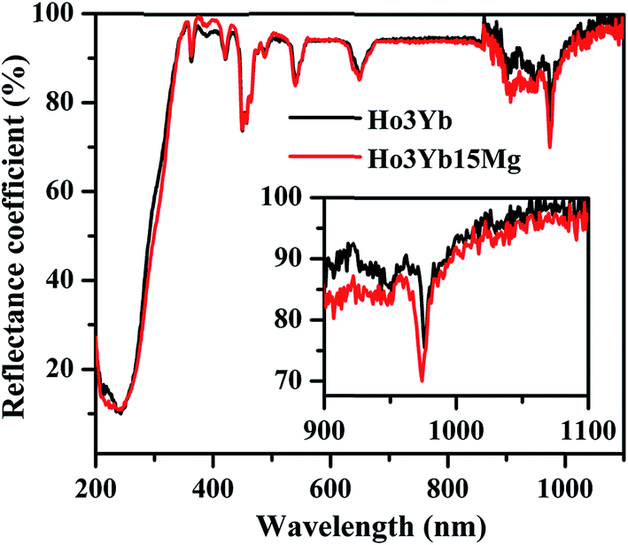 | ||
| Fig. 5 UV-vis-NIR diffuse reflectance spectra of the Ho3+/Yb3+ co-doped CaZrO3 phosphor in absence and presence of Mg2+ ions. | ||
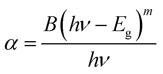
We have also calculated the optical band gap for the Ho3+/Yb3+ co-doped CaZrO3 phosphor in absence and presence of Mg2+ ions using Wood and Tauc formula39 as given below
 | ||
| Fig. 6 The (αhν)2 versus hν plots for the Ho3+/Yb3+ co-doped phosphor in absence and presence of Mg2+ ions. | ||
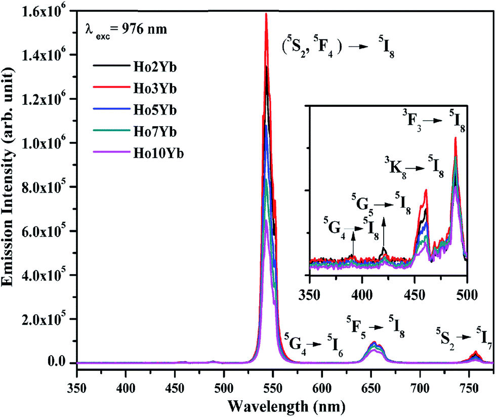 | ||
| Fig. 7 UC emission spectra of the Ho3+/Yb3+ co-doped CaZrO3 phosphors for different concentration of Yb3+ ions on excitation with 976 nm. The inset figure shows emissions in the UV and blue regions. | ||
This is due to the fact that at higher concentrations the inter-nuclear separation between the ions becomes smaller than the critical separation and the excitation energy migrates to the optical quenching centers. This causes a poor emission intensity of the co-doped phosphor. The peaks seen in the emission spectra matches very well for different concentration of Yb3+ ions. The inset figure shows a zoomed region of emissions in the UV and blue regions. They also show a variation in the emission intensity for different concentrations.
It well known that the Ho3+ ion contains large number of meta-stable states and can be easily excited for intense fluorescence. The Yb3+ ions can transfer their excitation energy to the different levels of Ho3+ ions and can enhance the emission intensity several times. Since the Yb3+ ion has very high absorption cross section for 976 nm, therefore, it efficiently absorbs 976 nm photons, and promoted to its excited (2F5/2) state. These excited ions transfer their excitation energy to the Ho3+ ions in the ground state (5I8). As a result, the Ho3+ ions are excited to 5I6 state through ground state absorption (GSA). These ions in 5I6 state absorb other 976 nm photons and are promoted to 5F4/5S2 excited states through excited state absorption (ESA). The ions in this state emit photons of 543 and 754 nm radiations via 5F4/5S2 → 5I8 and 5F4/5S2 → 5I7 transitions, respectively. Furthermore, some ions in this state relax non-radiatively to 5F5 state. The ions in 5F5 state give a transition to ground state with emission of a photon of 654 nm radiation. The ions in the 5F4 state further absorb other photons through excited state absorption (ESA) and are promoted to 3K7 excited state via energy transfer upconversion (ETU), which populates low lying 5G4, 5G5, 3K8 and 5F3 states. Finally, the ions in these states relax radiatively to emit different photons in UV and visible regions.11–16,19,20,38 The excitation processes involved in these emissions can be easily understood by schematic energy level diagram. The schematic energy level diagram for Ho3+ and Yb3+ ions are shown in Fig. 8 representing the involvement of different mechanisms such as GSA, ESA, ETU and CET in the excitation processes.
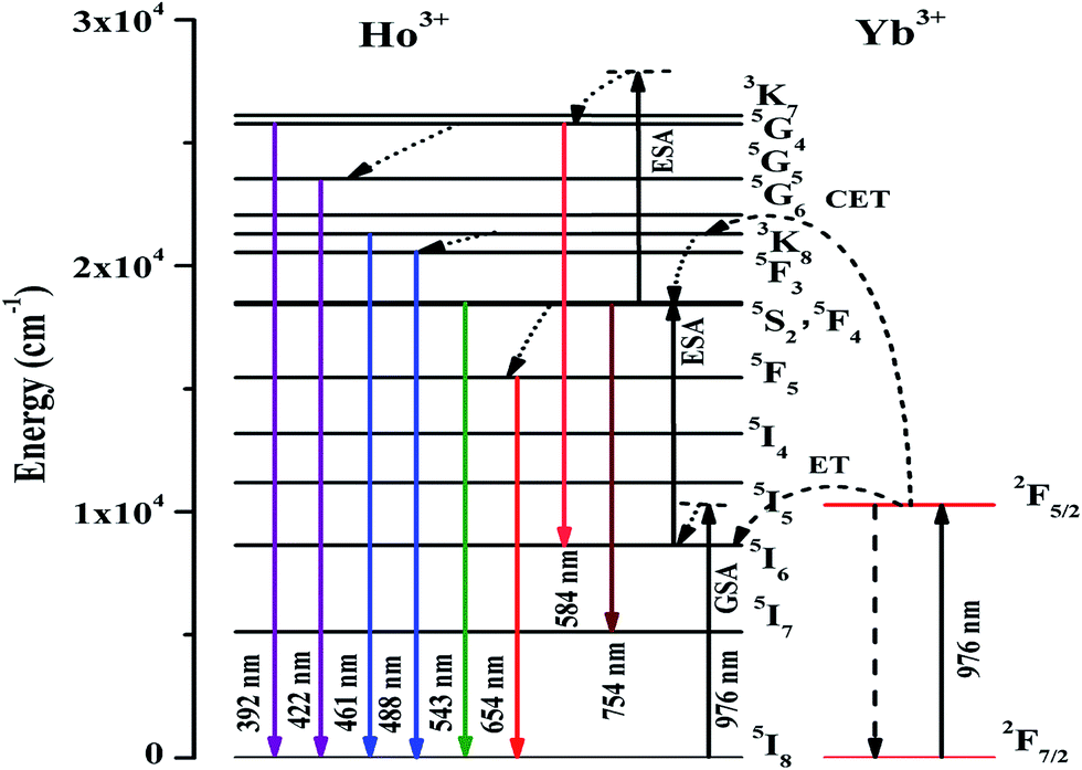 | ||
| Fig. 8 Schematic energy level diagram of Ho3+ and Yb3+ ions for different transitions on excitation with 976 nm. | ||
In order to confirm the number of photons involved in the upconversion emission, the power dependent measurements of the Ho3+/Yb3+ co-doped CaZrO3 phosphor on excitation with 976 nm have been carried out. The UC emission intensity of the Ho3+/Yb3+ co-doped CaZrO3 phosphor has been recorded at different pump power of 976 nm diode laser for 543 and 654 nm transitions. The dual logarithmic plot of input pump power versus emission intensity is found to be linear for these transitions, which is shown in Fig. 9. The values of the slopes (n) for these transitions have been calculated and are found to be 2.27 and 1.99.19,20 This confirms that the 5F4/5S2 and 5F5 levels of Ho3+ ions are populated by absorption of two NIR photons.
 | ||
| Fig. 9 Dual logarithmic plots for input pump power versus emission intensity of the Ho3+/Yb3+ co-doped CaZrO3 phosphor sample on excitation with 976 nm. | ||
The emission spectra of the Mg2+ activated Ho3+/Yb3+ co-doped CaZrO3 phosphor for different concentration of Mg2+ ions (i.e. 0, 5, 10, 15, 20 and 25 mol%) recorded in the range of 350–775 nm on excitation with 976 nm radiation are shown in Fig. 10. The emission intensity varies with the increase in the concentration of Mg2+ ions. Initially, the emission intensity increases from 5 to 15 mol% concentration of Mg2+ ions and it is optimum for 15 mol%. Finally, the emission intensity starts decreasing after this concentration due to concentration quenching. As is clear from the figure that the emission intensity of the Ho3+/Yb3+ co-doped CaZrO3 phosphor is found to be enhanced up to 4 times in presence of Mg2+ ions. As discussed earlier, the Mg2+ ion has smaller ionic radii than Ca2+ ion, it substitutes Ca2+ ion from the host and creates shrinkage in the lattice dimension; thereby increases the crystallinity. However, the effect of Mg2+ ion is reported in Ho3+/Yb3+ co-doped CaMoO4 phosphor by Dey et al. These workers have also found an enhancement in the emission intensity due to substitution and crystallinity of the host.5 Furthermore, Mg2+ doping results a relaxation in the crystal strain and the crystal strain relaxes with the increase in the concentration (i.e. 0.51, 0.48, 0.46 and 0.45 for 0, 5, 15 and 25 mol%). Interestingly, as the crystal strain decreases; the crystal defects present in the phosphor decreases simultaneously. As a result, the more ordered crystal structure is formed, which increases the emission intensity. The rate of decrease in the crystal strain is smaller for higher concentration. After 15 mol% of Mg2+ ions, the variation in the crystal strain seems to be saturated (i.e. from 0.46 to 0.45), however, the largest emission intensity is observed for 15 mol% concentration of Mg2+ ions. Therefore, the relaxation in the crystal strain is a consequence of large enhancement in the emission intensity.18
 | ||
| Fig. 10 Emission spectra of the Ho3+/Yb3+ co-doped CaZrO3 phosphor in absence and presence of different concentration of Mg2+ ions on excitation with 976 nm radiation. The insets are emissions in the UV and blue regions and an image of the sample. | ||
It is worth noting that the doping of Mg2+ does not influence the positions of emission peaks. A highly intense monochromatic green UC emission could become possible alongwith other emissions in the UV, blue, red and NIR regions. However, the peaks which are not present in the Ho3+/Yb3+ co-doped CaZrO3 phosphor are obtained with a larger intensity via Mg2+ doping (see inset figure). An additional peak centered at 392 nm is also observed in UV region due to 5G4 → 5I8 transition of Ho3+ ion, which arises mainly due to an increase in the crystallinity of the CaZrO3 phosphor. The color emitted by the sample is also shown as inset image in the figure. An enhancement in UC green emission intensity has been observed up to 4 times for Ho3+/Yb3+ co-doped CaZrO3 phosphor via Mg2+ doping. Therefore, the Ho3+/Yb3+/Mg2+ co-doped CaZrO3 phosphor can be suitable candidate for monochromatic green light and optical devices.
On the other hand, when the optimized concentration (15 mol%) of Mg2+ ions are added above the stoichiometric ratio in the Ho3+/Yb3+ co-doped CaZrO3 phosphor, the emission intensity is also found to be enhanced up to 1.35 times. However, this enhancement in the emission intensity is much smaller than that of doping the Mg2+ ions (15 mol%). It is expected that when the Mg2+ ions are added above the stoichiometric ratio, some of the Mg2+ ions occupy at the substitutional sites whereas remaining the interstitial sites.34 As a result, in this case a small shift in the XRD peaks is observed towards the lower angle side, which reflects an expansion in the lattice dimension. However, the crystal strain (0.57) is also higher in this case than the case of doping the Mg2+ ions (0.46), which is responsible for smaller emission intensity. A comparison of the emission intensity for 15 mol% doping and addition of Mg2+ ions in the Ho3+/Yb3+ co-doped CaZrO3 phosphors on excitation with 976 nm radiation is shown in Fig. 11. The inset in the figure is the emissions in the UV and blue regions of the sample. The ratio of the emission intensity of Mg2+ doping to its addition is almost 3:1 and it is due to change in crystallinity.34 Therefore; one can get larger emission intensity for doping the ions.
 | ||
| Fig. 11 Comparison of the emission intensity for 15 mol% doping and addition of Mg2+ ions in the Ho3+/Yb3+ co-doped CaZrO3 phosphors on excitation with 976 nm radiation. The inset shows emissions in the UV and blue regions of the sample. | ||
The visual perception realized by human eye is the only way to confirm the color emitted by the phosphor sample. However, it can be also expressed mathematically in two coordinate systems i.e. commission internationale de l'e'clairage (CIE) coordinates. The color emitted by a phosphor varies slightly with the concentration.18,19 The CIE plots for different concentration of Mg2+ in the Ho3+/Yb3+ co-doped CaZrO3 phosphors are shown in Fig. 12.
 | ||
| Fig. 12 CIE diagram for different concentration of Mg2+ doping in the Ho3+/Yb3+ co-doped CaZrO3 phosphors. | ||
The CIE coordinates for different concentration of Mg2+ in the Ho3+/Yb3+ co-doped CaZrO3 phosphors have been calculated and they are summarized in Table 1. The table shows the color coordinates have a small variation for Mg2+ doping and the Ho3+/Yb3+ co-doped CaZrO3 phosphors emit intense green emission in absence as well as presence of Mg2+ ions.
| Concentration of Mg2+ (mol%) | CIE coordinates (x, y) |
|---|---|
| 0 | (0.28, 0.70) |
| 5 | (0.28, 0.70) |
| 10 | (0.28, 0.70) |
| 15 | (0.28, 0.70) |
| 20 | (0.28, 0.71) |
| 25 | (0.28, 0.71) |
3.3 Lifetime measurements
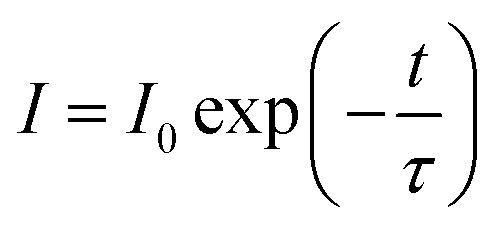
The lifetime measurements have been carried out in order to understand the factor responsible for an enhancement in the emission intensity via Mg2+ doping. The lifetime (τ) of the 5F4/5S2 level of Ho3+ ion is recorded on excitation with 976 nm radiation in absence and presence of Mg2+ ions and the decay curves thus obtained are shown in Fig. 13. The decay curves fit well with the single exponential profile with the following equation:where, I0 and I are the intensity at time 0 and t s, respectively and τ is the lifetime, which give lifetime of the 5F4/5S2 level of Ho3+ ion.
 | ||
| Fig. 13 Decay curves of 5F4/5S2 level of the Ho3+/Yb3+ co-doped CaZrO3 phosphor in absence (a) and presence of Mg2+ ions (b–f) on excitation with 976 nm radiation. | ||
The lifetimes thus obtained are given in the figure. It is found that the lifetime of the 5F4/5S2 level increases monotonically with Mg2+ doping up to 15 mol% and then decreases for higher concentration of Mg2+ ion.19 The enhancement in the lifetime of the 5F4/5S2 level of Ho3+ ion is a clear evidence for an increase in the emission intensity. The increase in the lifetime of the emitting levels may be due to an improvement in crystallinity, which enhances the rapid excitation, energy transfer and radiative transitions significantly.
4. Conclusions
The Ho3+/Yb3+ co-doped CaZrO3 phosphor has been synthesized through solid state reaction method. The XRD measurement confirms a shift in the peak position due to Mg2+ doping in the CaZrO3 phosphor. The scanning electron micrographs show an increase in the particle size via Mg2+ doping. The Ho3+/Yb3+ co-doped CaZrO3 phosphor gives intense monochromatic green upconversion emission at 543 nm due to 5F4/5S2 → 5I8 transition alongwith weak UV, blue, red and NIR emissions on excitation with 976 nm. The emission intensity is optimized with different concentrations of Yb3+ and it is optimum at 3 mol%. When Mg2+ ions are co-doped in the Ho3+/Yb3+ co-doped CaZrO3 phosphor the emission intensity of green band is enhanced up to 4 times. This enhancement is due to shrinkage in the lattice parameters and an increase in the crystallinity. The lifetime of 5F4/5S2 level increases in presence of Mg2+ ions, which favors to give large emission intensity. The intensity of other bands is negligibly small compared to green band. Therefore, the Ho3+/Yb3+/Mg2+ co-doped CaZrO3 phosphor can be a suitable candidate for monochromatic green light and optical devices.Acknowledgements
One of the author, Ms Akanksha Maurya, is thankful to ‘UGC’ – ‘India’ for providing research fellowship. The authors acknowledge to Prof. O. N. Srivastava, Department of Physics, Banaras Hindu University, Varanasi for XRD and SEM measurement facilities. We also acknowledge to ‘DST’ – ‘India’ for providing the financial assistance (Grant No. SR/S2/LOP-023/2012).References
- M. F. Joubert, Opt. Mater., 1999, 11, 181–203 CrossRef CAS.
- R. Scheps, Prog. Quantum Electron., 1996, 20, 271–358 CrossRef CAS.
- S. Fischer, J. C. Goldschmidt, P. Loper, G. H. Bauer, R. Bruggemann, K. Kramer, D. Biner, M. Hermle and S. W. Glunz, J. Appl. Phys., 2010, 108, 044912 CrossRef.
- R. V. Yadav, R. S. Yadav, A. Bahadur and S. B. Rai, RSC Adv., 2016, 6, 9049–9056 RSC.
- R. Dey, A. Kumari, A. K. Soni and V. K. Rai, Sens. Actuators, B, 2015, 210, 581–588 CrossRef CAS.
- M. K. Mahata, T. Koppe, T. Mondal, C. Brüsewitz, K. Kumar, V. K. Rai, H. Hofsäss and U. Vetter, Phys. Chem. Chem. Phys., 2015, 17, 20741 RSC.
- R. S. Yadav, R. K. Verma, A. Bahadur and S. B. Rai, Spectrochim. Acta, Part A, 2015, 142, 324–330 CrossRef CAS PubMed.
- G. Shan, R. Weissleder and S. A. Hilderbrand, Theranostics, 2013, 3(4), 267–274 CrossRef CAS PubMed.
- G. Ajithkumar, B. Yoo, D. E. Goral, P. J. Hornsby, A. L. Lin, U. Ladiwala, V. P. Dravid and D. K. Sardar, J. Mater. Chem. B, 2013, 1, 1561–1572 RSC.
- F. Auzel, Chem. Rev., 2004, 104(1), 139–174 CrossRef CAS PubMed.
- I. Etchart, I. Hernandez, A. Huignard, M. Berard, W. P. Gillin, R. J. Curry and A. K. Cheetham, J. Mater. Chem., 2011, 21, 1387–1394 RSC.
- A. Pandey, V. K. Rai, R. Dey and K. Kumar, Mater. Chem. Phys., 2013, 139, 483–488 CrossRef CAS.
- R. Wang, W. Zhang, Y. Xu and L. Xing, Opt. Laser Technol., 2014, 58, 52–55 CrossRef CAS.
- C. S. Lim, J. Phys. Chem. Solids, 2015, 78, 65–69 CrossRef.
- A. Pandey and V. K. Rai, Dalton Trans., 2013, 42, 11005–11011 RSC.
- X. Li, J. Zhu, Z. Man, Y. Ao and H. Chen, Sci. Rep., 2014, 4, 4446 Search PubMed.
- T. V. Gavrilović, D. J. Jovanović, L. V. Trandafilović and M. D. Dramićanin, Opt. Mater., 2015, 45, 76–81 CrossRef.
- R. S. Yadav, R. V. Yadav, A. Bahadur, T. P. Yadav and S. B. Rai, Mater. Res. Express, 2016, 3, 036201 CrossRef.
- R. S. Yadav, R. V. Yadav, A. Bahadur and S. B. Rai, RSC Adv., 2016, 6, 51768–51776 RSC.
- R. V. Yadav, S. K. Singh and S. B. Rai, RSC Adv., 2015, 5, 26321–26327 RSC.
- M. T. Lucchini, V. Babin, P. Bohacek, S. Gundacker, K. Kamada, M. Nikl, A. Petrosyan, A. Yoshikawa and E. Auffray, Nucl. Instrum. Methods Phys. Res., 2016, 816, 176–183 CrossRef CAS.
- S. Cui, Z. Tian, S. Pu and Y. Dai, RSC Adv., 2016, 6, 19957–19963 RSC.
- C. E. Cui, H. Liu, P. Huang and L. Wang, J. Lumin., 2014, 149, 196–199 CrossRef CAS.
- B. Wang, H. Lin, F. Huang, J. Xu, H. Chen, Z. Lin and Y. Wang, Chem. Mater., 2016, 28, 3515–3524 CrossRef CAS.
- A. S. H. Hameed, C. Karthikeyan, S. Sasikumar, V. S. Kumar, S. Kumaresan and G. Ravi, J. Mater. Chem. B, 2013, 1, 5950–5962 RSC.
- M. K. Chong, K. Pita and C. H. Kam, Appl. Phys. A, 2004, 79, 433–437 CrossRef CAS.
- H. Komatsu, T. Miki, D. Citterio, T. Kubota, Y. Shindo, Y. Kitamura, K. Oka and K. Suzuki, J. Am. Chem. Soc., 2005, 127(31), 10798–10799 CrossRef CAS PubMed.
- R. S. Yadav, R. K. Verma and S. B. Rai, J. Phys. D: Appl. Phys., 2013, 46, 275101 CrossRef.
- R. S. Yadav, R. K. Verma, A. Bahadur and S. B. Rai, Spectrochim. Acta, Part A, 2015, 137, 357–362 CrossRef CAS PubMed.
- J. Huang, L. Zhou, Y. Lan, F. Gong, Q. Li and J. Sun, Cent. Eur. J. Phys., 2011, 9(4), 975–979 CAS.
- N. Tiwari, R. K. Kuraria and S. R. Kuraria, Optik, 2015, 126, 3488–3491 CrossRef CAS.
- S. K. Gupta, P. S. Ghosh, N. Pathak and R. Tewari, RSC Adv., 2015, 5, 56526–56533 RSC.
- T. Orihashi, T. Nakamura and S. Adachi, RSC Adv., 2016, 6, 66130–66139 RSC.
- J. H. Chung, S. Y. Lee, K. B. Shim, S. Y. Kweon, S. C. Ur and J. H. Ryu, Appl. Phys. A, 2012, 108, 369–373 CrossRef CAS.
- T. H. D. Keijser, J. I. Langford, E. J. Mittemeijer and A. B. P. Vogels, J. Appl. Crystallogr., 1982, 15, 308–314 CrossRef.
- A. K. Parchur and R. S. Ningthoujam, Dalton Trans., 2011, 40, 7590–7594 RSC.
- L. V. Rij, L. Winnubst, L. Jun and J. Schoonman, J. Mater. Chem., 2000, 10, 2515–2521 RSC.
- J. Yu, Y. Yang, R. Fan, D. Liu, L. Wei, S. Chen, L. Li, B. Yang and W. Cao, Inorg. Chem., 2014, 53, 8045–8053 CrossRef CAS PubMed.
- D. L. Wood and J. Tauc, Phys. Rev. B: Solid State, 1972, 5, 3144–3151 CrossRef.
- P. Stoch, J. Szczerba, J. Lis, D. Madej and Z. Pedzich, J. Eur. Ceram. Soc., 2012, 32, 665–670 CrossRef CAS.
- A. Kumari, A. Pandey, R. Dey and V. K. Rai, RSC Adv., 2014, 4, 21844 RSC.
- S. Som, A. K. Kunti, V. Kumar, V. Kumar, S. Dutta, M. Chowdhury, S. K. Sharma, J. J. Terblans and H. C. Swart, J. Appl. Phys., 2014, 115, 193101 CrossRef.
| This journal is © The Royal Society of Chemistry 2016 |