DOI:
10.1039/C6RA23693F
(Paper)
RSC Adv., 2016,
6, 108393-108403
Corundum type indium oxide nanostructures: ambient pressure synthesis from InOOH, and optical and photocatalytic properties†
Received
23rd September 2016
, Accepted 7th November 2016
First published on 7th November 2016
Abstract
A simple, cost effective, surfactant free and scalable synthesis of rhombohedral In2O3 (rh-In2O3) nanostructures with controllable size and shape has been developed under ambient pressure by thermal dehydration of InOOH nanostructures. The InOOH nanostructures have been prepared by solvothermal reaction between indium nitrate hydrate with tetramethylammonium hydroxide (TMAH) in anhydrous methanol at 140 °C without any surfactant. The structure and morphology of the nanostructures have been characterized in detail by X-ray powder diffraction (XRD), Raman spectroscopy, and high-resolution transmission electron microscopy (HRTEM). The studies reveal that highly crystalline nanostructures of InOOH and rh-In2O3 with rice-grain type morphology are formed. The optical properties of the InOOH and rh-In2O3 nanostructures have been explored by UV-visible diffuse reflectance spectroscopy (UV-DRS) and room-temperature photoluminescence (PL) studies. The direct band gap of as-synthesized InOOH and rh-In2O3 nanostructures was estimated to be 3.75 and 2.95 eV, respectively, from the diffuse reflectance absorbance spectra. Both InOOH and rh-In2O3 nanostructures show intense blue emission under UV excitation which is attributed to the presence of oxygen vacancies. The thermal stability of the rh-In2O3 phase has been studied by differential scanning calorimetry (DSC), differential thermal analysis (DTA) and dilatometry of the as prepared sample. The potential of InOOH and rh-In2O3 nanostructures as photocatalytic materials for hydrogen generation from water/methanol (2![[thin space (1/6-em)]](https://www.rsc.org/images/entities/char_2009.gif) :1) mixtures under UV/vis irradiation has also been evaluated for the first time.
:1) mixtures under UV/vis irradiation has also been evaluated for the first time.
1. Introduction
Nanostructures of transparent conducting oxide (TCO) have attracted extensive attention in fundamental as well as applied research because of their transparency, range of crystal structures and technologically relevant properties.1–6 In2O3 is one of the most widely studied TCO mainly because of its transparency (Eg ≈ 3.75 eV), electrical conductivity (native n-type semiconductor), and high charge-carrier mobility.7 Because of these characteristics, the nanostructures of In2O3 have been widely investigated as optoelectronic devices, catalysts, sensors, and in solar cells.7–13 In2O3 is a polymorphic material and most of the literature studies are conducted on c-In2O3 which is the most stable phase thermodynamically and can easily be obtained at ambient pressure. It has a body-centered cubic bixbyite-type crystal structure (bcc-In2O3, I3a, No. 206) and consists of two types of In3+ ions (they are surrounded by oxygen in the octahedral and trigonal prismatic coordination alternatively).14 The other polymorph of In2O3 is metastable rh-In2O3 which is usually described as a high temperature–high pressure phase and transforms irreversibly into c-In2O3 upon heating.15 It has rhombohedral structure (rh-In2O3, R![[3 with combining macron]](https://www.rsc.org/images/entities/char_0033_0304.gif) c, No. 167) consisting of hexagonal close packed oxygen ions with In3+ ions filling up two-thirds of the six-coordinate C3v sites. The potential use of In2O3 in the form of low dimensional nanostructures has triggered interest in studying its metastable phases. Further, metastable rh-In2O3 phase has several interesting physical properties, such as more stable conductivity than its cubic counterpart and structural similarity with metal doped oxides like indium tin oxide (ITO) and it will be of great interest to study it for fundamental as well as applied science.
c, No. 167) consisting of hexagonal close packed oxygen ions with In3+ ions filling up two-thirds of the six-coordinate C3v sites. The potential use of In2O3 in the form of low dimensional nanostructures has triggered interest in studying its metastable phases. Further, metastable rh-In2O3 phase has several interesting physical properties, such as more stable conductivity than its cubic counterpart and structural similarity with metal doped oxides like indium tin oxide (ITO) and it will be of great interest to study it for fundamental as well as applied science.
The reversal of the phase stability in nanocrystalline materials has been associated with large surface to volume ratios in reduced dimensions. The high surface area of nanostructures can lead to the spontaneous stabilization of metastable phases, owing to the surface energy and/or surface stress contributions, both of which are dependent on size.16 In recent past there have been several reports on the synthesis of rh-In2O3 nanostructures under ambient pressure.17–27 The same rhombohedral structure can also be synthesized by the application of combined high-pressure and high-temperature, but this method is economically less efficient than ambient pressure chemical synthesis.28 The stabilization of rh-In2O3 under atmospheric pressure has been achieved by annealing of crystalline In(OH)3,18 or InOOH nano structures17,19–25 which were mainly prepared by the solution-based surfactant-assisted method. The literature reports on synthesis of rh-In2O3 reveal that the solvothermal method has been extensively used to prepare the intermediate InOOH and rh-In2O3 under mild conditions. However, the solvothermal methods employed use various organic/toxic solvents such as glycerol,25a ethylene glycol,25b triethylene glycol,26 dimethylformamide,19a diethylenetriamine25c etc. and various surfactants which might bring about great difficulties for the large-scale production and environmental protection. The use of surfactants to control the growth of nanoparticles may also influence the toxicity of the nanoparticles.29 Thus, the development of simple, mild surfactant free and economical synthetic pathways for the large-scale synthesis of well-controlled rh-In2O3 nanostructures under ambient conditions still remains a challenge.
In this study, we report a low temperature simple synthetic approach for controlled synthesis of corundum type indium oxide (rh-In2O3) nanostructures by dehydration of InOOH nanostructures at ambient pressure at 350 °C. Orthorhombic InOOH nanostructures have been prepared through a simple solvothermal reaction between indium nitrate hydrate and tetramethylammonium hydroxide at 140 °C without using any surfactant. The prepared nanostructures have been characterized in detail for their structure and morphology and their optical and photocatalytic properties for hydrogen generation via water splitting under UV-vis irradiation have been studied for the first time. In recent years, semiconductor-based photocatalytic solar-energy conversion by water splitting and CO2 reduction has been considered very promising to address the energy and environmental challenges.30 As wide band gap p-block semiconductors, both InOOH31–34 and rh-In2O3 (ref. 27, 35 and 36) nanostructures have been extensively used as photocatalysts for the degradation of organic pollutants, but to the best of our knowledge there is no report of these semiconductors being used as photocatalyst for hydrogen generation via water splitting.
2. Experimental details
Indium nitrate hydrate, In(NO3)3·xH2O (99.99%) and TMAH (25% solution in water) were procured from Aldrich. The exact water content in precursor indium nitrate was determined using thermogravimetric studies and value of x was found to be 2.55. Methanol (99.5%, S. D. Fine Chemicals, India) was dried and distilled over magnesium methoxide prior to use.
2.1 Synthesis of InOOH
In(NO3)3·2.55H2O (1.01 g, 2.91 mmol) was dissolved in 25 ml anhydrous methanol in beaker and 0.796 g (8.74 mmol) of TMAH solution was added drop wise to it over a period of fifteen minutes while sonicating the reaction mixture in an ultrasonic bath. After the addition was complete, the contents of the beaker were transferred to a Teflon lined stainless steel autoclave. The autoclave was heated to a temperature of 140 °C at the rate of 3 °C min−1 and maintained at this temperature for 4 h. It was then cooled to room temperature naturally. The resulting white product was retrieved by centrifugation and washed with anhydrous methanol (3 × 5 ml) and then dried under vacuum at room temperature for 1 hour to get InOOH nanopowder.
2.2 Synthesis of rh-In2O3
InOOH nanopowder obtained by above mentioned procedure was loaded in a quartz boat and heated in a tube furnace at 350 °C (heating rate 3 °C min−1) for 4 h in air. Then it was cooled to room temperature naturally. Thermal dehydration of InOOH readily afforded rh-In2O3 nanostructures.
2.3 Characterization
The crystal phases of the as-prepared products were characterized using a Philips X'Pert pro powder X-ray diffractometer with Cu-Kα radiation (λ = 1.5418 Å) operating at 40 kV and 30 mA. A standard Si single crystal was used for instrumental correction and calibration. Morphological and structural analyses of the samples were carried out by TEM and HRTEM (FEI Field Emission Gun (FEG) TECNAI G2 F20 S-TWIN high resolution transmission electron microscopes; accelerating voltage 200 kV). The TEM samples were prepared by dispersing the samples in ethanol, and placing a drop of the dispersion on a Cu TEM grid covered with a holey carbon film, which was then dried. The diffuse reflectance UV-vis spectra were recorded with a JASCO spectrophotometer (model: V-530, Japan). BET surface areas were measured with a Thermoscientific Surfer instrument with nitrogen as the adsorbing gas. The pore-size distribution and pore volume were calculated from desorption branch by using the Barret–Joyner–Halenda (BJH) method. Raman spectral studies were carried out using a micro-Raman spectrometer (model: STR-300, SEKI Technotron, Japan). The differential thermal analysis was carried out using a Setaram Setsys thermogravimetry analyzer under argon. DSC thermograms of nanopowders were recorded in a heat flux differential scanning calorimeter (model: DSC 823e, Mettler-Toledo GmBH, Switzerland). Samples (∼20 mg) were taken in platinum pan with covering lid and thermograms were recorded in the temperature range 0–675 °C under flowing argon atmosphere (∼60 ml min−1) with a heating rate of 10 °C min−1. Linear thermal dilation of as-prepared samples was measured in an integrated thermo mechanical analyzer (Model: Setsys TMA 1600, M/s. Setaram Instrumentation, France). As-prepared powder samples were pressed in the form of green pellets (6 mm diameter and 2–3 mm height) using a uniaxial hydraulic press at a pressing load of 2.5 tonnes. These pellets were used for linear dilation measurement under flowing argon atmosphere (∼60 ml min−1) with near zero load condition (load = 1 g) in temperature range 40–900 °C. Uniform heating/cooling rate (10 °C min−1) was used for all the experiments. Measurements were made during both heating and cooling cycles. Sample dilation was evaluated from the measured dilation curves by subtracting the blank, which was run under identical experimental conditions. All steady-state luminescence and lifetime measurements were carried out using an Edinburgh Instruments' FLSP 920 system with a 450 W Xe lamp, nanosecond hydrogen flash lamp as the excitation sources. EPR spectra was recorded at 100 K using Bruker X-band spectrometer (EMX series; EMM-1843) using a standard rectangular cavity ER4119HS operating at 9.60 GHz with a 100 kHz modulation frequency. Diphenyl picrylhydrazyl (DPPH) radical was used as reference for the calibration of g factor.
2.4 Photocatalytic activity under UV/vis irradiation
The photocatalytic activity of the InOOH and rh-In2O3 nanostructures was evaluated at room temperature with water/methanol (2:1 v/v, total volume 15 ml) mixtures in the presence of 0.1 g catalyst in a closed rectangular quartz cell (10 × 2.1 × 2.1 cm3) equipped with sampling and evacuation ports. The reaction mixture was evacuated before irradiation to provide air free conditions as oxygen acts as an electron scavenger and, on photo-adsorption, blocks the active sites for the reaction. The water/methanol mixtures with suspended photocatalyst were then irradiated under a SAIC UV-visible lamp. The reaction products were analyzed every 2 h by using a gas chromatograph (Netel Michro-1100, India) equipped with a thermal conductivity detector (TCD); a molecular sieves column with argon as the carrier was employed in the isothermal temperature mode at 50 °C oven temperature. The details of the reaction assembly and emission spectrum of the UV-visible lamp are shown in Fig. S1, ESI.†
3. Results and discussion
3.1 Synthesis
The solvothermal reaction between In(NO3)3·2.55H2O and TMAH in anhydrous methanol (solution pH ∼ 9) at 140 °C for 4 h readily affords InOOH nanoparticles. To understand how the solvothermal reaction between In(NO3)3·2.55H2O and TMAH unfolds, the reaction intermediates after immediate mixing of reactants, 1 and 2 h of the reaction were isolated. XRD analyses of the solid intermediates obtained reveal that InOOH is already formed after 1 h of the solvothermal reaction. No peaks due to In(OH)3 could be detected in the XRD patterns of the intermediates (Fig. S2, ESI†). Thermal dehydration of InOOH nanoparticles at 350 °C for 4 h readily affords rh-In2O3 nanostructures. It was noted that the use of fresh anhydrous methanol is essential to obtain InOOH nanostructures. During the preparation of InOOH by solvothermal route, it was observed that if commercially available methanol (contains 0.05% water) is used for the solvothermal reaction then a mixture of InOOH and In(OH)3 is obtained which on annealing gives a mixture of rh-In2O3 and bcc-In2O3. In some cases direct preparation of rh-In2O3 nanopowders involve calcinations of In(OH)3 intermediates formed,18 but in our study, we noted that direct thermal dehydration of In(OH)3 at ambient pressure produces only c-In2O3. Highly crystalline In(OH)3 nanostructures were obtained by the solvothermal reaction between In(NO3)3·2.55H2O and excess TMAH (pH > 9) at 140 °C for 3 h and their thermal dehydration at 300 °C for 2 h readily affords bcc-In2O3 nanostructures. The Bragg reflections observed in the XRD pattern shown in Fig. S3a and S3b† confirm the formation of body-centred cubic In(OH)3 (JCPDS card no. 01-076-1463) and cubic bixbyite In2O3 (JCPDS no. 88-2160, space group Ia3), respectively. No XRD peaks representing other crystalline phases were detected, indicating that the final products exhibited excellent crystallinity and high purity. It is clear from the low resolution TEM micrographs shown in Fig. S3c and S3d† that the materials presents cubic morphology with edge of the cube varying between 50–80 nm for In(OH)3 nanocubes. It is clear from the above observations that the water content in the reaction medium as well as pH of the reaction medium influences the phase and morphology of the products obtained. Yan et al.37 have also observed similar behavior while synthesizing InOOH via a solvothermal route using indium nitrate and a binary solution of water and N,N-dimethylformamide.
3.2 Structure and morphology
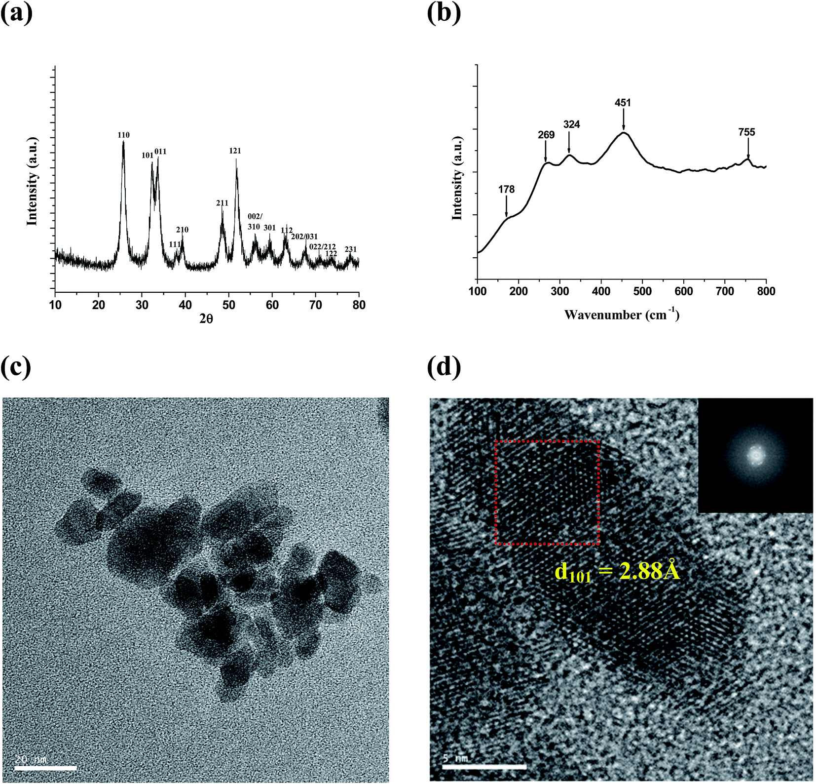
3.2.1 InOOH. Fig. 1a shows the XRD pattern of the white product prepared by the solvothermal reaction between In(NO3)3·2.55H2O and stoichiometric TMAH at 140 °C for 4 h. The reflection peaks can be indexed to orthorhombic InOOH (JCPDS card 71-2283). The peaks are broad indicating the nanocrystalline nature of the product. No characteristic peaks due to In(OH)3 are seen in the XRD pattern indicating the high purity of the product obtained. The major crystal planes are assigned to the diffraction peaks. The measured lattice parameters are found to be a = 5.252 Å, b = 4.578 Å and c = 3.254 Å and compare well with the literature values (JCPDS 71-2283, a = 5.260 Å, b = 4.560 Å, c = 3.270 Å). The phase purity of InOOH samples prepared has also been examined with Raman spectroscopy. InOOH has an orthorhombic crystal structure with space group symmetry P21nm containing two formula units per unit cell. The Raman spectrum of InOOH nanostructures (Fig. 1b) show bands at 178, 269, 324, 451, and 755 cm−1 consistent with literature reports.37,38
 |
| | Fig. 1 (a) XRD pattern, (b) room temperature Raman spectrum, (c) low resolution TEM image and (d) high resolution TEM image of InOOH nanostructures. Inset in (d) shows fast Fourier transform of the HR image. | |
Detailed information about the crystal structure and morphology of InOOH nanostructures has been studied by TEM. Fig. 1c shows the low magnification image of the InOOH nanostructures. Due to lack of any stabilizing surfactants, the particles are agglomerated but it is still possible to see some elongated grains with length varying between 8–20 nm and width varying between 4–8 nm. The high resolution (HR) TEM image of nanostructures (Fig. 1d) shows lattice spacing of 2.88 Å which corresponds to the (101) interplanar distance of orthorhombic InOOH. Fast Fourier transform of the HR image shown as inset in Fig. 1d clearly indicates the monocrystalline nature of the selected grain.
3.2.2 rh-In2O3. Fig. 2a shows the XRD patterns of the pale-yellow product obtained by annealing the InOOH precursor at 350 °C for 4 h. The reflection peaks in the XRD pattern can be indexed to rh-In2O3 (JCPDS card 22-0336) and no other impurities peaks are observed, indicating that the precursor InOOH is completely transformed into rh-In2O3 under the annealing conditions. The miller indices are indicated on each diffraction peak. The measured lattice parameters were found to be a = b = 5.485 Å and c = 14.53 Å, which are in good agreement with the reference (JCPDS card 22-0336, a = b = 5.487 Å, c = 14.510 Å). Room temperature Raman spectrum for the rh-In2O3 nanostructures is shown in Fig. 2b. Rhombohedral In2O3 structure belongs to space group Rc, D63d. On the basis of the group theory analysis, the optical modes of rh-In2O3 have the irreducible representation, as shown below:39
| Γopt = 2A1g + 5Eg + 2A1u + 2A2u + 3A2g + 4Eu |
where A1u, A2u, A2g, and Eu are infrared active or Raman inactive modes, and A1g and Eg are Raman active modes. The room temperature Raman spectrum of rh-In2O3 exhibits peaks at 166, 224, 274, 312, 386, 501 and 592 cm−1 in agreement with literature reports17,23,28 The peaks at 166 and 501 cm−1 are attributed to A1g modes while the Raman peaks at 224, 274, 312, 386 and 592 cm−1 are assigned to Eg modes. A weak and broad band observed at 450 cm−1 may be an overtone of band at 224 cm−1. Interestingly, the weak and broad peak observed at 312 cm−1 for the rh-In2O3 nanostructures prepared by us and reported by other authors also17,23 is not observed for the bulk rh-In2O3 sample prepared by high pressure and high temperature route.28 The reasons for such discrepancy can be related with the fact that nanoparticles prepared by chemical methods and samples prepared under extreme compression could be crystallographically slightly different.
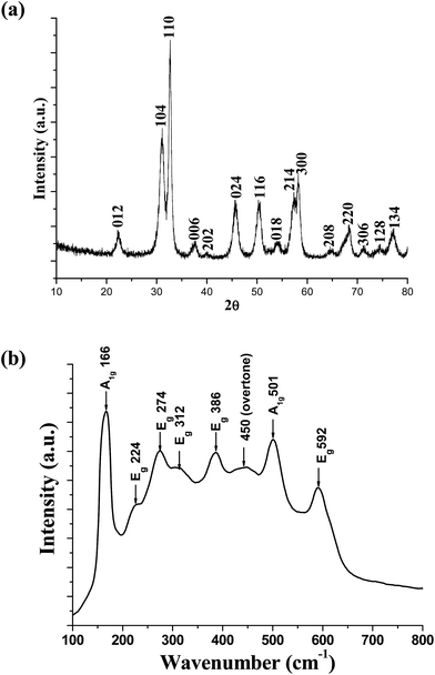 |
| | Fig. 2 (a) XRD pattern, and (b) room temperature Raman spectrum of rh-In2O3 nanostructures. | |
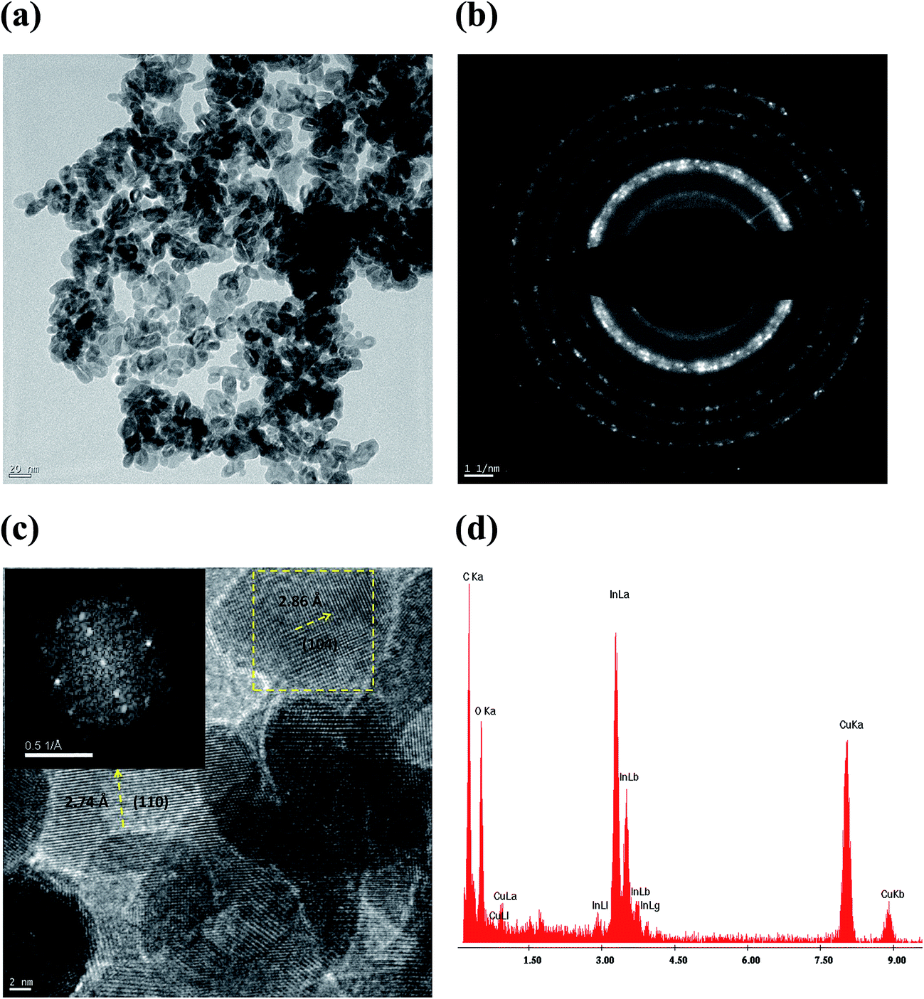
Fig. 3 shows TEM data for rh-In2O3 nanostructures. Fig. 3a shows low magnification image for the sample and it is clear from the image that the material presents rice grain type of morphology. Both the length and width of the nanoparticles are Gaussian distributed around 20 nm and 13 nm, respectively (scale bar is 20 nm), with standard deviations 5 nm for the length and 3 nm for the width. The minimum and maximum values observed for the length (width) are 10 nm (6 nm) and 30 nm (20 nm). The selected area diffraction (SAD) pattern of the nanopowder (Fig. 3b) features well-defined ring patterns indicating the polycrystalline nature of the sample. The measured interplanar distance (d-values) (from first ring to outer one for first 8 rings) are 3.968, 2.895, 2.739, 2.392, 1.994, 1.826, 1.604 and 1.394 Å, respectively, corresponding to the planes (012), (104), (110), (113), (024), (116), (214) and (220), consistent with the rhombohedral phase structure of In2O3. The HRTEM image of the nanostructures (Fig. 3c) shows well-defined lattice planes, indicating the single crystalline nature of the nanostructures. The lattice spacings of 2.74 and 2.86 Å observed in the HRTEM image correspond to (110) and (104) interplanar distances of rh-In2O3, respectively. Fast Fourier Transform (FFT) of the selected zone shown as inset in Fig. 3c confirms the monocrystalline structure of the selected nanograin. Fig. 3d shows a localized EDX spectrum of an individual In2O3 nanoparticle. The observed atom% values of In and O are well in agreement with the expected values for rh-In2O3 (expected atom%: In, 40; O, 60; observed: In, 44; O: 56% at) suggesting that the nanostructures are composed of In2O3.
 |
| | Fig. 3 TEM data for rh-In2O3 nanostructures. (a) Low resolution TEM micrograph. (b) SAD pattern. (c) High resolution TEM micrograph showing lattice fringes with FFT pattern shown as inset. (d) TEM-EDX spectrum. | |
3.3 Thermal analysis and phase stability of rh-In2O3 nanostructures
As mentioned earlier, there are several literature reports available in recent times on ambient pressure synthesis of metastable rh-In2O3, however, not many studies provide adequate information on stability aspects of rh-In2O3. In this report, we have investigated the stability of the rh-In2O3 phase using DSC, DTA and dilatometry.
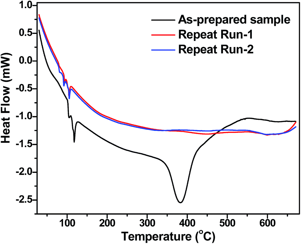
In the DSC thermogram one would expect an exothermic peak if metastable rh-In2O3 transforms to more stable c-In2O3 phase. DSC thermogram of as-prepared rh-In2O3 sample along with thermograms recorded on the same sample in repeated thermal cycling is shown in Fig. 4. The DSC thermogram shows two prominent endothermic peaks at 112.4 °C and 383 °C, respectively, along with a broad exothermic peak at ∼570 °C. However, during repeated thermal cycling only an endotherm around 90–100 °C is observed. The sample weight measured before and after DSC run indicated ∼2.8 wt% loss. The phase composition of the final material obtained after DSC experiment was verified by XRPD studies which confirmed that rh-In2O3 sample does not transform to cubic In2O3 (Fig. S4, ESI†). In the light of above observations, the observed DSC results are interpreted as follows: the endotherm at 112.4 °C has been assigned to the loss of adsorbed moisture on the surface of rh-In2O3 nanostructures, the endotherm seen at 383 °C is attributed to loss of residual surface functionalities (such as residual –OH groups) present on the surface of rh-In2O3 nanostructures. The presence of a broad exothermic peak at ∼570 °C may be due to heat of formation of water associated with the loss of surface –OH groups. Thermal cycling on the same sample (without exposure to ambient atmosphere) shows a weak endothermic peak between 100–110 °C. While we are not able to assign the exact reason for such an endotherm, one possibility is adsorption of moisture present in carrier gas (Ar) during DSC cooling cycle. Similar low temperature endotherms are observed for nanoparticles of different oxide materials studied in our lab (unpublished results). DTA is another thermo analytic technique which is used to study phase transitions in materials. Fig. S5† shows the data from DTA measurement on as prepared rh-In2O3 sample. An exothermic peak is seen at ∼735 °C clearly indicating the phase transition from rhombohedral to cubic phase of In2O3.
 |
| | Fig. 4 DSC thermograms recorded on as-prepared rh-In2O3 sample and thermally cycled samples. | |
3.3.1 Thermal expansion studies. The crystal structure of two polymorphs of In2O3 suggests that the transition from rh-In2O3 to c-In2O3 is likely to occur with volume expansion (theoretical densities: rh-In2O3 = 7.312 g cm−3; c-In2O3 = 7.123 g cm−3). This behavior can be easily observed in linear/volume expansion of the samples. Thus, linear thermal expansion of as-prepared sample (powder compacted to give green pellet) was measured in the temperature range 25–900 °C and Fig. 5 shows the dilation behavior recorded on the same sample in two successive heating–cooling cycles. It is clear from the Fig. 5a that the sample initially expands up to ∼540 °C, and then shrinks up to 675 °C. There is an abrupt but finite expansion between 675 and 700 °C, and thereafter the material expands nearly with the same rate as before 540 °C. The final material obtained after heating up to 900 °C when subjected to a re-run of TMA experiment, shows only expansion without any slope variation (Fig. 5b). These results can be interpreted as follows: initial expansion up to 540 °C is indicative of normal physical behavior of as-prepared material, after 540 °C, initial stage of sintering commences along with the loss of residual surface –OH groups and a net shrinkage is observed. The abrupt change in the slope of the dilation curve at ∼700 °C which indicates a sudden expansion (clearly indicated in the derivative curve, inset Fig. 5a) is quite interesting and suggests the onset of rh-In2O3 → c-In2O3 phase transition. The phase composition of the final material obtained after dilatometry experiments verified by PXRD studies reveals the presence of the cubic phase of In2O3 in the final product confirming the transformation of metastable rhombohedral phase to stable cubic phase. To corroborate this fact, X-ray powder diffraction patterns have been recorded for the rh-In2O3 nanopowders after annealing them at different temperatures from 650–800 °C in an electrical furnace (1 h at each temperature with a heating rate of 3 °C min−1). The results of the study are shown in Fig. S6, ESI,† and clearly reveal that rh-In2O3 phase is stable upto 700 °C, at 750 °C both rh-In2O3 and c-In2O3 phases coexist and at 800 °C, the transformation from rh-In2O3 to c-In2O3 phase is complete. Fig. S7, ESI† presents the detailed characterization data for the In2O3 nanoparticles obtained by heating InOOH nanoparticles at 800 °C for 2 h. Nearly spherical and highly crystalline nanoparticles of c-In2O3 were formed.
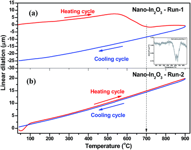 |
| | Fig. 5 (a) Linear thermal dilation profile of as prepared sample and (b) subsequent thermal cycling on the same sample. Inset in 5a shows the derivative plot of linear dilation profile recorded on as-prepared sample. Onset of phase transformation around 675–700 °C can be clearly seen in the derivative plot. | |
3.4 Optical properties
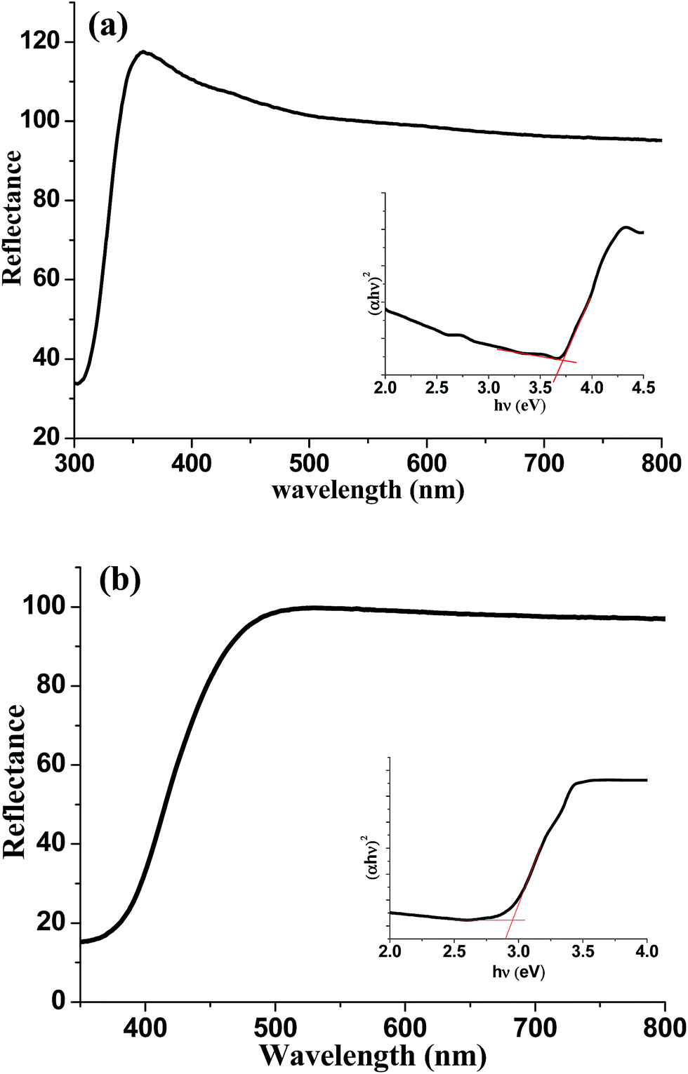
The optical properties of InOOH and rh-In2O3 nanostructures have been studied by UV-DRS and room temperature PL studies. Fig. 6 shows the diffuse reflectance UV-visible absorption spectra and plots of [αhν]2 versus hν (as insets) for InOOH and rh-In2O3 powder samples respectively. The Kubelka–Munk method was used to convert the diffuse reflectance to the absorbance (Y-axis). The band gaps were determined by the absorption edges extrapolated from the [αhν]2 and hν plots. The estimated direct band gaps for the as-prepared InOOH and rh-In2O3 nanostructures were 3.75, and 2.95 eV, respectively in accord with literature data.31,32b
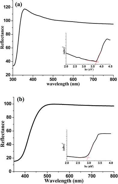 |
| | Fig. 6 UV-vis diffuse reflectance spectra for (a) InOOH and (b) rh-In2O3 nanostructures. Insets show the plots of (αhν)2 vs. hν derived from reflectance data showing band gap values. | |
Photoluminescence (PL) is a very useful tool to determine the structure, defect, and impurity in the nanocrystalline oxides. Literature studies on PL of InOOH and rh-In2O3 nanostructures reveal that PL of the nanostructures obtained by different synthetic procedures varies greatly. The reported luminescence peak positions range from 280 nm to 640 nm for InOOH21,40 and from 350 to 650 nm for rh-In2O3 (ref. 17, 19b, 21, 23 and 41) nanostructures depending upon the preparation method employed. Xu et al.21 observed PL emission peaks in the range 280–580 nm in both InOOH quantum dots and nanowires. Tang et al.40 studied the PL properties of InOOH nanowires synthesized at different heating rates and observed PL emission peaks at 456 and 496 nm for the samples synthesized at growth rate of 4 °C min−1 and 8 °C min−1, respectively. Yu17 Dong,23 and Lee et al.19b have observed PL peaks centered at 413 and 517 nm for rh-In2O3 microspheres, 378, 398 and 420 nm from rh-In2O3 nanofibres and 360, 400, 470 nm from rh-In2O3 nanocubes. The rh-In2O3 hierarchical microcrystals reported by Jiang et al.41 show PL emission peaks at 413, 460, 494 and 519 nm. It is clear from the above discussion that different preparation methods used produce nanostructures with different dimensionality, sizes, morphology, crystallinity and defect structure which influences the luminescence properties of the nanostructures prepared. Near band edge emissions can be favored by the high crystal quality and quantum confinement effect21 and emissions in the visible range are attributed to low crystallinity and structural defects which are mainly oxygen vacancies.
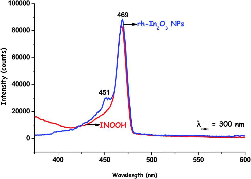
The PL properties of InOOH, and rh-In2O3 nanostructures prepared by us have been investigated at excitation wavelength of 300 nm. The room temperature PL spectra of InOOH and rh-In2O3 nanopowders are shown in Fig. 7. A very intense blue emission peak is seen at 468 nm for InOOH nanostructures, while two peaks at 451 and 469 nm are observed for rh-In2O3. As discussed above, photoluminescence in nanostructures of transparent conducting oxides is usually dominated by defect-based emission42–44 with a small or negligible contribution from band edge UV emission. InOOH and rh-In2O3 are both n-type semiconductors because of presence of oxygen vacancies. The oxygen vacancies act as the radiative center in the luminescence process by forming defect levels located inside the gap and trapping electrons from the valence band.45 In this study, we have prepared both InOOH and rh-In2O3 nanostructures under mild conditions at low temperatures of 140 °C and 350 °C respectively, thus, oxygen vacancies can easily be generated during the synthetic process. In particular, solution based synthetic methods lead to a high density of oxygen vacancies because of incomplete crystallization. Thus, the PL bands of InOOH and rh-In2O3 nanostructures observed in the blue region of visible spectrum are mainly attributed to the effect of oxygen vacancies or oxygen deficiencies, which are inevitable defects occurring during their synthetic procedure. Under the excitation of 300 nm irradiation, the electrons are excited from the valence band (VB) to the conduction band (CB). The electrons move freely in the CB and finally relax to the oxygen vacancies. The recombination of an electron occupying oxygen vacancies with a photo-excited hole yields the blue emission at 468 nm and at 451 and 469 nm for InOOH and rh-In2O3 nanostructures respectively. Electron paramagnetic resonance (EPR), is a powerful technique for characterizing the magnetic properties of defect centers on an atomic scale. Correlation of EPR and optical spectroscopies hence allows one to get a more complete insight into defect structures. The oxygen vacancies can be present in three different charge states in the nanocrystalline oxides: V0O, V+O, V++O. The presence of singly ionized oxygen vacancies can easily be observed by electron paramagnetic resonance spectroscopy as they are paramagnetic. Fig. 8 shows the results of EPR measurements carried out on InOOH and rh-In2O3 samples. A distinct EPR signal with g = 2.002 appears for the InOOH nanostructures (Fig. 8a), while a very broad and weak signal with g ≈ 2.1 appears for rh-In2O3 (Fig. 8b). The data suggests that both the nanostructures possess the same type of paramagnetic centers, i.e., singly ionized oxygen vacancies. These are assumed to be the recombination centers for the luminescence processes. The PL decay curves for InOOH and rh-In2O3 nanostructures at emission wavelength peak 468 nm (λexc = 300 nm) are shown in Fig. 9. Lifetime value is found to be 1.47 ns for InOOH and 1.14 ns for rh-In2O3 nanostructures indicating faster electron–hole recombination rate in the latter.
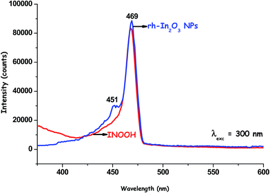 |
| | Fig. 7 Room temperature PL spectra for InOOH and rh-In2O3 nanostructures. | |
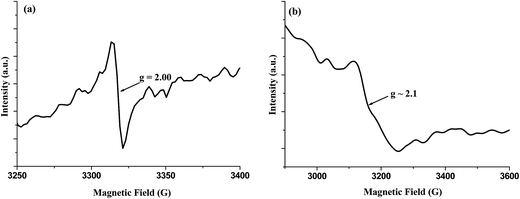 |
| | Fig. 8 EPR spectra of InOOH and rh-In2O3 nanostructures at 100 K. | |
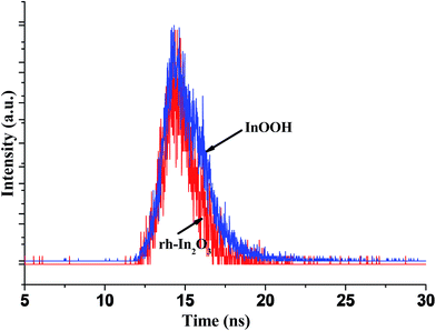 |
| | Fig. 9 Photoluminescence (PL) decay curves of InOOH and rh-In2O3 nanostructures at. emission wavelength of 468 nm. | |
3.5 Photocatalytic activity
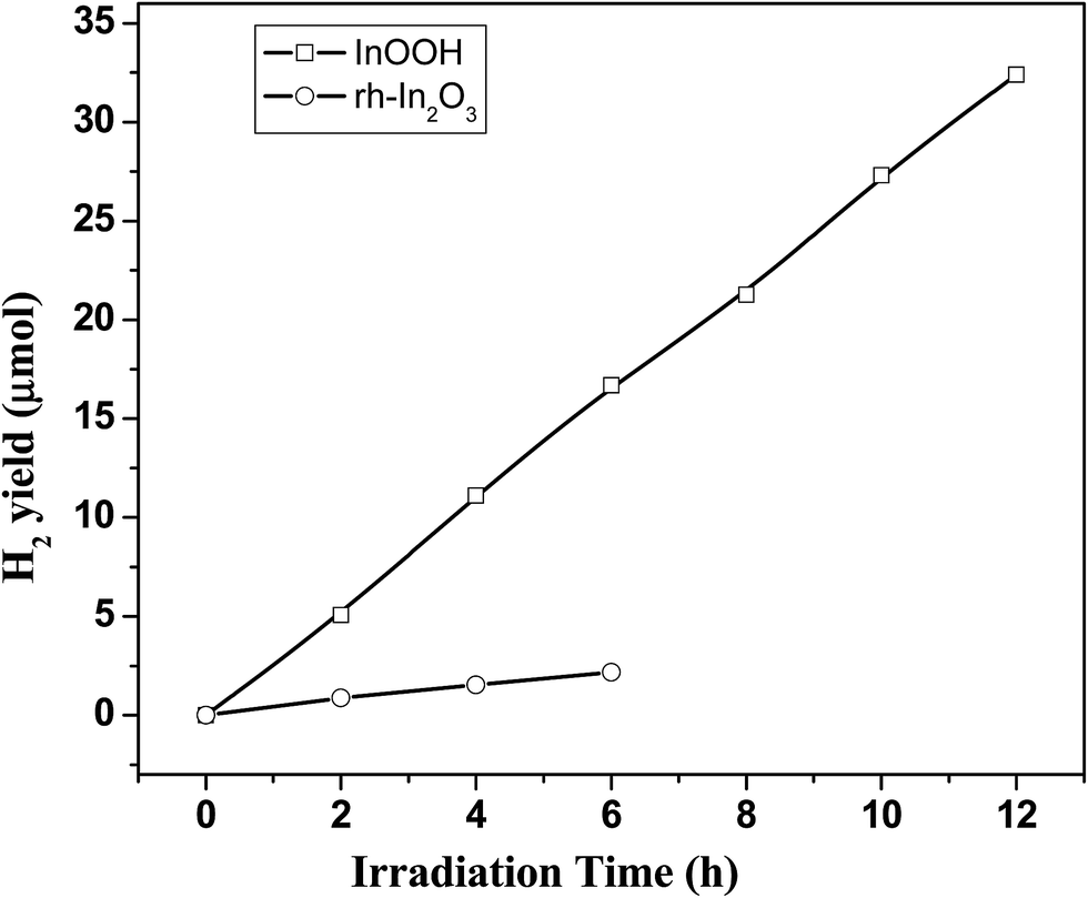
As p-block wide band gap semiconductors, both InOOH and rh-In2O3 are attractive candidates for photocatalytic studies under UV-vis irradiation. In case of InOOH the VB is composed of the O 2p orbitals, while the CB is composed of the hybridizations of the 5s and 5p orbitals of indium and is thus more dispersive because of dispersive characteristics of s orbitals.46 Similarly, In case of rh-In2O3 also the VB is composed mainly of oxygen 2p like states and CB consists mainly of indium 5s-like states.47 The more dispersive conduction band for both InOOH and rh-In2O3 will promote the mobility of the photoexcited electrons and enhance the charge separation. Further, the edge of conduction band (ECB) for InOOH is −0.4 V (vs. NHE), the edge of valence band (EVB) is 3.3 V (vs. NHE),31,48 and the energy value of calculated conduction band edge (ECB) for rh-In2O3 is −3.80 eV (vs. absolute vacuum scale).49 Thus, photogenerated charge carriers in both InOOH and rh-In2O3 nanostructures have strong enough redox abilities. In view of these observations, photocatalytic properties of InOOH and rh-In2O3 nanostructures have been explored for hydrogen generation from water/methanol (2:1) mixtures under UV/vis irradiation (16% UV + 84% visible). Fig. 10 shows the results of H2 evolution with InOOH and rh-In2O3 nanostructures without any cocatalyst. The observed hydrogen yields are low. InOOH nanostructures show H2 yield of 54 μmol g−1 h−1, while rh-In2O3 nanostructures show negligible activity. The apparent quantum efficiency (AQE) achieved using InOOH is only 0.1%. Thus, both InOOH and rh-In2O3 nanostructures are not efficient photocatalysts for H2 generation via water splitting. The intrinsic stability of InOOH nanostructures during the course of the photocatalytic experiment was confirmed by XRD of the sample after the photocatalytic reaction. The XRD pattern obtained for the spent photocatalyst was the same as that of fresh sample (Fig. S8, ESI†).
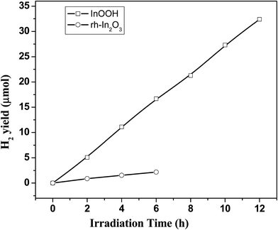 |
| | Fig. 10 Photocatalytic H2 yield (μmol) obtained from water/methanol mixtures under UV visible light for InOOH and rh-In2O3 nanostructures. | |
The photocatalytic activities of a semiconductor photocatalyst are governed by bulk (structure and electronic) and surface properties. The surface properties, viz., N2 BET surface areas, cumulative pore volumes and mean pore size diameter for InOOH and rh-In2O3 nanostructures are listed in Table 1. The pore size distribution and pore volumes are shown in Fig. S9, ESI.†
Table 1 Particle size, N2-BET surface area and porosity of InOOH and rh-In2O3 nanostructures
| S. No. |
Sample |
Particle size from TEM |
Specific surface area (m2 g−1) |
BJH pore size distribution |
Pore volume (cm3 g−1) |
| 1. |
InOOH |
Length: 8–20 nm |
53 |
Mesoporous in the range 20–150 Å |
0.175 |
| Width: 4–8 nm |
| 2. |
rh-In2O3 |
Length: 20 nm |
43 |
Mesoporous in the range 40–300 Å |
0.133 |
| Width: 10–15 nm |
It is clear from the data in Table 1 that, both surface area and pore volume decrease when InOOH sample is thermally annealed at 350 °C to obtain rh-In2O3 nanopowders. Also, both the materials show mesoporosity with mesoporous of varying sizes but rh-In2O3 has larger pores in a wide range as compared to InOOH nanopowders (Fig. S9, ESI†).
The photoactivity of InOOH is attributed to its appropriate electronic structure and its high surface area accompanied by mesoporosity. In general, a higher surface area is beneficial to the reduced probability of electron–hole recombination and promotes migration of photogenerated carriers. The more dispersive conduction band in InOOH will also promote the mobility of the photoexcited electrons and enhance the charge separation contributing to its photocatalytic activity. We believe that the difference in photoactivity of InOOH and rh-In2O3 nanostructures is most likely due to the difference in their bulk and surface properties. InOOH has a larger band gap than rh-In2O3. While this reduces the light that can be absorbed it may raise the valence band maximum to higher energy levels relative to redox potentials of adsorbed molecules. This increases the oxidation power of electrons and facilitates electron transfer from the InOOH to adsorbed molecules just like in case of anatase TiO2.50 Surface properties of a semiconductor may play a role in the adsorption of molecules and subsequent charge transfer to the molecule. InOOH has larger surface area as compared to rh-In2O3 nanostructures implying that it has more surface defects as compared to the latter. Surface defects are crucial for charge trapping and separation at the surface and thus InOOH with more surface defects will trap the charge carriers more efficiently as compared to rh-In2O3 leading to better photocatalytic activity. The supporting evidence for this comes from life time measurements carried out during PL studies on InOOH and rh-In2O3 nanostructures. Lifetime value is found to be 1.47 ns for InOOH and 1.14 ns for rh-In2O3 nanostructures indicating faster electron–hole recombination rate in the latter.
4. Conclusions
In conclusion, we have successfully developed a low temperature simple solution synthetic route to synthesize nanostructures of InOOH with controllable sizes and shape using inexpensive starting materials. The synthetic method is scalable and can be extended to get nanoparticles of In(OH)3 and c-In2O3 with cube like morphology by just varying the pH of the reaction mixture. Elongated rh-In2O3 nanostructures can be easily obtained by thermal annealing of InOOH nanostructures at 350 °C. XRD, HRTEM and Raman studies reveal that phase pure materials are obtained. Thermal studies carried out on as prepared rh-In2O3 samples reveal that on heating above 700 °C, metastable corundum type In2O3 transforms irreversibly into cubic bixbyite type In2O3. The nanostructures exhibit intense visible blue light emission which can be attributed to oxygen vacancies present in the materials synthesized. As the structural defects in TCOs are also responsible for electrical conductivity, the ability to tune their optical properties by size controlled synthesis of InOOH and rh-In2O3 nanostructures is promising for application in multifunctional optoelectronic materials and devices. The potential of InOOH and rh-In2O3 nanostructures as photocatalytic materials for hydrogen generation from water/methanol (2:1) mixtures under UV/vis irradiation has been evaluated for the first time. InOOH nanostructures show H2 yield of 54 μmol g−1 h−1, while rh-In2O3 nanostructures show negligible activity. The difference in their photocatalytic behavior is most likely due to their different bulk and surface properties.
Acknowledgements
The authors thank Dr A. Ballal, Molecular Biology Division, B. A. R. C. for carrying out low-resolution TEM studies on In(OH)3 and c-In2O3 samples. The authors also thank Dr R. M. Kadam and Dr V. Rane, Radiochemistry Division, B. A. R. C for carrying out EPR studies on InOOH and rh-In2O3 nanostructures. One of the authors, D. E. thanks the financial support to his research by Spanish Ministerio de Economıa y Competitividad (MINECO) under Grants No. MAT2013-46649-C04-01/02/03 and No. MAT2015-71070-REDC (MALTA Consolider). The authors also thank SC-SIE at Universitat de Valencia for support with the transmission electron microscope measurements.
References
- Special Issue of the MRS Bull.. 2000, 25, 15 Search PubMed.
- T. Minami, Semicond. Sci. Technol., 2005, 20, S35–S44 CrossRef CAS.
- P. D. C. King and T. D. Veal, J. Phys.: Condens. Matter, 2011, 23, 334214 CrossRef CAS PubMed.
- A. Stadler, Materials, 2012, 5, 661–683 CrossRef.
- J. Song, S. A. Kulinich, J. Li, Y. Liu and H. Zeng, Angew. Chem., Int. Ed., 2015, 54, 462–466 CAS.
- I. Hamberg and C. G. Granqvist, J. Appl. Phys., 1986, 60, R123 CrossRef CAS.
-
(a) O. Bierwagen, Semicond. Sci. Technol., 2015, 30, 024001 CrossRef;
(b) S. Y. Han, G. S. Herman and C. H. Chang, J. Am. Chem. Soc., 2011, 133, 5166–5169 CrossRef CAS PubMed.
- G. Shen, B. Liang, X. Wang, H. Huang, D. Chen and Z. L. Wang, ACS Nano, 2011, 5, 6148–6155 CrossRef CAS PubMed.
- V. P. Reddy, A. V. Kumar, K. Swapna and K. R. Rao, Org. Lett., 2009, 11, 1697–1700 CrossRef CAS PubMed.
- K. R. Reyes-Gil, E. A. Reyes-Garcia and D. Raftery, J. Phys. Chem. C, 2007, 111, 14579–14588 CAS.
- H. Yang, S. Wang and Y. Yang, CrystEngComm, 2012, 14, 1135–1142 RSC.
- E. Li, Z. Cheng, J. Xu, Q. Pan, W. Yu and Y. Chu, Cryst. Growth Des., 2009, 9, 2146–2151 CAS.
- R. Katoh, A. Furube, T. Yoshihara, K. Hara, G. Fujihashi, S. Takano, S. Murata, H. Arakawa and M. Tachiya, J. Phys. Chem. B, 2004, 108, 4818–4822 CrossRef CAS.
- S. Z. Karazhanov, P. Ravindran, P. Vajeeston, A. Ulyashin, T. G. Finstad and H. Fjellvåg, Phys. Rev. B: Condens. Matter Mater. Phys., 2007, 76, 075129 CrossRef.
-
(a) C. T. Prewitt, R. D. Shannon, D. B. Rogers and A. W. Sleight, Inorg. Chem., 1969, 8, 1985–1993 CrossRef CAS;
(b) A. Gurlo, S. Lauterbach, G. Miehe, H. J. Kleebe and R. Riedel, J. Phys. Chem. C, 2008, 112, 9209–9213 CrossRef CAS.
- H. Zhang and J. F. Banfield, J. Mater. Chem., 1998, 8, 2073–2076 RSC.
- D. Yu, S. H. Yu, S. Zhang, J. Zuo, D. Wang and Y. Qian, Adv. Funct. Mater., 2003, 13, 497–501 CrossRef CAS.
-
(a) M. Epifani, P. Siciliano, A. Gurlo, N. Barsan and U. Weimar, J. Am. Chem. Soc., 2004, 126, 4078–4079 CrossRef CAS PubMed;
(b) V. D. Ashok and S. K. De, J. Phys. Chem. C, 2011, 115, 9382–9392 CrossRef CAS.
-
(a) C. Chen, D. Chen, X. Jiao and C. Wang, Chem. Commun., 2006, 4632–4634 RSC;
(b) C. H. Lee, M. T. Kim, A. Kim, J. Paek, J. W. Lee, S.-Y. Choi, K. Kim, J.-B. Park and K. Lee, J. Am. Chem. Soc., 2006, 128, 9326–9327 CrossRef CAS PubMed.
-
(a) Z. Zhuang, Q. Peng, J. Liu, X. Wang and Y. Li, Inorg. Chem., 2007, 46, 5179–5187 CrossRef CAS PubMed;
(b) J. Q. Xu, Y. P. Chen, Q. Y. Pan, Q. Xiang, Z. X. Cheng and X. W. Dong, Nanotechnology, 2007, 18, 115615 CrossRef;
(c) Y. Fan, Z. Li, L. Wang and J. Zhan, Nanotechnology, 2009, 20, 285501 CrossRef PubMed.
- X. Xu and X. Wang, Inorg. Chem., 2009, 48, 3890–3895 CrossRef CAS PubMed.
- L.-Y. Chen, Y. Liang and Z.-D. Zhang, Eur. J. Inorg. Chem., 2009, 903–909 CrossRef CAS.
- H. Dong, Z. Chen, L. Sun, L. Zhou, Y. Ling, C. Yu, H. H. Tan, C. Jagadish and X. Shen, J. Phys. Chem. C, 2009, 113, 10511–10516 CAS.
-
(a) H. Yang, L. Liu, H. Liang, J. Wei and Y. Yang, CrystEngComm, 2011, 13, 5011–5016 RSC;
(b) S. S. Farvid and P. V. Radovanovic, J. Am. Chem. Soc., 2012, 134, 7015–7024 CrossRef CAS PubMed.
-
(a) P. Li, H. Fan, Y. Cai, M. Xu, C. Long, M. Li, S. Lei and X. Zou, RSC Adv., 2014, 4, 15161–15170 RSC;
(b) L.-Y. Chen, Z.-X. Wang and Z.-D. Zhang, New J. Chem., 2009, 33, 1109–1115 RSC;
(c) Y. Tang and J. Ma, RSC Adv., 2014, 4, 25692–25697 RSC.
- W.-H. Zhang and W.-Z. Zhang, J. Phys. Chem. Solids, 2013, 74, 1271–1275 CrossRef CAS.
- M. Wu, C. Wang, Y. Zhao, L. Xiao, C. Zhang, X. Yu, B. Luo, B. Hu, W. Fan and W. Shi, CrystEngComm, 2015, 17, 2336–2345 RSC.
- B. García-Domene, J. A. Sans, F. J. Manjón, S. V. Ovsyannikov, L. S. Dubrovinsky, D. Martinez-Garcia, O. Gomis, D. Errandonea, H. Moutaabbid, Y. Le Godec, H. M. Ortiz, A. Muñoz, P. Rodríguez-Hernández and C. Popescu, J. Phys. Chem. C, 2015, 119, 29076–29087 Search PubMed.
- A. Hoshino, K. Fujioka, T. Oku, M. Suga, Y. F. Sasaki, T. Ohta, M. Yasuhara, K. Suzuki and K. Yamamoto, Nano Lett., 2004, 4, 2163–2169 CrossRef CAS.
-
(a) W. Ong, L. Tan, S. Chai, S.-T. Yong and A. R. Mohamed, Nano Res., 2014, 7, 1528–1547 CrossRef CAS;
(b) W. Ong, L. Tan, S. Chai and S. Yong, Dalton Trans., 2015, 44, 1249–1257 RSC;
(c) Y. Wei, J. Su, X. Wan, L. Guo and L. Vayssieres, Nano Res., 2016, 9, 1561–1569 CrossRef CAS;
(d) M. Zeng, Z. Chai, X. Deng, Q. Li, S. Feng, J. Wang and D. Xu, Nano Res., 2016, 9, 2729–2734 CrossRef CAS;
(e) T. Sun, J. Song, J. Jia, X. Li and X. Sun, Nano Energy, 2016, 26, 83–89 CrossRef CAS;
(f) F. Li, L. Zhang, J. Tong, Y. Liu, S. Xu, Y. Cao and S. Cao, Nano Energy, 2016, 27, 320–329 CrossRef CAS.
- Z. Li, Z. Xie, Y. Zhang, L. Wu, X. Wang and X. Fu, J. Phys. Chem. C, 2007, 111, 18348–18352 CAS.
-
(a) L. Song, C. Chen, S. Zhang and Q. Wei, Catal. Commun., 2011, 12, 1051–1054 CrossRef CAS;
(b) S. Ge, B. Wang, J. Lin and L. Zhang, CrystEngComm, 2013, 15, 721–728 RSC.
- Y. Song, L. Xu, W. Shi and J. Guan, J. Nanopart. Res., 2014, 16, 2295 CrossRef.
- S. Yang, C.-Y. Xu, S.-P. Hu, W.-S. Wang, J. Yu and L. Zhen, Bull. Korean Chem. Soc., 2016, 37, 522–528 CrossRef CAS.
- Y. S. Cho and Y. D. Huh, Bull. Korean Chem. Soc., 2010, 31, 1769–1772 CrossRef CAS.
- J. Yin and H. Cao, Inorg. Chem., 2012, 51, 6529–6536 CrossRef CAS PubMed.
- T. Yan, X. Wang, J. Long, H. Lin, R. Yuan, W. Dai, Z. Li and X. Fu, New J. Chem., 2008, 32, 1843–1846 RSC.
- X. Xue and M. Kanzaki, J. Phys. Chem. B, 2007, 111, 13156–13166 CrossRef CAS PubMed.
- J. R. Ferraro and K. Nakamoto, Introductory Raman Spectroscopy, Academic Press, Boston, 1994 Search PubMed.
- S. Tang, J. Zhang, S. Wu, C. Hu, Y. Li, L. Jiang and Q. Cui, J. Phys. Chem. C, 2014, 118, 21170–21176 CAS.
- H. Jiang, L. Zhao, L. Gai, L. Ma, Y. Ma and M. Li, CrystEngComm, 2013, 15, 7003–7009 RSC.
- L. Zhang, L. Yin, C. Wang, N. Iun, Y. Qi and D. Xiang, J. Phys. Chem. C, 2010, 114, 9651–9658 CAS.
- H. M. Xiong, D. G. Shchukin, H. Mohwald, Y. Xu and Y. Y. Xia, Angew. Chem., Int. Ed., 2009, 48, 2727–2731 CrossRef CAS PubMed.
- A. van Dijken, E. A. Meulenkamp, D. Vanmaekelbergh and A. Meijerink, J. Lumin., 2000, 90, 123–128 CrossRef CAS.
- H. Cao, X. Qiu, Y. Liang and Q. Zhu, Appl. Phys. Lett., 2003, 83, 761–763 CrossRef CAS.
- J. Sato, H. Kobayashi, K. Ikarashi, N. Saito, H. Nishiyama and Y. Inoue, J. Phys. Chem. B, 2004, 108, 4369–4375 CrossRef CAS.
- P. D. C. King, T. D. Veal, F. Fuchs, C. Y. Wang, D. J. Payne, A. Bourlange, H. Zhang, G. R. Bell, V. Cimalla, O. Ambacher, R. G. Egdell, F. Bechstedt and C. F. McConville, Phys. Rev. B: Condens. Matter Mater. Phys., 2009, 79, 205211 CrossRef.
- M. A. Butler and D. S. Ginley, J. Electrochem. Soc., 1978, 125, 228–232 CrossRef CAS.
- Y. Xu and M. A. A. Schoonen, Am. Mineral., 2000, 85, 543–556 CrossRef CAS.
- M. Batzill, Energy Environ. Sci., 2011, 4, 3275–3286 CAS.
Footnote |
| † Electronic supplementary information (ESI) available: Diagram of reaction assembly and the emission spectrum of lamp in UV-visible wavelengths used for photocatalytic activity studies, XRD patterns of the reaction intermediates isolated, characterization data for In(OH)3 and c-In2O3 nanocubes obtained by the solvothermal reaction between In(NO3)3·2.55H2O and excess TMAH, XRD patterns for the material obtained after DSC experiment, DTA measurement data for the rh-In2O3 sample, XRD patterns for the rh-In2O3 nanostructures heated in the temperature range 650–800 °C, characterization data for the c-In2O3 nanoparticles obtained by thermal annealing of InOOH nanostructures, XRD pattern of the used catalyst, pore size distribution data for InOOH and rh-In2O3 nanostructures. See DOI: 10.1039/c6ra23693f |
|
| This journal is © The Royal Society of Chemistry 2016 |
Click here to see how this site uses Cookies. View our privacy policy here.