Green preparation of D-tryptophan imprinted self-supported membrane for ultrahigh enantioseparation of racemic tryptophan

Zhiyong Zhou, Ke Cui, Yu Mao, Wenshuai Chai, Nian Wang and Zhongqi Ren*
Beijing Key Laboratory of Membrane Science and Technology, College of Chemical Engineering, Beijing University of Chemical Technology, Beijing 100029, People's Republic of China. E-mail: renzq@mail.buct.edu.cn; Tel: +86-10-64433872

Received 22nd September 2016 , Accepted 31st October 2016

First published on 8th November 2016


Abstract

Enantioseparation of chiral compounds is important for pharmaceutical production. Chiral resolution of racemic tryptophan has attracted increasing attention. Traditional separation methods have the disadvantages of being high cost, energy intensive and environmentally unfriendly. Membrane permeation seems promising for application in the enantioseparation of racemic tryptophan with the advantages of energy efficiency and no additives. However, lots of organic compounds are commonly used for the preparation of various membranes for enantioseparation of racemic tryptophan. Therefore, we prepared a molecularly imprinted self-supported membrane (MISM) using a green and clean method with natural polymer sodium alginate (SA) as the functional polymer, water as the solvent and CaCl2 as the crosslinking agent. The crosslinking by CaCl2 was confirmed by attenuated total reflection Fourier transform infrared (ATR-FTIR) and X-ray photoelectron spectroscopy (XPS) analyses. The surface and internal structures of the MISM and non-imprinted self-supported membrane (NISM) were observed by atomic force microscope (AFM) and scanning electron microscope (SEM). The addition of a template molecule can significantly improve the MISM surface roughness. The compact structures of MISM and NISM were confirmed by the cross-sectional images. The thermo stability of MISM was studied. In addition, the effects of pH and crosslinking time on the swelling behavior of MISM were investigated. A high concentration of OH in alkali solution can remarkably weaken the electrostatic interaction between Ca2+ and COO. The effects of preparation conditions, permeation conditions and concentration polarization on the pressure-driven permeation performance of MISM were investigated. Ultrahigh enantioseparation performance in 99% ee of the permeation solution can be obtained under mild conditions. Reducing the concentration polarization by increasing the environmental temperature and continuous stirring is beneficial to obtaining ultrahigh ee. Comparison with previous studies on pressure-driven permeation performance indicates that the proposed green and clean preparation method for a molecularly imprinted self-supported membrane is not only environmentally friendly but also efficient in obtaining ultrahigh ee.

Introduction

As is well known, chiral compounds are very important to the function of the human body. Nucleic acids, amino acids, polysaccharides, proteins and enzymes, which are significantly related to body functions, are almost all chiral compounds. Furthermore, a lot of synthetic chemical products, such as pharmaceutical molecules, are also chiral. The enantiomers of chiral compounds have almost the same physical and chemical properties, but show different biological activities and pharmacological effects. For instance, R-thalidomide can be used to cure vomiting, rheumatic autoimmune disease and tumor proliferation, but S-thalidomide results in teratogenesis. In the 1960s, thousands of infants suffered from developmental deformities after taking S-thalidomide.1 In the 1990s, the FDA issued some regulations to supervise chiral drugs.2 Therefore, the development of chiral resolution technology is important for drug safety and obtaining high economic value.

Chiral resolution has attracted more and more attention. Various kinds of methods, such as crystallization,3 chromatography,4,5 enzymatic kinetic resolution,6 extraction7 and membrane separation,8 have been widely applied in the separation of chiral compounds. Among these methods, membrane separation is a promising technology with advantages of green preparation, low cost, continuous operation and facile scale up,9 which attracts ever-increasing attention. Many studies have been carried out to realize the resolution of racemic tryptophan with membrane separation technology,10–13 which is often divided into chiral selective membrane and achiral selective membrane coupled with affinity dialysis.10 For achiral selective membranes, the effective chiral recognition molecules, such as human serum albumins, are very expensive,14 which is not beneficial for industrial application. Thus, in this study, a chiral selective membrane was selected as the resolution membrane for racemic tryptophan.

Molecularly imprinted membranes have the advantages of membrane separation technology and molecularly imprinted technology. Molecularly imprinted technology first appeared in 1972 (ref. 15) and went through a rapid development stage after the report of a theophylline molecularly imprinted polymer by Mosbach.16 Molecularly imprinted technology introduces molecular recognition abilities into polymeric materials and membranes, which endows the polymer or membrane with two important characteristics: specific recognition and the “gate effect”. Specific recognition is beneficial to the enhancement of membrane selectivity.17 The “gate effect” can increase the permeability of the template molecule.18 It is worth mentioning that both selectivity and permeability are the main performance indices of membrane separation. Hence, membrane separation performance can be greatly improved by molecularly imprinted technology. In addition, molecularly imprinted technology also exhibits excellent environmental stability and low cost,19 which is beneficial to the application of molecularly imprinted membranes.

Many studies have been conducted to separate various chiral enantiomers with molecularly imprinted membranes.20,21 Good adsorption selectivity22,23 or concentration-driven permeation selectivity with24,25 can be obtained with molecularly imprinted membranes. However, there are few publications about pressure-driven permeation resolution, which can significantly increase mass transfer flux.

Sodium alginate (SA) is a natural, non-toxic, biocompatible and biodegradable polysaccharide26 that has been widely used in chiral resolution,25,27 drug release,28 membrane separation,29 water purification30,31 and biochemical processes.32 Thus, SA is an environmentally friendly polymer for membrane preparation. However, the traditional crosslinking agents for SA like epichlorohydrin33 and glutaric dialdehyde34 are environmentally unfriendly. Therefore, a water soluble inorganic crosslinking agent may be a better choice.

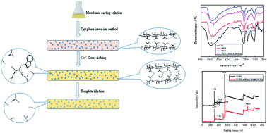
To explore a green and clean way to obtain high permeation resolution of racemic tryptophan, we prepared a molecularly imprinted self-supported membrane (MISM) with D-tryptophan as the template molecule, SA as the functional polymer, CaCl2 as the crosslinking agent and deionized water as the solvent, as shown in Fig. 1. CaCl2 is always selected as a nontoxic crosslinking agent for sodium alginate microsphere,35 but it is seldom used for membrane preparation. Thus, in this study, CaCl2 aqueous solution was selected as the crosslinking agent to replace the conventional organic crosslinking agent for green fabrication of the SA membrane. No organic solvents or crosslink agents with very high toxicity were introduced into the preparation process for the D-tryptophan imprinted self-supported membrane.


image file: c6ra23555g-f1.tif
Fig. 1 Preparation of D-tryptophan imprinted self-supported membrane.

The crosslinking with CaCl2 was confirmed by Fourier transform infrared (FT-IR) and X-ray photoelectron spectroscopy (XPS) analyses. The surface and internal structures of the MISM and non-imprinted self-supported membrane (NISM) were observed by atomic force microscope (AFM) and scanning electron microscope (SEM). In addition, the thermostability of the MISM was studied. The effects of temperature and pH on the degree of swelling of the MISM were investigated. The effects of preparation conditions (template molecule content, membrane thickness and crosslinking time), permeation conditions (pressure, feed concentration and pH value) and concentration polarization on the permeation performance of the MISM were investigated.

Results and discussion

Characteristics of D-tryptophan membranes

Fig. 2a shows the ATR-FTIR spectra of the pure SA, eluted MISM, NISM and eluted MISM without crosslinking.27 The characteristic peaks ranging from 800 to 890 cm−1 are ascribed to the polysaccharide structure in SA. The characteristic peaks at 1409 cm−1 and 1593 cm−1 are ascribed to the symmetric and asymmetric stretching vibrations of COO, respectively. Comparing spectra b with c, it can be seen that the characteristic peak of the symmetric stretching vibration for COO blue shifts after crosslinking. On the contrary, the characteristic peak for the asymmetric stretching vibration of COO red shifts after crosslinking. The characteristic peak at 1080 cm−1 is ascribed to the stretching vibration of C–O–C. The characteristic peak at 1036 cm−1 is ascribed to the deformation vibration of C–O. The characteristic peak at 1732 cm−1 ascribed to the stretching vibration of C[double bond, length as m-dash]O can be observed in spectra a and c, which is the main characteristic peak of membrane cross-linked with Ca2+. However, no appearance of the characteristic peak of the stretching vibration of C[double bond, length as m-dash]O can be found in spectra b and d, in accordance with the absence of crosslinking with Ca2+.
image file: c6ra23555g-f2.tif
Fig. 2 (a) ATR-FTIR spectra of membranes; (b) X-ray photoelectron spectra of the MISM after crosslinking and the MISM without crosslinking.

Fig. 2b shows the XPS analysis for the surfaces of the MISM and the MISM without crosslinking. It can be seen that the characteristic peak of sodium atom at 1071 eV on the MISM surface without crosslinking (Na% = 4.5, Table 1) disappears on the MISM surface (Na% = 0, Table 1). However, the characteristic peak of calcium atom at 346 eV appears on the MISM surface (Ca% = 2.1, Table 1) with no appearance on the MISM without crosslinking (Ca% = 0, Table 1), indicating that the crosslinking with CaCl2 was realized by the coordination of two carboxyl groups in SA and one Ca2+ through ion exchange with Na+, which agrees with the results of the ATR-FTIR spectra.

Table 1 Elemental compositions (atom%) determined by XPS for the membrane surface
Membrane C% O% Ca% N% Na%
MISM 55.7 40.3 2.1 2.0 0
MISM without crosslinking 60.5 32.7 0 2.1 4.5


AFM analysis can directly display the surface roughness of membranes. Fig. 3 shows the AFM images of MISM and NISM before and after crosslinking. Both the root-mean-square (RMS) roughnesses of the MISM before (110.0 nm) and after crosslinking (77.3.0 nm) are much larger than those of the NISM before (16.1 nm) and after crosslinking (28.1 nm), indicating that the addition of template molecule can significantly increase the MISM's surface roughness, which is beneficial to the increase of the effective area and flux of mass transfer of the MISM.


image file: c6ra23555g-f3.tif
Fig. 3 AFM analyses of the MISM (a) and NISM (b) without crosslinking, and the MISM (c) and NISM (d) after crosslinking.

Fig. 4c and d show the surface morphologies of MISM and NISM after crosslinking. It is obvious that lots of protuberances can be seen on the surface of the MISM, but no protuberances appear on the NISM's surface. Meanwhile, the protuberances also appear on the surface of the MISM without crosslinking, as shown in Fig. 4a. The protuberances on the MISM's surface can be attributed to the dissolving-out behavior of the template molecule D-tryptophan from the membrane solution during the preparation process, which is consistent with the AFM analyses. Furthermore, the internal structures of the MISM and NISM were also analyzed. As shown in Fig. 4e and f, the cross-sectional images of the MISM and NISM after crosslinking confirm the compact structure of the prepared membranes.


image file: c6ra23555g-f4.tif
Fig. 4 (a) and (b) SEM images of the surfaces of the MISM and NISM without crosslinking. (c) and (d) SEM images of the surfaces of the MISM and NISM after crosslinking. (e) and (f) SEM images of the cross-sectional structures of the MISM and NISM after crosslinking.

The thermostability of the D-tryptophan imprinted self-supported membrane was determined by thermal gravimetric analysis (TGA). As shown in Fig. 5, the thermolysis can be divided into two stages: 65 °C to 121 °C and 193 °C to 301 °C. The first stage (65 °C to 121 °C) represents the evaporation of adsorptive water and the weight loss is 9.39%. The breaking, decomposition and carbonization of the polymer chains of SA occur during the second stage of thermal decomposition (193 °C to 301 °C), resulting in a 40.52% weight loss from the self-supported membrane.


image file: c6ra23555g-f5.tif
Fig. 5 TGA curve of D-tryptophan imprinted self-supported membrane.

The effect of pH on the swelling behavior of the MISM was investigated and is shown in Fig. 6a. When the pH is 12, the MISM swells seriously and the DS reaches up to 280% at swelling equilibrium. However, when the MISM is placed in aqueous solution at pH 2 or deionized water, the DS is only approximately 142%, which is nearly half of the DS value at pH 12. The crosslinking of the SA polymer chain depends on the electrostatic interaction between Ca2+ and COO, which can be weakened by a high concentration of OH in alkali solution. Therefore, the DS value obtained in alkali solution is higher than that obtained in neutral or acidic solution. Fig. 6b shows the effect of crosslinking time on the DS value of the MISM. The DS value first decreases and then remains constant with increasing crosslinking time, indicating that the crosslinking of the membrane has been accomplished at 60 min.


image file: c6ra23555g-f6.tif
Fig. 6 (a) Effect of pH on degree of swelling of the MISM. (b) Effect of crosslinking time on degree of swelling of the MISM.

Effects of preparing conditions on permeation performance of MISM

As shown in Fig. 7, the enantiomeric excess (ee%) first keeps steady and then decreases to 88.85% with increasing the mass ratio of the template molecule to SA. In fact, when the mass ratio of the template molecule to SA is zero, the membrane is a non-imprinted self-supported membrane. It can be seen that when the mass ratio is 0.1, the permeation flux of D-tryptophan for the MISM (5.80 × 10⁻⁵ mol m−2 h−1) is nearly two times of that for the NISM (2.91 × 10⁻⁵ mol m−2 h−1). It is worth mentioning that the enantiomeric excess stays above 98% with increasing permeation flux of D-tryptophan (the mass ratio is less than 0.1), which reveals the advantage of molecularly imprinted technology that the MISM can greatly increase the permeability owing to the gate effect.18 The D-tryptophan imprinted site in the MISM is beneficial for template molecule D-tryptophan to “pass” the membrane, but it is not suitable for other solutes in the feed solution, such as L-tryptophan. The molecularly imprinted technology not only gains a large mass transfer flux but also retains good separation performance. Therefore, a mass ratio of the template molecule to SA of 0.1 was selected in this article.
image file: c6ra23555g-f7.tif
Fig. 7 Effect of template molecule content on permeation flux of the MISM and enantiomeric excess (membrane thickness: 45 μm, crosslinking time: 1.5 h, pressure: 0.2 MPa, feed concentration: 0.5 mmol L−1).

Membrane thickness is an important factor for self-supported membrane properties, such as mass transfer resistance and chiral resolution ability. Fig. 8 shows the effect of membrane thickness on permeation flux and enantiomeric excess. The solute flux decreases with increasing membrane thickness. However, no obvious change for enantiomeric excess with the increase of membrane thickness can be observed. In other words, membrane thickness only has an impact on the permeation fluxes of D-tryptophan and L-tryptophan, but doesn't work on the chiral resolution performance. Although both large flux and high selectivity are the goals for membrane separation,36 a thinner membrane is not beneficial to the strength of the membrane. Thus, a thickness of 0.045 mm was selected for the membrane preparation in this study.


image file: c6ra23555g-f8.tif
Fig. 8 Effect of membrane thickness on permeation flux of the MISM and enantiomeric excess (crosslinking time: 1.5 h, pressure: 0.2 MPa, feed concentration: 0.5 mmol L−1).

As discussed previously, when the crosslinking time is longer than 60 min, no obvious change of the DS value with increasing crosslinking time can be observed, indicating that the crosslinking of the membrane has been accomplished at 60 min. Fig. 9 shows the effect of crosslinking time on the permeation flux of the MISM and enantiomeric excess. The enantiomeric excess first increases and then remains above 98% with increasing crosslinking time, which agrees well with the previous results. The crosslinking time has a direct impact on the membrane crosslinking degree and swelling degree, which makes a difference to the membrane permeation performance. To ensure good permeation performance, the crosslinking time of 1.5 h was selected in this article.


image file: c6ra23555g-f9.tif
Fig. 9 Effect of crosslinking time on permeation flux of the MISM and enantiomeric excess (membrane thickness: 45 μm, pressure: 0.2 MPa, feed concentration: 0.5 mmol L−1).

Effects of permeation conditions on the permeation performance of the MISM

The effect of feed concentration on the permeation flux of the MISM and enantiomeric excess was investigated, as shown in Fig. 10. The permeation flux of the solute increases with the increase of feed concentration. Furthermore, the permeation flux of L-tryptophan increases more sharply than that of D-tryptophan, resulting in the decrease of enantiomeric excess with increasing feed concentration. According to solution-diffusion theory,37 the concentration difference between the feed side and the permeate side produces a chemical potential gradient across the membrane when the pressure is constant. The increase of the concentration difference increases the solute flux, which leads to decreased enantiomeric selectivity. Therefore, the enantiomeric excess of the permeate decreases with increasing feed concentration, which agrees with the “trade-off” effect in membrane separation. The feed concentration should be lower than 0.5 mmol L−1.
image file: c6ra23555g-f10.tif
Fig. 10 Effect of feed concentration on permeation flux of the MISM and enantiomeric excess (membrane thickness: 45 μm, crosslinking time: 1.5 h, pressure: 0.2 MPa).

The effect of operating pressure on the permeation flux of the MISM and the enantiomeric excess was studied and is shown in Fig. 11. It can be seen that the permeation flux of D-tryptophan increases slightly with increasing operating pressure. However, the permeation flux of L-tryptophan increases sharply with the increase of operating pressure, resulting in the decrease of enantiomeric excess. In this study, pressure was used as the driving force in the membrane permeation process. The increase of operating pressure accelerates the movement of solution, leading to the decreased diffusion selectivity and adsorption selectivity.38 Although higher pressure can provide a larger permeation flux, taking consideration of the decrease of enantiomeric excess, 0.2 MPa was selected as the operating pressure in this study.


image file: c6ra23555g-f11.tif
Fig. 11 Effect of operating pressure on the permeation flux of the MISM and enantiomeric excess (membrane thickness: 45 μm, crosslinking time: 1.5 h, feed concentration: 0.5 mmol L−1).

The pH is also a key factor in the permeation performance of the MISM. As shown in Fig. 12, when the pH value is higher than the isoelectric point of tryptophan (5.89), the enantiomeric excess keeps above 99%. However, when the pH is lower than the isoelectric point of tryptophan (5.89), the enantiomeric excess is approximately zero, indicating that the MISM shows nearly no enantiomeric selectivity. When the pH is lower than the isoelectric point, the tryptophan molecule is positively charged, which is beneficial to the electrostatic interaction with COO in the SA molecule. However, when the pH is higher than the isoelectric point, the tryptophan molecule is negatively charged, resulting in the disappearance of electrostatic interaction between the tryptophan molecule and COO in the SA molecule. The electrostatic interaction between the tryptophan molecule and the COO in the SA molecule may change the chiral environment of the MISM, resulting in the disappearance of the affinity differences for D-tryptophan and L-tryptophan with SA. Thus, no obvious differences of adsorbed rate on the membrane surface and diffusion rate in the membrane exist, leading to a sharp decrease of the enantiomeric selectivity of MISM when the pH is lower than the isoelectric point. In addition, the comparison of the enantioseparation permeation performances of various membranes reported previously with the MISM for racemic tryptophan is listed in Table 2. It can be seen that the % ee value obtained in this work with the MISM prepared with a totally green and clean method (>99%) is much higher than those in the literature, which indicates that the proposed green and clean method is a considerable method with advantages of being environmentally friendly with an ultrahigh % ee.


image file: c6ra23555g-f12.tif
Fig. 12 Effect of pH of feed solution on the permeation flux of the MISM and enantiomeric excess (membrane thickness: 45 μm, crosslinking time: 1.5 h, pressure: 0.2 MPa, feed concentration: 0.5 mmol L−1).
Table 2 Comparison of enantioseparation permeation performances of various membranes for racemic tryptophan
Membrane Solvent Crosslinking agent Driving force Permeation performance Reference
β-Cyclodextrin glutaraldehyde crosslinked polysulfone membrane N,N-Dimethyl formamide Glutaraldehyde Pressure 49% ee 8
Chitosan functionalized cellulose acetate membrane Acetone, xylene and dioxane Glutaraldehyde Pressure 66% ee 12
Sodium alginate membranes Acetone Glutaraldehyde + HCl Pressure 54% ee 27
1.2 × 10⁻⁴ mol m−2 h−1
BSA immobilized membrane Dimethylformamide Glutaraldehyde Pressure 30.8% ee 39
7.5 × 10⁻⁵ mol m−2 h−1
MISM Water CaCl2 Pressure >99% ee This work
8.0 × 10⁻⁵ mol m−2 h−1


Concentration polarization is a common phenomenon in membrane separation. The selective filtering of the membrane prevents a part of the solute from passing through the membrane, causing an increase in the surface concentration of the membrane.40 Therefore, a concentration gradient perpendicular to the membrane surface is formed by the concentration boundary. Owing to the formation of concentration polarization, the volume flux of the solvent decreases with the increase of the permeation pressure on the membrane surface. What is worse, a solute gel layer may be formed by the concentration polarization.41

Environmental temperature is an important factor for concentration polarization. However, the effect of environmental temperature on concentration polarization is very comprehensive. The change of environmental temperature will lead to a variation in the mass transfer coefficient, the boundary layer, the diffusion coefficient, the solute resistivity and the permeability,42 all of which are associated with concentration polarization. As shown in Fig. 13a, the permeation flux of D-tryptophan increases slightly and the permeation flux of L-tryptophan decreases sharply with increasing environmental temperature, resulting in increased enantiomeric excess. When the temperature is higher than 16 °C, the enantiomeric excess stays above 99%. In addition, the concentration polarization also has a big influence on the water flux. As shown in Fig. 13b, water flux increases gradually with the increase of environmental temperature. Low temperature aggravates the concentration polarization, which increases the surface concentration of the membrane and the permeation pressure, leading to the decrease of water flux. Moreover, environmental temperatures higher than 20 °C (25 to 35 °C, realistic outdoor temperature) are beneficial to reducing the concentration polarization, resulting in maintaining the high % ee (>99%). Thus, a realistic outdoor temperature (25 to 35 °C) was selected as the optimal environmental temperature.


image file: c6ra23555g-f13.tif
Fig. 13 (a) Effect of environmental temperature on permeation flux of the MISM and enantiomeric excess. (b) Effect of environmental temperature on water flux (membrane thickness: 45 μm, crosslinking time: 1.5 h, pressure: 0.2 MPa, feed concentration: 0.5 mmol L−1).

It is known that stirring provides a tangential velocity to the membrane surface in a dead-end filtration membrane module, which is beneficial to increasing the mass transfer coefficient of the solute and decreasing the concentration on the membrane surface,43 resulting in weakening the influence of concentration polarization. As shown in Fig. 14, the water flux increases and the enantiomeric excess stays above 99% with time passing with stirring. However, the water flux decreases with time passing without stirring. Moreover, the enantiomeric excess begins to decrease after 36 h without stirring. Both the concentration on the membrane surface and permeation pressure increase without stirring. In other words, no stirring aggravates the concentration polarization, resulting in the decrease of water flux and enantiomeric excess.


image file: c6ra23555g-f14.tif
Fig. 14 Comparison of the water flux of the MISM and enantiomeric excess with stirring and without stirring (membrane thickness: 45 μm, crosslinking time: 1.5 h, pressure: 0.2 MPa, feed concentration: 0.5 mmol L−1).

Conclusions

In summary, a D-tryptophan imprinted self-supported membrane was prepared using a green and clean method with D-tryptophan as the template molecule, SA as the functional polymer, water as the solvent and CaCl2 as the crosslinking agent. The ATR-FTIR and XPS spectra analyses confirm the crosslinking with CaCl2. As observed in AFM and SEM images, the MISM surface roughness can be greatly improved by the addition of the template molecule and the prepared membrane is compact. The prepared self-supported membrane is quite stable at temperatures lower than 193 °C. Neutral and acidic environments are beneficial to the crosslinking of the MISM. The optimal preparation conditions for the MISM were obtained as: mass ratio of the template molecule to SA of 0.1, thickness of 0.045 mm and crosslinking time of 1.5 h. Ultrahigh enantioseparation ability in 99% ee of the permeation solution within a range of mild conditions, such as operation pressure lower than 0.3 MPa, feed concentration lower than 0.5 mmol L−1 and pH higher than pI (5.89), was obtained in pressure-driven permeation experiments. The molecularly imprinted technology can greatly increase the permeability due to the gate effect. High environmental temperature and continuous stirring are beneficial to the reduction of concentration polarization to maintain high ee%. This study demonstrates that the proposed green and clean membrane preparation method is feasible and efficient.

Experimental section

Material

D-Tryptophan (purity > 99%), L-tryptophan (purity > 99%) and racemic tryptophan (purity > 99%) were purchased from J&K Scientific in Beijing, China. L-Leucine (purity > 99%) was obtained from Aladdin in Shanghai, China. Sodium alginate was purchased from Sinopharm Chemical Reagent Co. Ltd. in Beijing, China. CaCl2 was purchased from Jinke Chemical Reagent Plant in Tianjin, China. Glacial acetic acid and anhydrous copper sulfate were purchased from Beijing Chemical Factory, China. All the above chemical reagents were of analytical grade and were used without further purification. Methanol was chromatographic grade and was purchased from Siyou Chemical Reagent Co. Ltd. in Tianjin, China. Deionized water prepared by filtration through an ultrapure purification system (Shanghai SKYLARK) was used to prepare all solutions.

Preparation of D-tryptophan imprinted self-supported membrane

A certain amount of template molecule D-tryptophan and functional polymer SA were dissolved in deionized water at 60 °C for 8 h. Then the SA membrane casting solution was treated by ultrasonic wave for 20 min and centrifuged at 4000 rpm for 20 min to completely remove any air bubbles. A certain weight of membrane casting solution was poured into a flat organic glass disc and then dried at 60 °C by dry phase inversion method for the formation of the membrane. The membrane was cross-linked in 5% CaCl2 aqueous solution for 1.5 h. The template molecule D-tryptophan was eluted by ultrasonic wave in a mixture of glacial acetic acid, methanol and water (1[thin space (1/6-em)]:[thin space (1/6-em)]5[thin space (1/6-em)]:[thin space (1/6-em)]5, v/v/v) for 3 h. After that the D-tryptophan imprinted self-supported membrane was stored in 50% methanol aqueous solution. The same preparation procedure was carried out for the non-imprinted self-supported membrane only without adding the template molecule (D-tryptophan).

Characterization of membrane

Attenuated total reflection Fourier transform infrared spectroscopy (ATR-FTIR). FTIR spectra were obtained by the attenuated total reflection (ATR) technique on a Nicolet 8700 Fourier transform infrared spectrometer (Fisher Scientific, USA). The scanning wavelength ranged from 500 cm−1 to 4000 cm−1.
X-ray photoelectron spectroscopy (XPS). The chemical characterization of the membrane surface was conducted on an ESCALAB 250 X-ray photoelectron spectrometer (Fisher Scientific, USA) using monochromated Al K-alpha 150 W as the X-ray excitation source.
Scanning electron microscopy (SEM). An S-4700 scanning electron microscope (Hitachi, Japan) was used to investigate the imprinted and non-imprinted membrane surfaces and cross-sectional morphologies. For SEM characterization of cross-sectional morphology, the membrane was frozen and fractured in liquid nitrogen. All the membrane samples were covered with a thin layer of gold.
Atomic force microscopy (AFM). The surface morphology and roughness of the imprinted and non-imprinted membranes were characterized using a Dimension FastScan atomic force microscope (Bruker, Germany).
Thermogravimetric analysis (TGA). The thermogravimetric analysis of the D-tryptophan imprinted self-supported membrane was carried out on an STA 409 PC/PG simultaneous thermal analyzer (NETZSCH, Germany). The membrane samples were analyzed under an N2 atmosphere with a heating rate of 10 °C min−1. The temperature ranged from 20 to 800 °C.
Swelling degree analysis. Since SA is soluble in water, membranes prepared with SA are easy to swell. The prepared membrane was used in aqueous solution during the permeation process and membrane swelling has a big influence on the membrane permeation performance. Thus, it is very important to investigate the MISM swelling. In this study, the swelling degree (DS) of the prepared MISM was determined in deionized water. DS is calculated as follows:
 
image file: c6ra23555g-t1.tif(1)
where mi represents the wet weight of the membrane (g) after swelling and md represents the dry weight of the membrane (g) before swelling.
Pressure-driven permeation procedure. The permeation device is a self-designed dead-end filtration module driven by pressure with N2 gas, as shown in Fig. 15. The device pressure ranges from 0.1 MPa to 1.0 MPa and the total volume of the permeate is 500 mL. The effective permeation area of the prepared membrane is 19.16 cm2.
image file: c6ra23555g-f15.tif
Fig. 15 Schematic diagram of pressure-driven permeation device.

The prepared membrane was first swelled fully in deionized water for 1 h and then fixed onto a support plate in the membrane device. A feed solution with a certain concentration of D,L-tryptophan was poured into the membrane device. The pressure was set by adjusting the switch on the N2 cylinder. The permeate sample was collected from the permeate side in a fixed period of time.

Sample analysis. The permeate concentration was determined by an LC-20AT high performance liquid chromatograph (Shimadzu, Japan). Preliminary experiments on determination of the permeate concentration indicated that the deviations of the measured values of permeate concentration were within ±3%. The chromatography column was a Zorbax Extend C18 (250 mm × 4.6 mm, 5 μm, Agilent, USA). The detection wavelength of the UV spectrophotometer (SPD-20AT detector) was 289 nm. The chiral mobile phase is a mixture of methanol and aqueous solution (1[thin space (1/6-em)]:[thin space (1/6-em)]3, v/v). The aqueous solution contains 5 mmol L−1 CuSO4 and 10 mmol L−1 L-leucine.

The solute permeation flux JS,i, water permeation flux JV and enantiomeric excess % ee were calculated as follows:

 
image file: c6ra23555g-t2.tif(2)
 
image file: c6ra23555g-t3.tif(3)
where i represents D-tryptophan or L-tryptophan, V is the total permeate volume, C is the concentration of the permeate, S is the effective membrane area, and t is the running time.
 
image file: c6ra23555g-t4.tif(4)
where CD and CL represent the concentrations of D-tryptophan and L-tryptophan in the permeation solutions, respectively.

Acknowledgements

This work was supported by the National Natural Science Foundation of China (21276012 and 21576010), National Science and Technology Major Project of the Ministry of Science and Technology of China (2013ZX09202005 and 2014ZX09201001-006-003), Fundamental Research Funds for the Central Universities (BUCTRC-201515), BUCT Fund for Disciplines Construction and Development (XK1508) and Higher Education and High-quality and World-class Universities (PY201607). The authors gratefully acknowledge these grants.

References

  1. B. Knoche and G. Blaschke, J. Chromatogr. A, 1994, 666, 235–240 CrossRef CAS.
  2. W. H. D. Camp, Chirality, 1989, 1, 2–6 CrossRef PubMed.
  3. H. Chen, J. Zhou and J. Deng, Polym. Chem., 2016, 7, 125–134 RSC.
  4. H. Noguchi, M. Takafuji, V. Maurizot, I. Huc and H. Ihara, J. Chromatogr. A, 2016, 1437, 88–94 CrossRef CAS PubMed.
  5. I. J. Stavrou and Z. S. Breitbach, Electrophoresis, 2015, 36, 3061–3068 CrossRef CAS PubMed.
  6. J. G. Ferreira, C. R. Princival, D. M. Oliveira, R. X. Nascimento and J. L. Princival, Org. Biomol. Chem., 2015, 13, 6458–6462 CAS.
  7. J. Wang, H. Yang, J. Yu, X. Chen and F. Jiao, Chem. Eng. Sci., 2016, 143, 1–11 CrossRef CAS.
  8. K. Singh, P. Ingole, H. C. Bajaj and H. Gupta, Desalination, 2012, 298, 13–21 CrossRef CAS.
  9. M. Ulbricht, Polymer, 2006, 47, 2217–2262 CrossRef CAS.
  10. Z. Zhou, Y. Xiao, T. A. Hatton and T. S. Chung, AIChE J., 2011, 57, 1154–1162 CrossRef CAS.
  11. Z. Zhou, Y. Xiao, T. A. Hatton and T. S. Chung, J. Membr. Sci., 2009, 339, 21–27 CrossRef CAS.
  12. Z. Zhou, J. H. Cheng, T. S. Chung and T. A. Hatton, J. Membr. Sci., 2012, 389, 372–379 CrossRef CAS.
  13. J. K. Yong, Y. Xiao and T. S. Chung, J. Chromatogr. A, 2010, 1217, 1904–1911 CrossRef CAS PubMed.
  14. H. Wang, Y. Li and T. S. Chung, AIChE J., 2009, 55, 2284–2291 CrossRef CAS.
  15. G. Wulff, W. Vesper, R. Grobe-Einsler and A. Sarhan, Die Makromolekulare Chemie, 1977, 178, 2799–2816 CrossRef CAS.
  16. G. Vlatakis, L. I. Andersson, R. Muller and K. Mosbach, Nature, 1993, 361, 645–647 CrossRef CAS PubMed.
  17. S. A. Piletsky, T. L. Panasyuk, E. V. Piletskaya, I. A. Nicholls and M. Ulbricht, J. Membr. Sci., 1999, 157, 263–278 CrossRef CAS.
  18. M. Ulbricht, J. Chromatogr. B: Anal. Technol. Biomed. Life Sci., 2004, 804, 113–125 CrossRef CAS PubMed.
  19. Y. Chen, L. Chen, R. Bi, L. Xu and Y. Liu, Anal. Chim. Acta, 2012, 754, 83–90 CrossRef CAS PubMed.
  20. Z. Jiang, Y. Yu and H. Wu, J. Membr. Sci., 2006, 280, 876–882 CrossRef CAS.
  21. Y. Sueyoshi, A. Utsunomiya, M. Yoshikawa, G. P. Robertson and M. D. Guiver, J. Membr. Sci., 2012, 401–402, 89–96 CrossRef CAS.
  22. H. H. Yang, S. Q. Zhang, W. Yang, X. L. Chen, Z. X. Zhuang, J. G. Xu and X. R. Wang, J. Am. Chem. Soc., 2004, 126, 4054–4055 CrossRef CAS PubMed.
  23. S. Fireman-Shoresh, D. Avnir and S. Marx, Chem. Mater., 2003, 15, 3607–3613 CrossRef CAS.
  24. Y. Sueyoshi, C. Fukushima and M. Yoshikawa, J. Membr. Sci., 2010, 357, 90–97 CrossRef CAS.
  25. H. Wu, Y. Zhao, M. Nie and Z. Jiang, Sep. Purif. Technol., 2009, 68, 97–104 CrossRef CAS.
  26. J. Yang and W. He, Int. J. Biol. Macromol., 2012, 50, 428–431 CrossRef CAS PubMed.
  27. J. H. Kim, J. Jegal, J. H. Kim, K. H. Lee and Y. Lee, J. Appl. Polym. Sci., 2003, 89, 3046–3051 CrossRef CAS.
  28. C. V. Liew, W. C. Lai, L. C. Ai and P. W. S. Heng, Int. J. Pharmaceut., 2006, 309, 25–37 CrossRef CAS PubMed.
  29. R. S. Xing, F. S. Pan, J. Zhao, K. T. Cao, C. Y. Gao, S. Yang, G. H. Liu, H. Wu and Z. Y. Jiang, RSC Adv., 2016, 6, 14381–14392 RSC.
  30. R. Karthik and S. Meenakshi, Int. J. Biol. Macromol., 2015, 72, 711–717 CrossRef CAS PubMed.
  31. T. Lu, T. Xiang, X. L. Huang, C. Li, W. F. Zhao, Q. Zhang and C. S. Zhao, Carbohydr. Polym., 2015, 133, 587–595 CrossRef CAS PubMed.
  32. L. Wang, R. M. Shelton, P. R. Cooper, M. Lawson, J. T. Triffitt and J. E. Barralet, Biomaterials, 2003, 24, 3475–3481 CrossRef CAS PubMed.
  33. X. L. Chen, D. Wang, W. W. Wang, Y. Su and C. J. Gao, Desalin. Water Treat., 2011, 30, 146–153 CrossRef CAS.
  34. J. M. Yang, N. C. Wang and H. C. Chiu, J. Membr. Sci., 2014, 457, 139–148 CrossRef CAS.
  35. J. H. Lin, C. K. Chen, S. P. Wen and C. W. Lou, Mater. Sci. Eng., C, 2015, 52, 111–120 CrossRef CAS PubMed.
  36. S. Y. Yang, I. Ryu, H. Y. Kim, J. K. Kim, S. K. Jang and T. P. Russell, Adv. Mater., 2006, 18, 709–712 CrossRef CAS.
  37. D. Fierro, A. Boschetti-de-Fierro and V. Abetz, J. Membr. Sci., 2012, 413–414, 91–101 CrossRef CAS.
  38. L. Z. Flores-López, J. Caloca, E. Rogel-Hernández and H. Espinoza-Gomez, Cellulose, 2014, 21, 1987–1995 CrossRef.
  39. K. Singh, H. C. Bajaj, P. Ingole and A. Bhattacharya, Sep. Sci. Technol., 2010, 45, 346–354 CrossRef CAS.
  40. S. K. Zaidi and A. Kumar, Sep. Purif. Technol., 2004, 36, 115–130 CrossRef CAS.
  41. S. S. Sablani, M. Goosen, R. Al-Belushi and M. Wilf, Desalination, 2001, 141, 269–289 CrossRef CAS.
  42. K. Touati, F. Tadeo, C. Hänel and T. Schiestel, Desalin. Water Treat., 2016, 57, 10477–10489 CrossRef CAS.
  43. R. Bian, K. Yamamoto and Y. Watanabe, Desalination, 2000, 131, 225–236 CrossRef CAS.

This journal is © The Royal Society of Chemistry 2016
Click here to see how this site uses Cookies. View our privacy policy here.