Phytofabricated metallic nanoparticles and their clinical applications

Anupriya Baranwal a, Kuldeep Mahato a, Ananya Srivastava b, Pawan Kumar Maurya c and Pranjal Chandra *a
aDepartment of Biosciences and Bioengineering, Indian Institute of Technology Guwahati, Guwahati-781039, Assam, India. E-mail: pchandra13@iitg.ernet.in; Fax: +91-361-258-2249; Tel: +91-361-258-3207
bDepartment of Chemistry, Indian Institute of Technology Delhi, New Delhi-110016, India
cInter-disciplinary Laboratory of Clinical Neuroscience (LiNC), Department of Psychiatry, Universidade Federal de Sao Paulo-UNIFESP, Sao Paulo, Brazil

Received 20th September 2016 , Accepted 26th October 2016

First published on 27th October 2016


Abstract

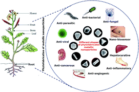
Metallic nanoparticles (MNPs) have seen myriad applications in various fields of science and technology. Green chemistry based NP synthesis offers several advantages over conventional approaches. In addition to being inexpensive, the biological approach is a facile, rapid, and environmentally benign method to synthesize NPs. Characterization of NPs is generally based on their dimension, surface area, and dispersity and the most common techniques used for the purpose are UV-Vis, DLS, AFM, SEM, FTIR, XRD, TEM, Raman spectroscopy, etc. These techniques are considered very useful as they provide valuable information which helps these materials to be used for diverse biological and environmental applications. Until now, numerous plant sources have been utilized to generate copious MNPs, such as silver, gold, copper, titanium, platinum, palladium, zinc, and iron NP. Among these MNPs, the noble ones have attracted significant researchers' attention because they are non-corrosive and resistant to oxidation in moist air. They have been useful in providing remedies with less or no adverse effects for several acute diseases like cancer, malaria, HIV, and hepatitis. Herein, we present a systematic review, focusing on phytofabricated MNPs and their wide range of applications, such as sensors, diagnostics and therapeutics, antimicrobial agents, anti-inflammatory agents, cancer treatment, etc.


image file: c6ra23411a-p1.tif

Anupriya Baranwal

Miss Anupriya Baranwal is currently pursuing M.Tech. in the Department of Biosciences and Bioengineering at the Indian Institute of Technology Guwahati. She received her B. Tech. degree in Biotechnology from Anand Engineering College, Agra, India. Her current research interest mainly includes nanobiotechnology and nanomedicine.

image file: c6ra23411a-p2.tif

Kuldeep Mahato

Mr Kuldeep Mahato is currently a Ph.D. student in the Department of Biosciences and Bioengineering at the Indian Institute of Technology Guwahati, India. He received his B. Tech. degree in Biotechnology from the National Institute of Technology Durgapur, India. His current research interest includes nanobiosensor, paper based biomedical devices, and nanobiotechnology.

image file: c6ra23411a-p3.tif

Ananya Srivastava

Dr Ananya Srivastava has done her Ph.D. from the Department of Chemistry Indian Institute of Technology Delhi, India. She has completed her masters of science in chemistry with a gold medal from D.D.D. University Gorakhpur, India. Her current research interest lies in synthetic organic chemistry, material science, and nanomedicine.

image file: c6ra23411a-p4.tif

Pawan Kumar Maurya

Dr Pawan Kumar Maurya is currently working as a researcher at Universidade Federal de Sao Paulo – UNIFESP, working on biochemical diagnostics, nanomedicine, and clinical biochemistry. He has done a Ph.D. from the University of Allahabad (A Central University), India & Post-Doc from National Taiwan University (NTU), Taipei Medical University (TMU), Taiwan. He is involved in teaching courses of Advanced Biochemistry and Metabolic Regulations at Amity University, India. He has published 48 research papers to date.

image file: c6ra23411a-p5.tif

Pranjal Chandra

Dr Pranjal Chandra is currently employed as Assistant Professor in the Department of Biosciences and Bioengineering, Indian Institute of Technology Guwahati. He earned his Ph.D. from Pusan National University, South Korea and did post-doctoral training at Technion-Israel Institute of Technology, Israel. He has published 50 papers/chapters/review in reputed journals and edited a book. Pranjal's research contributions are highly interdisciplinary, spanning a wide range in nanobiotechnology, nanobiosensors, lab-on-chip systems for biomedical diagnostics, and nanomedicine. He is the recipient of many prestigious awards and fellowships, including Ramanujan fellowship (DST, Gov. of India), BK-21 and NRF fellowship of South Korea, Technion post-doctoral fellowship, Israel. He is an editorial board member of World Journal of Methodology.


1. Introduction

Nanotechnology is the creation and careful manipulation of materials on an atomic or molecular level, and the use of these materials at the nano level for various purposes.1 “Nano” is acquired from a Greek word ‘nanos’ which means dwarf i.e. extremely small. In nanotechnology, the word nano indicates one billionth (10−9) of a meter and NPs are defined as groups of atoms or molecules in the range of 1–100 nm. NPs can be divided into two different groups, inorganic NPs and organic NPs. Inorganic NPs include magnetic NPs (iron oxide), noble metal NPs (silver and gold), and semi-conductor NPs (titanium oxide and zinc oxide), while organic NPs include carbon nanotubes, liposomes, and dendrimers. Extensive research in nanotechnology has opened several highly advanced and practically applicable research frontiers, such as nanobiotechnology, nanomedicine, and theranostics. These researches integrate the fundamental and applied aspects of nanotechnology and biology, where nanomaterials are synthesized either by using traditional physicochemical methods or by using biosynthetic routes mediated by microbes, whole plants, plant tissue, algae, etc.2,3 NPs are of particular importance because of their minuscule size and large surface area to volume ratio which imparts them with properties that are significantly different from those of the same material in bulk. Due to these unique properties, NPs are used for various applications which extend from catalysis, bio-imaging, bio-sensing,4 diagnostics,5 wound dressings, medical implants,6 gene/drug delivery, to the environment, cosmetics, food and feed, mechanics, and electronic components.1,7 The use of NPs in the medical field has seen an exponential increase because NP-bound drugs are claimed to have certain advantages which are not seen in traditional forms of drug.8 NP-linked drugs have a greater in vivo half-life, longer persistence in the body and can convey higher concentrations of drug to target locations.9 Targeted drug delivery using aptamer and antibody functionalization has been used to deliver drug molecules using NPs as carriers in recent years.10 Among all the noble MNPs, AgNPs and AuNPs are of greater interest due to their unique characteristics, such as chemical stability, good conductivity, catalytic activity, and ability to conjugate various biomolecules.11,12

NPs can be synthesized by following two basic approaches – ‘top down’ or ‘bottom up’.13 ‘Top down’ fabrication requires a huge amount of material and reduces large-size material to the nanoscale level, whereas the ‘bottom up’ approach creates NPs by adding atoms and molecular-scale components. The top down approach has a shortcoming of generating defects in the NP's surface structure, which affects the surface chemistry and physical characteristics of synthesized NPs. Being an effective approach, bottom up is preferred to avoid such drawbacks; however, it is more time consuming. Traditionally, physical, chemical, and physicochemical methods have been used to generate and stabilize the MNPs.14 Laser ablation,15 ion sputtering, mechanical milling,16 colloidal lithography,17 and high energy irradiation18 are the physical approaches and are of gold standard, whereas chemical methods, such as electrochemical and photochemical reduction, offer a simplistic approach to preparing MNPs.19 These methods are valuable, but issues related to carcinogenicity, and cellular and environmental toxicity are challenging, limiting MNP production if they are to be used for invasive biomedical applications.20 Therefore, there is need for clean, reliable, non-toxic, environmentally benign approaches to synthesize NPs.21 To address these issues, green synthesis of MNPs involving a bottom up approach came into the spotlight. This method is considered highly effective, because it offers an inexpensive, easily scalable, and environmentally friendly approach for NP production. More importantly, less or no requirement of high temperature, pressure, energy, and toxic chemicals make it a method of choice. However, the detailed mechanism behind NP synthesis is still under investigation.22

In the last few decades, there has been extensive research in green chemistry based NPs synthesis and several reports have been published to date. These reports focus on the exploitation of living entities, such as bacteria,23 fungus,24 plants,25 and viruses,26 for NP production. Extracts from plants such as Allium sativum,27 Aloe vera,28 Curcuma longa,29 Eclipta prostrata,30 Emblica officinalis,31 Azadirachta indica,32 Piper longum,33 Terminalia catappa34 and various others35 have been utilized for generating noble MNPs. Biogenesis wards off the problems faced by the traditional physicochemical approach and results in NPs of varied shapes, like spherical,36 hexagonal,37 tetrahedral,38 quasi spherical,36 decahedral,39 triangular,28 cubic,40 spheroidal,41 sizes, and composition. The biosynthetic route is proficient not only because it reduces metallic salts into their respective MNPs, but also because it can be used to generate MNPs of well-defined shape and size at a large scale.42 Among various biological routes of MNP production, microbe-mediated synthesis has not gained much importance due to its complex and special requirements of highly aseptic conditions, strain isolation, culture preparation and maintenance. However, the use of plant extracts for MNP synthesis has become a better method of choice due to its ease of improvement and lack of special requirements.25 Extracts from different plants contain different concentrations and combinations of reducing agents (alkaloids, phenolic acids, terpenoids, flavones, steroids, polysaccharides, amino acids, oximes, tannins, polyols, etc.) and due to these compounds plant extracts as a whole have been effectively utilized for the synthesis of MNPs.43

Over the past few decades, reviews focusing exclusively on green MNP synthesis have been published with great emphasis on different biological sources;2,35 however, their diverse application has not been well documented. We therefore present this review with the aim of giving a brief description of the green synthesis of MNPs using natural biological factories, especially plant extracts, techniques used for their characterization, and the potential clinical and therapeutic applications of these MNPs.

2. Plant-mediated synthesis of metallic nanoparticles

Plants have long been known to accumulate specific metals by reducing their ionic forms in different organs and tissues away from the penetration site of the ions, and later these metals can be extracted in an approach called phytomining. Detailed study of metal bioaccumulation has shown that these metals get accumulated in the form of nano-sized particles.44 This study paved the way for researchers to develop plant mediated nanoparticle synthesis. Until now, not only the whole plant but different plant parts such as leaves, fruit, peel, roots, seeds, callus, stems, and flowers have been utilized to synthesize MNPs like silver, gold, platinum, palladium, magnetite, zinc, copper, titanium oxide, nickel, etc.45 The general steps of plant-mediated MNP synthesis are shown in Fig. 1.
image file: c6ra23411a-f1.tif
Fig. 1 Basic steps involved in plant-mediated metallic nanoparticle synthesis.

The mechanism of plant-mediated MNP synthesis is shown in Fig. 2, indicating the general biosynthetic steps common to various types of plant materials. The MNP formation mechanism can be divided into three key stages. First is the activation stage during which metallic ions get reduced into metal atoms, followed by their nucleation. Second is the growth stage during which small nanoparticles aggregate spontaneously to form large-sized MNPs, and the duration of this stage decides which morphology the nanoparticle would ultimately achieve. Not only this, the growth stage is also followed by an increase in the thermodynamic stability of the MNPs. Third is the termination stage during which the plant extract helps to stabilize freshly prepared MNPs by letting them acquire the most energetically favourable conformation.


image file: c6ra23411a-f2.tif
Fig. 2 General schematic representation of MNP synthesis via plant extracts. The metallic ions (Me+) interact with bio-reductants present in the plant extract and get reduced to their respective metallic atoms (Me0). Stabilizing agents present in the plant extract bind with the reduced atoms to provide stability. The reduced metal atoms and biomolecule complex interacts with adjacent similar complexes to generate small MNPs, followed by growth and aggregation forming large-sized MNPs.

2.1. Role of plant metabolites in metallic nanoparticle synthesis

There are several plant biomolecules which play a crucial role in the reduction of metal salts to form nanoparticles and stabilize them by acting as capping agents (Fig. 3). A few commonly known biochemicals present in plant extracts and their probable mechanisms in MNP synthesis are discussed as follows:
image file: c6ra23411a-f3.tif
Fig. 3 Different chemical constituents of plant extracts which induce MNP formation. Carotene (A), quercetin (B), luteolin (C), eugenol (D), tyrosine (E), ferulic acid (F), and tryptophan (G).
Terpenoids. Terpenoids belong to a diverse organic polymer class which displays strong anti-oxidant activity. They are produced in plants from 5 carbon isoprene units and have been demonstrated to be associated with MNP production. The data from FTIR spectroscopy laid a platform to describe the mechanism of terpenoid-mediated metal reduction into MNPs. Singh et al. in their work suggested that deprotonation of the eugenol–OH group leads to the formation of resonance structures which can undergo a further oxidation process, which is eventually followed by active reduction of metal salts to form MNPs.46
Sugars. Sugars present in plant extracts can also assist in MNP synthesis. The linear aldehyde-containing monosaccharides, such as glucose, have been known to act as reducing agents. However, keto groups containing monosaccharides, such as fructose, can act as reducing agents only after they have undergone a series of tautomeric transitions. According to studies, the aldehyde group present in sugar gets oxidized into a –COOH group through nucleophilic addition of a hydroxyl group which in turn results in metal ion reduction and, thus, MNP formation.44 For instance, Cinnamon camphora leaf extract has been utilized for the formation of AgNPs and AuNPs.47
Flavonoids. Flavonoids belong to a large group of polyphenolic compounds which consists of numerous classes, such as flavonols, isoflavonoids, chalcones, flavones, and anthocyanins. Researchers have found that the enolic groups in flavonoids can undergo tautomeric transitions to give keto groups by releasing a reactive hydrogen atom which can reduce metal ions into their respective MNPs. For instance, the transformation of an enol group of luteolin flavonoid and rosmarinic acid present in Ocimum basilicum extract into a keto group played a crucial role in the reduction of silver ions into AgNPs.39
Proteins/peptides. Proteins/peptides are polymers of amino acids, organic compounds with both acidic (–COOH) and basic (–NH2) functional groups present in them. According to FTIR spectroscopy data, nascent MNPs are very often found in association with proteins/peptides which are present in plant extracts. Both carbonyl and/or amino groups of the main chain or side chain can mediate the binding of metallic ions such as the amino group of lysine, arginine, and histidine or the carboxyl group of aspartate and glutamate. Other groups which can bind metal ions include the thioether of methionine, the thiol of cysteine, and the hydroxyl group of serine, threonine, and tyrosine. It has been experimentally proven that these amino acids chelate metallic ions and reduce them to form MNPs.44 In one example, a cyclic peptide present in the latex of Jatropha curcas has been utilized for the formation of AgNPs.48
Tannins. Tannins belong to a class of hydrophilic polyphenolic compounds and are found in different parts of plants, such as pods, bark, roots, leaves, and plant galls. They act as powerful reducing agents (anti-oxidants) by donating electrons to the reactive oxygen species (ROS). Moreover, they are capable of chelating metal ions, leading to MNP formation.49 For example, tannins present in a leaf extract of Calliandra haematocephala have assisted in the formation of AgNP by providing electrons to ionic silver (Ag+).50

2.2. Factors influencing the synthesis and geometry of metallic nanoparticles

There are several factors like pH, temperature, reactant concentration, and reaction time which are known to influence the nucleation and formation of MNPs. These parameters are extremely important to discuss since they directly influence the geometry and stability of MNPs and the reproducibility of the biosynthetic method.
Impact of pH and temperature. The pH and temperature of the reaction mixture play a pivotal role in MNP synthesis from plant extracts. The varying hydrogen ion concentration and fluctuating temperature can generate MNPs of variable size, shape, and texture.29 Studies have shown that the lower the pH value the larger the size of the MNPs produced and vice versa. For instance, rod-shaped AuNP produced from the biomass of the Avena sativus plant were larger when the reaction mixture was kept at a pH value of 2, whereas on increasing this value to 3 and 4 the MNP size became relatively smaller.51 This study suggested that in the pH range 3 and 4 more functional groups were available in the reaction mixture for particle nucleation whereas at pH 2, the availability of functional groups was less and hence, agglomeration of particles led to the formation of larger AuNPs.

Green technology based MNP synthesis usually requires a temperature below 100 °C. Raju et al. studied AuNP synthesis from Semecarpus anacardium leaf extract over a range of temperatures52 and it was concluded that higher temperature will lead to highly spherical NPs whereas lower temperature supports nanotriangle formation.53 Rapid NP growth rate is observed at high incubation temperatures. AgNP synthesis from Chrysosporium tropicum over a temperature range revealed that with an increase in temperature from 25 to 30 °C, there was an increase in absorption and NP production. When the incubation temperature was kept at 25 °C, most of the AgNPs were of smaller size, whereas with subsequent increment in temperature to 30 °C, the formation of larger particles with a well-defined shape was reported. This observation suggests that there is a direct and distinct relationship between absorbance and the reaction mixture temperature.54

Influence of reduction time. Incubation time plays a vital role in determining the shape and quality of MNPs. Prathna et al. studied the effect of incubation time on AgNP synthesis from Azadirachta indica leaf extract and revealed that with an increase in incubation time, there is an increase in particle size.32 Variation in length of incubation time can influence particle properties in several ways; first, incubation for a long duration can lead to aggregation of NPs, which eventually results in large-sized particles; second, NPs may have a varying shelf life; and, third, in some cases their size may also decrease due to prolonged incubation time.
Influence of reactant concentration. Different plant extracts tend to have different biomolecular concentrations and studies have revealed that varying the concentration of biomolecules influences the geometry of NPs. It has been found that by increasing the concentration of Aloe vera extract in the reaction mixture there is an increase in spherical AuNP population and the percentage of gold nanotriangle formation was reduced.28 Moreover, AgNPs of various, shapes such as hexagonal, triangular, and spherical, have been synthesized by varying the amount of Plectranthus amboinicus plant extract in the reaction mixture.55

Based on the above discussion, it is evident that these factors greatly influence the size, shape, texture, and stability of MNPs. However, it cannot be denied that these experimental factors are prone to change with different plant extracts, as they contain different biochemicals and, hence, should be optimized for each synthetic procedure.

3. Characterization of metallic nanoparticles

Synthesis of MNPs is followed by their characterization based on properties like size, shape, surface area, and dispersity. In the case of both chemical and green approaches, the metal ions in aqueous metal salts get reduced and a color change is observed in the reaction mixture as a result, which is the very first indication that MNPs are being formed. However, a detailed characterization is required to ensure their formation.

The most commonly used techniques for characterization of MNPs are as follows: UV-visible spectroscopy (UV-Vis), dynamic light scattering (DLS), atomic force microscopy (AFM), scanning electron microscopy (SEM), Fourier transformation infrared spectroscopy (FTIR), transmission electron microscopy (TEM), powder X-ray diffraction (XRD), Raman spectroscopy, Auger electron spectroscopy (AES), photo correlation spectroscopy (PCS), inductively coupled plasma spectrometry (ICP), scanning probe electron microscopy (SPM), X-ray photoelectron spectroscopy (XPS), low energy ion scattering (LEIS), scanning tunneling microscopy (STM), time of flight secondary ion mass spectrometry (TOF-SIMS), and energy dispersive X-ray spectroscopy (EDS).

UV-visible spectroscopy is usually the first technique which is used to confirm MNP formation. It covers the range of both UV and visible radiations of the electromagnetic spectrum, where UV radiation lies in the range of 190–380 nm and visible radiation extends from 380 to 800 nm. This technique utilizes visible light of wavelength range 300–800 nm for identification, characterization and analysis of MNPs of various sizes.35 For instance, AgNPs and AuNPs show absorption in wavelength ranges of 400–450 nm and 500–550 nm, respectively.56

DLS is usually used to determine the surface charge, quality, size distribution and polydispersity index of prepared MNPs suspended in a liquid medium. The mean hydrodynamic radius of MNP is calculated by using the Stokes–Einstein equation:

 
image file: c6ra23411a-t1.tif(1)
where Rh is the hydrodynamic radius of NP, k is the Boltzmann constant, T is absolute temperature, η is the viscosity of the solvent, and D is the translational diffusion coefficient.57 However, the determination of attached functional groups, such as hydroxyls, carbonyls, amines, and the surface chemistry study of prepared MNPs are depicted by FTIR. Fig. 4 shows different important techniques used for the characterization of MNPs.


image file: c6ra23411a-f4.tif
Fig. 4 Different techniques used for the characterization of metallic nanoparticles.

TEM and SEM are electron microscopy techniques that are routinely used to determine the surface topology and other sub-micron-level characteristics of MNPs.58 TEM is used for the morphological characterization of MNPs and has thousand-fold higher resolution compared to SEM, whereas SEM provides morphological information at submicron level and elemental information when integrated with EDS.59

XRD is used in phase identification: i.e. examination of the overall oxidation state of particles as a function of time and determination of crystal structure of MNPs.60 Penetration of X-rays into the nanomaterial results in a diffraction pattern which is matched with standards to obtain structural data.35 XRD allows calculation of average MNP size (D) with the use of the Debye–Scherer equation:

 
image file: c6ra23411a-t2.tif(2)
where K is the Scherer constant with a value which ranges from 0.9 to 1, λ is the X-ray wavelength; B is the broadening of the diffraction line; θ is Bragg's diffraction angle. The line width of the (111) XRD peak gives the MNP size.61 Measurement of zeta potential enables a determination of the stability of prepared MNPs and greater values of zeta potential represent highly stable MNPs.58

AES, XPS, SPM and TOFSIMS are the techniques which allow primary surface analysis of MNPs. XPS and AES assist in the study of the composition and depth of the coating on particles, surface enrichment and depletion at NP surfaces. XPS analysis offers an advantage over other techniques as it can be used to determine the NP size when other techniques can't be used due to inappropriate conditions. Like FTIR, TOFSIMS is also used to attain molecular information about MNPs' surface chemistry.58

Thermal Gravimetric Analysis (TGA) and Differential Thermal Analysis (DTA) help in the analysis of the thermal stability of prepared MNPs and the identification of crystalline conditions. However, inductively coupled plasma spectrometry (ICP) allows the characterization of MNPs under actual environmental conditions. It helps in the quantification and precise size measurement of MNPs in the range 10–200 nm.62

AFM, STM and other scanning probe microscopy allow the characterization of MNPs under realistic conditions.63 AFM helps in the three-dimensional characterization of MNPs with subnanometer resolution and offers unique advantages over other techniques: viz. the determination of MNPs' physical properties, such as electrical, mechanical, and magnetic properties and, direct visualization of MNPs in hydrated form.

Raman spectroscopy helps in the detection of the vibrational modes of molecules, and therefore it has been widely used to determine the vibrational signals of various chemical groups linked to the MNP surface during the process of particle fabrication. For instance, surface enhanced Raman scattering (SERS) is useful in measuring single molecular attachment on an NP's surface.64

The low energy ion scattering (LEIS) process involves the scattering of low energy ionic beams, typically obtained from an inert gas (Ar, Rd), from the surface of particles, which leads to an energy loss in the ions. This loss in energy during the scattering process is examined and used for elemental analysis of the outermost surface of the material. LEIS is particularly useful because it offers high sensitivity towards the outermost layers of the MNP.65

Complete characterization of synthesized MNPs with the aforementioned techniques is considered very useful because it allows a proper understanding of the different aspects of MNPs, which play an important role in their widespread applications.

4. Metallic nanoparticles: types and their applications

The following section briefly discusses different types of phytofabricated MNPs and their widespread biomedical applications:

4.1. Silver nanoparticle

Silver is a rare but naturally occurring noble metal which has been widely used for various purposes across civilizations. For many years it has been used as jewellery and fine cutlery in many societies and it was believed that Ag offered health benefits to the users. The medicinal and preservative properties of silver have been known since ancient times. Out of all the metals with antimicrobial properties, silver was found to have the most effective antibacterial action and as result AgNPs have been synthesized by using plant sources like Tribulus terrestris L., Abutilon indicum, Cymbopogon citratus, O. tenuiflorum, S. tricobatum, Syzygium cumini, Centella asiatica, and Citrus sinensis and used as antimicrobials in numerous commercially available formulations.66 Another application of AgNPs has been seen in crop management and to provide safety from plant diseases, thereby improving crop yield.67 Recently, Logeswari et al. revealed the fungicidal effect of AgNPs and it was found that they can control phytopathogens in much a safer way compared to traditional fungicides.68 Huang et al. studied the anti-cancerous and anti-protozoal activities exhibited by AgNPs.47

4.2. Gold nanoparticle

Gold is another noble metal, very well known for exerting remedial properties against numerous diseases. There are several reports in history where a colloidal gold solution was used as a potable solution in order to cure various infections.69 Engineered AuNPs are of particular interest because they offer several unique properties, such as biocompatibility, high surface reactivity, minuscule size, varied shape, and resistance to oxidation.70 Albeit AuNPs are biologically inactive, they can be modified to have photochemical and chemical functionality.71 Owing to the unique properties offered by AuNPs they have found applications in numerous fields, such as biosensors,72 antimicrobial agents,73 theranostics,74 cancer hyperthermia,28 and gene and drug delivery platforms.75 The unique physicochemical properties of AuNPs have been exploited to transport and unload pharmaceuticals. AuNPs (cages, spheres, and rods) with their characteristic absorption in the near infra-red region could be used for the destruction of bacteria and cancerous cells via a photo-thermal heating mechanism.76 Synthesis of AuNPs has been achieved by conventional methods, but using plants as natural factories has provided a better, environmentally benign approach.1 For example, Aloe vera plant extract has been utilized to obtain gold nanotriangles of a size between 20 to 50 nm.28 Das et al. exploited Nyctanthes arbortristis flower extract to synthesize spherical AuNPs of a diameter of approximately 20 nm.77 There are several independent reports of AuNPs synthesis of various shapes (spherical, triangular, quasispherical, cubic hexagonal, decahedral, icosahedral, and rod shaped)35 by using a variety of plant sources, such as Anacardium occidentale,78 Camelia sinensis,79 Cymbopogon sp.,80 Geranium,81 Vitex negundo L.,82 Terminalia catappa,34 Memecylon edule,83 and Cinnamomum camphor.84

4.3. Copper and copper oxide nanoparticles

CuNPs possess noteworthy bactericidal activity against common pathogens such as E. coli and S. aureus85 and cytotoxic activity.86 Furthermore, CuO-NPs have been found to exhibit antioxidant and bactericidal behaviour.87 The fact that both CuNP and CuO-NPs possess strong antibacterial activity has enabled them to be used as an effective antibacterial coating on hospital equipment. Extracts from plants such as Syzygium aromaticum88 and Euphorbia nivulia89 have been utilized to synthesize CuNP, and CuO-NP synthesis was made possible by using Sterculia urens extract.85

4.4. Titanium dioxide and zinc oxide nanoparticles

Titanium is an abundant and widely distributed element in the earth's crust and its natural metallic form doesn't exist as it exhibits great affinity for oxygen and other metals. Its unique properties, like potential oxidation strength, high photo-stability, optical properties, anti-corrosive and photo-catalytic properties, and non-toxicity, have resulted in potential applications of TiO2-NPs in various fields, such as photo-catalysis, dermatological therapies, cosmetics, and skin care products. Currently preparation of TiO2-NP is under investigation as a novel treatment for dermatitis, such as acne vulgaris, hyper-pigmented skin lesions, atopic dermatitis, and recurrent condyloma accuminata.90 The antiseptic and bactericidal properties of TiO2-NPs have been studied against A. hydrophila, P. mirabilis, E. coli, S. aureus, and P. aeruginosa pathogens and the NPs were found to be highly effective against E. coli and S. aureus.91 TiO2-NPs have been synthesized using different plant extracts, such as Psidium guajava,91 Aloe vera,92 Nyctanthes arbortristis, and Citrus sinensis.93,94

ZnO nano formulation has also been studied recently as an important component in biomedical and cosmetic products1 and it has been synthesized using diverse plants sources, such as a flower extract of Cassia auriculata48 and leaf extracts of Hibiscus rosa-sinensis,95 Pongamia pinnata, Camellia sinensis,96 and Aloe vera.97 ZnO-NPs exhibit potent bactericidal action, and have been employed in waste water treatment, food packaging, and personal care products. Vimala et al. studied ZnO-NP as a drug delivery platform for doxorubicin and observed cytotoxicity against breast (MCF-7) and colon (HT-29) cancer cells.98

4.5. Palladium and platinum nanoparticles

Palladium is a noble metal with remarkable optical, catalytic, mechanical and electronic properties. PdNPs have been widely used in catalysis,99 drug manufacture and environmental pollutant processing,100 electrochemical applications,101 and/or as a sensor for the detection of various analytes. The medicinal application of PdNP is seen in dental treatment, target-based pro-drug activation, photo-thermal therapy, anti-tumor and antibacterial agents, and for prostate cancer and choroidal melanoma brachytherapy.102 However, PtNPs produced from Ocimum sanctum extract were found to have a catalytic property and therefore have the potential to be used in the production of hydrogen fuel components and chemical sensing.103

4.6. Indium oxide nanoparticle

Indium oxide (In2O3) is an important n-type semiconductor with several unique properties, like the ability to form strong interactions with poisonous gas molecules, high visible light transparency and electrical conductivity.104 In2O3-NPs have been produced using Aloe vera extract and extensively studied as promising materials for gas-sensing applications.105 But, to the best of our knowledge, plants other than Aloe vera have not been exploited for its synthesis; therefore, more research work needs to be focused in this direction.

5. Clinical applications of metallic nanoparticles

The following section briefly discusses clinical applications of phytofabricated MNPs. Since AgNP and AuNP are the most commonly synthesized MNPs by biological approaches, we have therefore described their applications in particular; however, applications of other MNPs synthesized by using green approaches have also been listed. Information about biological source, type of synthesized MNP and its biomedical application in various biological models are comprehensively described in Table 1.
Table 1 Summarizes the bactericidal, fungicidal, anti-viral, anti-parasitic and anti-cancerous activities of biosynthesized metallic nanoparticles
S. no. Biological source Salt used Test microorganism/cell line Impact References
(A) Antibacterial activities of metallic nanoparticles
1 Tribulus terrestris L. AgNO3 S. pyogens, B. subtilis, P. aeruginosa, E. coli and S. aureus Effective against test microorganisms 107
2 Abutilon indicum AgNO3 S. paratyphi, K. pneumoniae, B. subtilis, and P. aeruginosa Highly effective against test microorganisms 108
3 Cymbopogon citratus AgNO3 E. coli, P. milabilis, P. aeruginosa, S. flexneri, S. somenei, and K. pneumoniae AgNPs caused strong inhibition of the test microorganisms 109
4 O. tenuiflorum, S. tricobatum, Syzygium cumini, Centella asiatica, and Citrus sinensis AgNO3 S. aureus, P. aeruginosa, E. coli and K. pneumoniae AgNPs synthesized from O. tenuiflorum and S. tricobatum have highest anti-microbial activity against S. aureus and E. coli, respectively 68
5 Trianthema decandra AgNO3, HAuCl4 S. aureus, E. coli, S faecalis, P. aeruginosa, P. vulgaris, E. faecalis, B. subtilis, and Y. enterocolitica Both AgNP and AuNPs showed strong inhibition of test microorganisms 110
6 Salix alba HAuCl4 K. pneumonia, B. subtilis and S. aureus Highly effective against Staphylococcus aureus 111
7 Aloe vera Zn(NO3)2 S. aureus, S. marcescens, P. mirabilis and C. freundii Effective against test microorganisms 97
[thin space (1/6-em)]
(B) Anti-fungal activities of metallic nanoparticles
8 Sinapis arvensis AgNO3 Neofusicoccum parvum AgNPs showed significant antifungal activity against the microorganism 62
9 Brassica rapa L. AgNO3 Gloeophyllum abietinum, G. trabeum, Chaetomium globosum, Phanerochaete sordida AgNPs exhibited broad spectrum antifungal activity against the test microorganisms 112
10 Hypnea musciformis AgNO3 Candida albicans, Candida parapsilosis, Aspergillus niger AgNPs showed potent antifungal activity against test fungal strains 113
11 Rubus fruticosus (raspberry) AgNO3 Cladosporium cladosporioides, Aspergillus niger AgNPs exhibited significant inhibition of test fungal strains 114
12 Kluyveromyces marxianus, Candida utilis AgNO3 Candida albicans, Candida glabrata, Candida krusei AgNPs caused effective inhibition of test microorganisms 115
13 Salix alba HAuCl4 Alternaria solani, Aspergillus niger, Aspergillus flavus AuNPs exhibited good antifungal activity 111
14 Nepenthes khasiana HAuCl4 Aspergillus niger, Candida albicans AuNPs showed significant antifungal activity 116
15 Adansonia digitata AgNO3 Alternaria solani, Aspergillus flavus, Aspergillus niger, Penicillium chrysogenum, Trichoderma harzianum AgNPs acted as an effective antifungal agent 117
[thin space (1/6-em)]
(C) Anti-viral activity of metallic nanoparticles
16 Cinnamomum cassia AgNO3 Avian influenza virus subtype H7N3 AgNPs exhibited effective antiviral activity against test microorganism 118
17 Alternaria species, F. oxysporum, Curvularia species, C. indicum, and Phoma species AgNO3 Herpes simplex virus, Human parainfluenza virus type 3 AgNPs inhibited infectivity of tested viral strains 119
18 Aspergillus species AgNO3 Bacteriophage AgNPs showed viral inactivation with increasing concentration 120
[thin space (1/6-em)]
(D) Anti-parasitic activity of metallic nanoparticles
19 Catharanthus roseus AgNO3 Plasmodium falciparum Synthesized AgNPs were effective against tested malarial parasite 121
20 Azadirachta indica, Saraca asoca AgNO3 Plasmodium falciparum AgNPs caused effective inhibition of Plasmodium falciparum 122
21 Eclipta prostrata C4H6O4Pd Plasmodium berghei PdNPs inhibited growth of Plasmodium berghei 123
[thin space (1/6-em)]
(E) Anti-cancerous activity of metallic nanoparticles
22 Psidium guajava, Syzygium aromaticum AgNO3, HAuCl4 HT-29, HEK-293, K-562, HeLa cell line AuNPs produced from clove extract inhibited growth of HT-29, HEK-293 and, HeLa at moderate concentrations 124
23 Gelidiella sp. AgNO3 Hep-2 cell line AgNPs showed cytotoxic effect against test cell line 125
24 Corallina officinalis HAuCl4 MCF-7 cell line AuNPs exhibited anti-tumor activity which was supposed to be due to polyphenol content in algal extract 126
25 Olea europaea AgNO3 MCF-7 cell line AgNPs in olive extract possessed better cytotoxic effect against test cell lines 127
26 Sargassum muticum Zn(Ac)2·2H2O CT-26, WEHI-3, 4T1, and CRL-1451 (cancer cell lines), 3T3 (normal cell line) Synthesized ZnO-NPs exhibited cytotoxicity activity against WEHI-3 cell line without any effect on 3T3 normal cell lines 128


5.1. Antibacterial properties of metallic nanoparticles

Several bacterial species pose a serious threat to humankind and are ineffectively treated with current medications. Therefore, there is a strong requirement for new bactericides which can overcome this serious issue. AgNPs of varied shapes (spherical, rod-shaped, truncated/irregular, and hexagonal)1 and sizes have been prepared by green chemistry which prove to work as effective broad-spectrum biocides against Gram positive and Gram negative bacteria (Table 1A).106 The exact mechanism of toxicity caused by AgNPs is still under investigation. However, positively charged Ag ions are considered very crucial for bactericidal activity. AgNPs can penetrate cells by causing serious damage to the cell wall or cell membrane. It was found that once Ag ions are inside the cell they intercalate between the nitrogenous base pairs, avoiding hydrogen bond formation, which eventually leads to DNA damage. The bactericidal effect of these MNPs exhibited dosage dependence; however, it showed independence from acquisition of resistance against antibiotics.25 Increasing concentrations of AgNPs are seen to have faster membrane permeability than lower concentrations and consequently rupture the bacterial cell wall. In addition to AgNPs, the antibacterial effect of AuNPs, ZnO-NPs, and TiO2-NPs on clinically isolated pathogens has been studied and it was found that they can act as effective bactericidal agents (Table 1A).129

5.2. Anti-fungal properties of metallic nanoparticles

In the last few years there have been numerous reports of dermatitis caused by fungal species, Trichophyton and Candida.130 Fungal infections are commonly observed in patients who are immune-compromised due to cancer chemotherapy, HIV infections or disease like severe combined immunodeficiency. This rising trend of fungal infection is concerning not only because a limited number of medications are available, but also because treatment with antifungals has led to the emergence of resistant fungal strains. Not only this, commercially available antibiotics have several side effects. For example, amphotericin B has deleterious effects on the kidney, can cause renal failure, fever, nausea, trembling, and diarrhoea after being administered, and the antibiotic fluconazole causes liver toxicity and stops testosterone synthesis. Therefore, there is an inevitable requirement for novel anti-fungal medications.131 Plant-derived AgNPs have shown tremendous potential as anti-fungal agents and hence can be used in place of commercial antibiotics such as amphotericin and fluconazole. Fungal cells have ergosterol in their membranes and form various gradients across cytoplasmic membranes which allow them to maintain their membrane potential ability. AgNPs disrupt this gradient and destroy the membrane conformity, which eventually leads to cell death.132 Gradient disruption and loss of membrane conformity have also been correlated with inhibition of budding in yeast.133 AgNPs also offer promising activity against spore-producing fungus and effectively destroy the fungal growth.45 Similar to AgNPs, other MNPs like AuNPs and ZnO-NPs, have been studied for their antifungal activity and they were found to act as potent antifungal agents against several pathogenic strains like A. solani, P. graminis tritici, A. flavus, A. niger, C. albicans, T. harzianum, and R. stolonifer (Table 1B).97,115

5.3. Anti-viral activity of metallic nanoparticles

Viral infections pose a serious global threat to human health, being one of the many causes of deaths worldwide. Vaccination programs have significantly reduced the disease burden, but the emergence of resistance in viral strains and the deleterious side effects of extended anti-viral medications have brought about the imperative need for the development of safe and effective substitutes to traditional anti-viral drugs.134 Biosynthesized MNPs have proven to be an alternative drug for treating and controlling the viral pathogens due to their exceptional physical and chemical characteristics and large surface area to volume ratio.135 AgNPs can act as potent anti-viral agents because they inhibit virus entry into the host by interfering in the recognition of receptors. It is also unlikely that the virus will develop resistance against AgNPs as they attack a wide range of targets on the pathogen (Table 1C).136 Chemically produced AgNPs have shown potential anti-viral activity against cell-free viruses and cell-associated viruses. However, Fatima et al. observed similar activity in phytofabricated AgNPs against Avian influenza virus subtype H7N3, showing the promising impact of phytofabricated AgNPs in clinical research.118

5.4. Anti-parasitic activity of metallic nanoparticles

Parasitic diseases such as malaria, trypanosomiasis, and leishmaniasis are one of the many major health issues across the globe and the only choice to treat these diseases is with anti-parasitic chemotherapy, because parasitic infections do not elicit a prominent immune response, and thus efficacious vaccination against these infections is not possible.121 Keeping these problems in mind, there is an urgent need for potent and affordable anti-parasitic medications to control these infections. Despite intensive endeavours to eradicate malaria, it continues to be one of the greatest menaces to human health due to the rapid emergence of resistance in malarial parasites. Plant-derived drugs have been successfully employed to cure malaria for several years and, according to published reports, their anti-plasmodial activity is associated with the presence of certain chemical compounds, such as xanthones, terpenes, flavonoids, steroids, lignans, anthraquinones, and phenolic acid.137 Recent studies have shown that biologically synthesized MNPs, such as silver, platinum, and palladium, are effective in controlling the malarial population.45 Rajakumar et al. studied the anti-plasmodial activity of biologically synthesized PdNPs in Swiss albino mice and found that these PdNPs are effective in controlling the growth of Plasmodium berghei.123 More examples of anti-parasitic activity of MNPs are provided in Table 1D.

5.5. Anti-cancerous activity of metallic nanoparticles

In recent years there has been a surge in cancer cases and most of the time they end up being fatal due to the unavailability of satisfactory drugs or drug carriers.138 Sakamoto et al. highlighted the role of nanotechnology in cancer treatment by allowing early detection, accurate diagnosis, and targeted drug delivery.139 Recently, studies have reported that plant-derived MNPs can prove to be novel anti-cancer agents, as they have great potential to arrest uncontrolled cell growth and regulate the systematic cell cycle mechanism in cancerous cells. Researchers believed that this improved cytotoxic potential is a result of the presence of secondary metabolites and other non-metal compositions in the synthesizing medium.140 Several MNPs have been investigated for their cytotoxic effects, showing different degrees of cytotoxicity (Table 1E). The apoptotic activity of AgNP was found to be mediated by JNK- and ROS-dependent mechanisms, including the mitochondrial pathway in NIN3T3 cells.141,142 AgNPs regulate the cell cycle and enzymes in the blood stream and relatively control free radical formation in the cell.143

In 2012, the cytotoxic effect of AuNP was studied and it was found that AuNPs at moderate concentrations induced apoptosis in malignant cells without causing any damage to normal cells.144 Raghunandan et al. investigated the impact of biosynthesized AuNPs on four different cancerous cell lines – viz. HT-29, HEK-293, K-562, and HeLa cell lines – and found that AuNPs produced from clove extract inhibited the growth of HEK-293, HeLa and HT-29 cell lines.124 Similarly, green chemistry based AgNPs possessed remarkable cytotoxicity in HeLa cell lines compared to commercially available drugs.145

5.6. Anti-inflammatory activities of metallic nanoparticles

Inflammation is defined as a cascade of physiological responses to numerous abuses, such as injuries, harmful pathogens, and damaged cells. There are several potential inflammatory molecules – viz. enzymes, monoamines, cytokines, and chemokines – secreted by immune cells such as leukocytes, macrophages, mast cells, T-cells, and NK cells.33 It is a well-known fact that ROS play a pivotal role in modifying the inflammatory response during acute inflammation, which eventually causes cell injury and tissue damage. Hence, the inflammatory response must be terminated when its purpose has been achieved to prevent unnecessary tissue damage.146 For years, steroidal and non-steroidal anti-inflammatory drugs have been used as the main therapeutic agent to treat inflammation but associated side effects have limited their use. Therefore, the development of an alternative drug with no side effects with similar potential is required. In this context, researchers synthesized AgNPs using Sambucus nigra and Piper nigrum extracts and reported that the synthesized NPs have promising potential to act as anti-inflammatory agents in both in vitro and in vivo conditions.147,148

5.7. Hepatocurative properties of metallic nanoparticles

Being involved in the detoxification process, the liver is highly susceptible to injuries which can alter its metabolic functions and lead to chronic and acute diseases.149 Hepatotoxins which can result in such disorders are microbial metabolites, minerals, chemotherapeutic agents, and environmental pollutants.150 Biotransformation of these toxins in the liver leads to free radical formation. The malfunctioning activity of anti-oxidant enzymes causes oxidative degradation of lipids by assisting free radicals to interact with sulfhydryl groups such as glutathione and protein thiols, which eventually results in cell death.151 Phytofabricated AgNPs via. Andrographis paniculata leaf extract have shown a strengthening of the anti-oxidant defence system by allowing efficient free radical scavenging which cures the affected liver.152 Until now, the hepatocurative activity of plant-mediated MNP has rarely been attempted; therefore future work could be performed in this direction.

5.8. Anti-angiogenic properties of metallic nanoparticles

Angiogenesis is a crucial yet complex physiological phenomenon involved in blood vessel formation and plays a significant role in the normal growth and wound healing process. Gene products expressed from multiple genes in different cell types contribute to a cascade of events which culminates in performing controlled angiogenesis. The most important growth factors involved in this process are vascular endothelial growth factor (VEGF) and fibroblast growth factors (FGF).153 Any disparity in the normal levels of these growth factors may result in neovascularization, which can cause serious health problems, such as malignant, ocular, and inflammatory diseases. Antibody engineering and modification technology has been used to generate modified antibodies such as Lucentis, an anti-VEGF antibody fragment which proved to be an effective anti-angiogenic agent.154 In spite of its being effective, lack of efficiency limits its usage, so there is a need to develop an alternative anti-angiogenic agent which is not only effective but also efficient in its action. Studies have shown the significance of plant-mediated MNPs as powerful anti-angiogenic agents. For example, AgNPs exhibited a significant inhibitory effect on blood capillary formation in response to VEGF signalling, endothelial cell viability, cell migration and cell growth. The anti-angiogenic property of nanosilver is not only limited to these aforesaid applications, but it can also inhibit in vivo angiogenesis.155

5.9. Metallic nanoparticles in drug delivery

A number of drugs are seen to be associated with problems of poor stability, low selectivity, water insolubility and toxicity, so there is an immense need to develop a better drug delivery system which can overcome these problems. Nanoparticle-mediated drug delivery aims for enhanced delivery or uptake by target cells and thus reduces the toxic effects of a free drug to non-target organs. This results in an increase in the therapeutic index. Singh et al. developed a drug delivery system for controlled treatment of cancer by using AuNPs synthesized from Padina gymnospora leaf extract.156 Not only this, phytofabricated AuNPs from Jasminum sambac leaf extract have also been reported to deliver antimicrobial drugs, such as kanamycin and ampicillin, very efficiently.157 It has also been debated that these MNPs are highly biocompatible: i.e. exert no/less toxic effects during in vitro and in vivo investigations. To the best of our knowledge, not much work has been done in the field of green nanoparticles (phytofabricated) mediated drug and biomolecule delivery; therefore future work could be performed in this direction.

5.10. Anti-oxidant property of metallic nanoparticles

An anti-oxidant, as the name suggests, is a molecule that prevents the oxidation of other molecules. Oxidation of molecules leads to free radical formation, which initiates several chain reactions that may cause cellular damage.158 Free radicals, such as reactive oxygen species (ROS), are frequently generated as a natural by-product of normal mitochondrial oxidative metabolism and play an important role in cell signalling and homeostasis.159 Maintenance of ROS at a low concentration is strictly recommended to ward off oxidative stress conditions. Catalase, superoxide dismutase, and glutathione peroxidase are enzymes involved in free radical scavenging, providing an antioxidant defence mechanism inside the cell. This scavenging power of anti-oxidants plays a pivotal role in the management of various chronic diseases, such as diabetes, cancer, metabolic disorders, nephritis, and neurodegenerative disorders. In the green approach, ZnO-NPs synthesized from Polygala tenuifolia root extract showed an anti-oxidant effect and the effect was found to be dose dependent: i.e. a high concentration of NPs showed increase in anti-oxidant activity.146 Similarly, leaf extracts of Terminalia species and Chenopodium murale have been used to synthesize AgNPs and their anti-oxidant effect was examined by free radical scavenging of 1,1-diphenyl-2-picryl hydroxyl radical (DPPH),160 implying the potential use of MNPs in oxidative stress monitoring and medical diagnosis.

5.11. Metallic nanoparticles as nano-biosensors

MNP-based biosensors offer new prospects in diagnostics and clinical research due to their unique physicochemical properties.161,162 According to the reports, these biosensors could assist in disease diagnosis, marker analysis, cell tracking, therapy and disease pathogenesis monitoring, and for imaging.163,164 Plant-mediated MNPs have been used for the detection of clinically relevant molecules. For instance, in 2012, Kaur et al. synthesized AuNPs using Syzygium aromaticum extract for the colorimetric detection of urea in a simple, inexpensive, and disposable setting. It was further applied to detect urea in milk samples, showing the real application of these AuNPs in food safety.165 Raja et al. and Farhadi et al. used Calliandra haematocephala and Chlorogalum sp. extract to synthesize AgNP and applied it to the detection of hydrogen peroxide and mercuric ions, respectively.50,166 The literature has shown that most of the phytofabricated nano-sensors reported to date have not been genuinely analyzed for real samples and most of them are being used for chemical compound sensing. On the other hand, to the best of our knowledge, no work has been done on bio-functionalized phytofabricated MNPs for biosensing applications.

6. Conclusion and future perspectives

Nanotechnology as a pioneering technology has been anticipated to give hope for different fields of science, especially in biomedicine and therapy. The green synthesis of MNPs using plant tissue has been extensively studied in the last couple of decades, and it is still a fascinating area of research. The crude plant extract induces MNP production in an inexpensive, facile, clean, and non-toxic manner, with an ease of scalability. Utilization of these MNPs has been attempted for the treatment of several infectious and life-threatening diseases like malaria, HIV, hepatitis, cancer, etc. However, there has not been much work in this field at the pilot or commercial scales. Therefore, more research should be directed towards developing facile methods by understanding the kinetics and mechanism of MNP for large-scale synthesis using plant sources. Nanomedicine safety is not yet well understood, so for the continual use of MNPs in medical therapeutics like oncology there is an inevitable need to understand the safety and risk factors associated with the nanoparticles. In future, more research should be performed towards the use of phytofabricated MNPs for the functionalization of various biomolecules (antibodies, peptides, aptamers, etc.) and drugs for theranostic applications. It is also anticipated that these MNPs might have lower or no toxicity. Hence, they could be good candidates for advanced clinical research.

Conflict of interest

Authors report no conflict of interest in this work.

Acknowledgements

This work is supported DST-ECRA research grant (ECR/2016/000100) and DST Ramanujan Fellowship (SB/S2/RJN-042/2015) awarded to Dr Pranjal Chandra.

References

  1. M. Shah, D. Fawcett, S. Sharma, S. K. Tripathy and G. E. J. Poinern, Materials, 2015, 8, 7278–7308 CrossRef .
  2. S. Iravani, Green Chem., 2011, 13, 2638–2650 RSC .
  3. T. Luangpipat, I. R. Beattie, Y. Chisti and R. G. Haverkamp, J. Nanopart. Res., 2011, 13, 6439–6445 CrossRef CAS .
  4. M. Choudhary, P. Yadav, A. Singh, S. Kaur, J. Ramirez-Vick, P. Chandra, K. Arora and S. P. Singh, Electroanalysis, 2016, 28, 1–11 CrossRef .
  5. P. Chandra, J. Anal. Bioanal. Tech., 2015, 6, 1–2 Search PubMed .
  6. P. Chandra, A. Prashad, K. Mahato, S. N. Joshi, P. K. Maurya and A. Srivastava, J. Mol. Eng. Mater., 2016, 4, 1–22 Search PubMed .
  7. K. A. Khalil, H. Fouad, T. Elsarnagawy and F. N. Almajhdi, Int. J. Electrochem. Sci., 2013, 8, 3483–3493 Search PubMed .
  8. V. Wagner, A. Dullaart, A.-K. Bock and A. Zweck, Nat. Biotechnol., 2006, 24, 1211–1217 CrossRef CAS PubMed .
  9. S. Sahoo, S. Parveen and J. Panda, Nanomed. Nanotech. Biol. Med., 2007, 3, 20–31 CrossRef CAS PubMed .
  10. Y. Chen, V. J. Mohanraj, F. Wang and H. A. Benson, AAPS PharmSciTech, 2007, 8, 131–139 CrossRef PubMed .
  11. P. Chandra, H.-B. Noh, M.-S. Won and Y.-B. Shim, Biosens. Bioelectron., 2011, 26, 4442–4449 CrossRef CAS PubMed .
  12. R. Pallela, P. Chandra and H.-B. Noh, Biosens. Bioelectron., 2016, 85, 883–890 CrossRef CAS PubMed .
  13. K. N. Thakkar, S. S. Mhatre and R. Y. Parikh, Nanomed. Nanotech. Biol. Med., 2010, 6, 257–262 CrossRef CAS PubMed .
  14. P. Chandra, J. Singh, A. Singh, A. Srivastava, R. N. Goyal and Y.-B. Shim, J. Nanopart. Res., 2013, 1–12 CrossRef .
  15. F. Mafuné, J.-Y. Kohno, Y. Takeda and T. Kondow, J. Phys. Chem. B, 2001, 105, 9050–9056 CrossRef .
  16. P. Pardha-Saradhi, G. Yamal, T. Peddisetty, P. Sharmila, S. Nagar, J. Singh, R. Nagarajan and K. S. Rao, PLoS One, 2014, 9, e106715 Search PubMed .
  17. G. Zhang and D. Wang, J. Am. Chem. Soc., 2008, 130, 5616–5617 CrossRef CAS PubMed .
  18. M. Treguer, C. de Cointet, H. Remita, J. Khatouri, M. Mostafavi, J. Amblard, J. Belloni and R. De Keyzer, J. Phys. Chem. B, 1998, 102, 4310–4321 CrossRef CAS .
  19. S.-Y. Won, P. Chandra, T. S. Hee and Y.-B. Shim, Biosens. Bioelectron., 2013, 39, 204–209 CrossRef CAS PubMed .
  20. J. Ai, E. Biazar, M. Jafarpour, M. Montazeri, A. Majdi, S. Aminifard, M. Zafari, H. R. Akbari and H. G. Rad, Int. J. Nanomed., 2011, 6, 1117–1127 CAS .
  21. N. Kulkarni and U. Muddapur, J. Nanotechnol., 2014, 2014, 1–8 CrossRef .
  22. S. Dhuper, D. Panda and P. Nayak, NanoTrends: A Journal of Nanotechnology and Its Applications, 2012, 13, 16–22 Search PubMed .
  23. M. Husseiny, M. A. El-Aziz, Y. Badr and M. Mahmoud, Spectrochim. Acta, Part A, 2007, 67, 1003–1006 CrossRef CAS PubMed .
  24. P. Mukherjee, A. Ahmad, D. Mandal, S. Senapati, S. R. Sainkar, M. I. Khan, R. Parishcha, P. Ajaykumar, M. Alam and R. Kumar, Nano Lett., 2001, 1, 515–519 CrossRef CAS .
  25. S. Ahmed, M. Ahmad, B. L. Swami and S. Ikram, J. Adv. Res., 2016, 7, 17–28 CrossRef CAS PubMed .
  26. A. Merzlyak and S.-W. Lee, Curr. Opin. Struct. Biol., 2006, 10, 246–252 CrossRef CAS PubMed .
  27. M. Ahamed, M. M. Khan, M. Siddiqui, M. S. AlSalhi and S. A. Alrokayan, Phys. E, 2011, 43, 1266–1271 CrossRef CAS .
  28. S. P. Chandran, M. Chaudhary, R. Pasricha, A. Ahmad and M. Sastry, Biotechnol. Prog., 2006, 22, 577–583 CrossRef CAS PubMed .
  29. M. Sathishkumar, K. Sneha and Y.-S. Yun, Bioresour. Technol., 2010, 101, 7958–7965 CrossRef CAS PubMed .
  30. A. K. Jha, K. Prasad, V. Kumar and K. Prasad, Biotechnol. Prog., 2009, 25, 1476–1479 CrossRef CAS PubMed .
  31. B. Ankamwar, C. Damle, A. Ahmad and M. Sastry, J. Nanosci. Nanotechnol., 2005, 5, 1665–1671 CrossRef CAS PubMed .
  32. T. Prathna, N. Chandrasekaran, A. M. Raichur and A. Mukherjee, Colloids Surf., A, 2011, 377, 212–216 CrossRef CAS .
  33. S. J. P. Jacob, J. Finub and A. Narayanan, Colloids Surf., B, 2012, 91, 212–214 CrossRef PubMed .
  34. B. Ankamwar, Eur. J. Chem., 2010, 7, 1334–1339 CAS .
  35. A. K. Mittal, Y. Chisti and U. C. Banerjee, Biotechnol. Adv., 2013, 31, 346–356 CrossRef CAS PubMed .
  36. J. Kasthuri, S. Veerapandian and N. Rajendiran, Colloids Surf., B, 2009, 68, 55–60 CrossRef CAS PubMed .
  37. S. S. Shankar, A. Rai, A. Ahmad and M. Sastry, J. Colloid Interface Sci., 2004, 275, 496–502 CrossRef CAS PubMed .
  38. J. Gardea-Torresdey, J. Parsons, E. Gomez, J. Peralta-Videa, H. Troiani, P. Santiago and M. J. Yacaman, Nano Lett., 2002, 2, 397–401 CrossRef CAS .
  39. N. Ahmad, S. Sharma, M. K. Alam, V. Singh, S. Shamsi, B. Mehta and A. Fatma, Colloids Surf., B, 2010, 81, 81–86 CrossRef CAS PubMed .
  40. J. Y. Song and B. S. Kim, Korean J. Chem. Eng., 2008, 25, 808–811 CrossRef CAS .
  41. T. Prathna, N. Chandrasekaran, A. M. Raichur and A. Mukherjee, Colloids Surf., B, 2011, 82, 152–159 CrossRef CAS PubMed .
  42. J. E. Hutchison, ACS Nano, 2008, 2, 395–402 CrossRef CAS PubMed .
  43. K. Mukunthan and S. Balaji, Int. J. Green Nanotechnol., 2012, 4, 71–79 CrossRef CAS .
  44. V. Makarov, A. Love, O. Sinitsyna, S. Makarova, I. Yaminsky, M. Taliansky and N. Kalinina, Acta Naturae, 2014, 6, 35–44 CAS .
  45. P. Kuppusamy, M. M. Yusoff, G. P. Maniam and N. Govindan, Saudi Pharm. J., 2014, 24, 473–484 CrossRef PubMed .
  46. A. K. Singh, M. Talat, D. P. Singh and O. N. Srivastava, J. Nanopart. Res., 2010, 12, 1667–1675 CrossRef CAS .
  47. J. Huang, Q. Li, D. Sun, Y. Lu, Y. Su, X. Yang, H. Wang, Y. Wang, W. Shao and N. He, Nanotechnology, 2007, 18, 1–11 Search PubMed .
  48. P. Ramesh, A. Rajendran and M. Meenakshisundaram, J. Nanosci. Nanotechnol., 2014, 2, 41–45 Search PubMed .
  49. H. Křížová and J. Wiener, in Conference proceeding: Nanocon, 2013, pp. 600–605 Search PubMed .
  50. S. Raja, V. Ramesh and V. Thivaharan, Arabian J. Chem., 2015 DOI:10.1016/j.arabjc.2015.06.023 .
  51. V. Armendariz, I. Herrera, M. Jose-Yacaman, H. Troiani, P. Santiago and J. L. Gardea-Torresdey, J. Nanopart. Res., 2004, 6, 377–382 CrossRef CAS .
  52. D. Raju, U. J. Mehta and S. Hazra, Trees, 2011, 25, 145–151 CrossRef .
  53. J. Mittal, A. Batra, A. Singh and M. M. Sharma, Adv. Nat. Sci.: Nanosci. Nanotechnol., 2014, 5, 043002 CrossRef .
  54. N. Soni and S. Prakash, Am. J. Nanotechnol., 2011, 2, 112–121 CrossRef CAS .
  55. K. B. Narayanan and N. Sakthivel, Mater. Charact., 2010, 61, 1232–1238 CrossRef CAS .
  56. R. Devika, S. Elumalai, E. Manikandan and D. Eswaramoorthy, Sci. Rep., 2012, 1, 12 Search PubMed .
  57. N. Ahmad, S. Bhatnagar, S. S. Ali and R. Dutta, Int. J. Nanomed., 2015, 10, 7019–7030 CAS .
  58. S. Roy and T. Das, International Journal of Plant Biology and Research, 2015, 3, 1–11 Search PubMed .
  59. A. S. Eppler, G. Rupprechter, E. A. Anderson and G. A. Somorjai, J. Phys. Chem. B, 2000, 104, 7286–7292 CrossRef CAS .
  60. S. Sun, C. Murray, D. Weller, L. Folks and A. Moser, Science, 2000, 287, 1989–1992 CrossRef CAS PubMed .
  61. J. M. Ashraf, M. A. Ansari, H. M. Khan, M. A. Alzohairy and I. Choi, Sci. Rep., 2016, 6, 20414 CrossRef CAS PubMed .
  62. M. Khatami, S. Pourseyedi, M. Khatami, H. Hamidi, M. Zaeifi and L. Soltani, Bioresources and Bioprocessing, 2015, 2, 1–7 CrossRef .
  63. P. Klapetek, M. Valtr, D. Nečas, O. Salyk and P. Dzik, Nanoscale Res. Lett., 2011, 6, 1–9 CrossRef PubMed .
  64. G. E. J. Poinern, A Laboratory Course in Nanoscience and Nanotechnology, CRC Press, Boca Raton, 2014, pp. 207–219 Search PubMed .
  65. D. R. Baer, D. J. Gaspar, P. Nachimuthu, S. D. Techane and D. G. Castner, Anal. Bioanal. Chem., 2010, 396, 983–1002 CrossRef CAS PubMed .
  66. P. Asha Rani, G. Low Kah Mun, M. P. Hande and S. Valiyaveettil, ACS Nano, 2008, 3, 279–290 CrossRef PubMed .
  67. L. R. Khot, S. Sankaran, J. M. Maja, R. Ehsani and E. W. Schuster, Crop Prot., 2012, 35, 64–70 CrossRef CAS .
  68. P. Logeswari, S. Silambarasan and J. Abraham, J. Saudi Chem. Soc., 2015, 19, 311–317 CrossRef .
  69. M.-C. Daniel and D. Astruc, Chem. Rev., 2004, 104, 293–346 CrossRef CAS PubMed .
  70. S. K. Ghosh and T. Pal, Chem. Rev., 2007, 107, 4797–4862 CrossRef CAS PubMed .
  71. P. Prasson and B. Chittaranjan, International Journal of Scientific and Research Publications, 2012, 2, 1–4 Search PubMed .
  72. H.-B. Noh, P. Chandra, J. O. Moon and Y.-B. Shim, Biomaterials, 2012, 33, 2600–2607 CrossRef CAS PubMed .
  73. M. Ahire, K. Pardesi, J. Bellare, D. D. Dhavale, A. Jabgunde and A. Balu, Int. J. Nanomed., 2012, 7, 483–496 Search PubMed .
  74. P. Chandra, D. Das and A. A. Abdelwahab, Digest Journal of Nanomaterials & Biostructures, 2010, 5 Search PubMed .
  75. G. F. Paciotti, L. Myer, D. Weinreich, D. Goia, N. Pavel, R. E. McLaughlin and L. Tamarkin, Drug Delivery, 2004, 11, 169–183 CrossRef CAS PubMed .
  76. H. A. Salam, P. Rajiv, M. Kamaraj, P. Jagadeeswaran, S. Gunalan and R. Sivaraj, Int. Res. J. Biol. Sci., 2012, 1, 85–90 Search PubMed .
  77. R. K. Das, N. Gogoi and U. Bora, Bioprocess Biosyst. Eng., 2011, 34, 615–619 CrossRef CAS PubMed .
  78. D. Sheny, J. Mathew and D. Philip, Spectrochim. Acta, Part A, 2011, 79, 254–262 CrossRef CAS PubMed .
  79. A. R. Vilchis-Nestor, V. Sánchez-Mendieta, M. A. Camacho-López, R. M. Gómez-Espinosa, M. A. Camacho-López and J. A. Arenas-Alatorre, Mater. Lett., 2008, 62, 3103–3105 CrossRef CAS .
  80. S. S. Shankar, A. Rai, A. Ahmad and M. Sastry, Chem. Mater., 2005, 17, 566–572 CrossRef CAS .
  81. S. S. Shankar, A. Ahmad and M. Sastry, Biotechnol. Prog., 2003, 19, 1627–1631 CrossRef CAS PubMed .
  82. M. Zargar, A. A. Hamid, F. A. Bakar, M. N. Shamsudin, K. Shameli, F. Jahanshiri and F. Farahani, Molecules, 2011, 16, 6667–6676 CrossRef CAS PubMed .
  83. T. Elavazhagan and K. D. Arunachalam, Int. J. Nanomed., 2011, 6, 1265–1278 CrossRef CAS PubMed .
  84. X. Yang, Q. Li, H. Wang, J. Huang, L. Lin, W. Wang, D. Sun, Y. Su, J. B. Opiyo and L. Hong, J. Nanopart. Res., 2010, 12, 1589–1598 CrossRef CAS .
  85. V. V. T. Padil and M. Černík, Int. J. Nanomed., 2013, 8, 889–898 Search PubMed .
  86. M. Valodkar, R. N. Jadeja, M. C. Thounaojam, R. V. Devkar and S. Thakore, Mater. Chem. Phys., 2011, 128, 83–89 CrossRef CAS .
  87. D. Das, B. C. Nath, P. Phukon and S. K. Dolui, Colloids Surf., B, 2013, 101, 430–433 CrossRef CAS PubMed .
  88. I. Subhankari and P. Nayak, World J. Nano Sci. Eng., 2013, 2, 14–17 Search PubMed .
  89. M. Valodkar, P. S. Nagar, R. N. Jadeja, M. C. Thounaojam, R. V. Devkar and S. Thakore, Colloids Surf., A, 2011, 384, 337–344 CrossRef CAS .
  90. A. Wiesenthal, L. Hunter, S. Wang, J. Wickliffe and M. Wilkerson, Int. J. Dermatol., 2011, 50, 247–254 CrossRef CAS PubMed .
  91. T. Santhoshkumar, A. A. Rahuman, C. Jayaseelan, G. Rajakumar, S. Marimuthu, A. V. Kirthi, K. Velayutham, J. Thomas, J. Venkatesan and S.-K. Kim, Asian Pac. J. Trop. Med., 2014, 7, 968–976 CrossRef CAS PubMed .
  92. K. G. Rao, C. Ashok, K. V. Rao, C. S. Chakra and P. Tambur, International Journal of Advanced Research in Physical Science, 2015, 2, 28–34 Search PubMed .
  93. M. Sundrarajan and S. Gowri, Chalcogenide Lett., 2011, 8, 447–451 CAS .
  94. K. G. Rao, C. Ashok, K. V. Rao, C. S. Chakra and V. Rajendar, International Journal of Multidisciplinary Advanced Research Trends, 2015, 2, 1–9 Search PubMed .
  95. R. S. Devi and R. Gayathri, International Journal of Current Engineering and Technology, 2014, 4, 2444–2446 Search PubMed .
  96. S. Senthilkumar and T. Sivakumar, Int. J. Pharmacol. Pharm. Sci., 2014, 6, 461–465 Search PubMed .
  97. S. Gunalan, R. Sivaraj and V. Rajendran, Prog. Nat. Sci., 2012, 22, 693–700 CrossRef .
  98. K. Vimala, S. Sundarraj, M. Paulpandi, S. Vengatesan and S. Kannan, Process Biochem., 2014, 49, 160–172 CrossRef CAS .
  99. D. Astruc, Inorg. Chem., 2007, 46, 1884–1894 CrossRef CAS PubMed .
  100. B. Karimi, S. Abedi, J. H. Clark and V. Budarin, Angew. Chem., Int. Ed., 2006, 45, 4776–4779 CrossRef CAS PubMed .
  101. P. R. West, S. Ishii, G. V. Naik, N. K. Emani, V. M. Shalaev and A. Boltasseva, Laser Photonics Rev., 2010, 4, 795–808 CrossRef CAS .
  102. A. Dumas and P. Couvreur, Chem. Sci., 2015, 6, 2153–2157 RSC .
  103. C. Soundarrajan, A. Sankari, P. Dhandapani, S. Maruthamuthu, S. Ravichandran, G. Sozhan and N. Palaniswamy, Bioprocess Biosyst. Eng., 2012, 35, 827–833 CrossRef CAS PubMed .
  104. K. Gopchandran, B. Joseph, J. Abraham, P. Koshy and V. Vaidyan, Vacuum, 1997, 48, 547–550 CrossRef CAS .
  105. S. Maensiri, P. Laokul, J. Klinkaewnarong, S. Phokha, V. Promarak and S. Seraphin, J. Optoelectron. Adv. Mater., 2008, 10, 161–165 Search PubMed .
  106. C. Marambio-Jones and E. M. Hoek, J. Nanopart. Res., 2010, 12, 1531–1551 CrossRef CAS .
  107. V. Gopinath, D. MubarakAli, S. Priyadarshini, N. M. Priyadharsshini, N. Thajuddin and P. Velusamy, Colloids Surf., B, 2012, 96, 69–74 CrossRef CAS PubMed .
  108. R. Mariselvam, A. Ranjitsingh, A. U. R. Nanthini, K. Kalirajan, C. Padmalatha and P. M. Selvakumar, Spectrochim. Acta, Part A, 2014, 129, 537–541 CrossRef CAS PubMed .
  109. N. Geetha, T. Geetha, P. Manonmani and M. Thiyagarajan, Aust. J. Basic Appl. Sci., 2014, 8, 324–331 CAS .
  110. R. Geethalakshmi and D. Sarada, Int. J. Nanomed., 2012, 7, 5375–5384 CrossRef CAS PubMed .
  111. N. U. Islam, K. Jalil, M. Shahid, A. Rauf, N. Muhammad, A. Khan, M. R. Shah and M. A. Khan, Arabian J. Chem., 2015 DOI:10.1016/j.arabjc.2015.06.025 .
  112. K. B. Narayanan and H. H. Park, Eur. J. Plant Pathol., 2014, 140, 185–192 CrossRef CAS .
  113. J. S. Devi and B. V. Bhimba, Biosci., Biotechnol. Res. Asia, 2014, 11, 235–238 CrossRef CAS .
  114. J. Pulit, M. Banach, R. Szczygłowska and M. Bryk, Acta Biochim. Pol., 2013, 60, 795–798 Search PubMed .
  115. S. Ashur, Int. J. Curr. Microbiol. Appl. Sci., 2014, 3, 384–396 Search PubMed .
  116. B. Bhau, S. Ghosh, S. Puri, B. Borah, D. Sarmah and R. Khan, Adv. Mater. Lett., 2015, 6, 55–58 CrossRef .
  117. C. M. K. Kumar, P. Yugandhar and N. Savithramma, J. Appl. Pharm. Sci., 2015, 5, 082–089 Search PubMed .
  118. M. Fatima, N. Zaidi, D. Amraiz and F. Afzal, J. Microbiol. Biotechnol., 2016, 26, 151–159 CrossRef CAS PubMed .
  119. S. Gaikwad, A. Ingle, A. Gade, M. Rai, A. Falanga, N. Incoronato, L. Russo, S. Galdiero and M. Galdiero, Int. J. Nanomed., 2013, 8, 4303–4314 Search PubMed .
  120. G. Narasimha, H. Khadri and M. Alzohairy, Pharm. Lett., 2012, 4, 649–651 CAS .
  121. S. Ponarulselvam, C. Panneerselvam, K. Murugan, N. Aarthi, K. Kalimuthu and S. Thangamani, Asian Pac. J. Trop. Biomed., 2012, 2, 574–580 CrossRef CAS PubMed .
  122. A. Mishra, N. K. Kaushik, M. Sardar and D. Sahal, Colloids Surf., B, 2013, 111, 713–718 CrossRef CAS PubMed .
  123. G. Rajakumar, A. A. Rahuman, I.-M. Chung, A. V. Kirthi, S. Marimuthu and K. Anbarasan, Parasitol. Res., 2015, 114, 1397–1406 CrossRef PubMed .
  124. D. Raghunandan, B. Ravishankar, G. Sharanbasava, D. B. Mahesh, V. Harsoor, M. S. Yalagatti, M. Bhagawanraju and A. Venkataraman, Cancer Nanotechnol., 2011, 2, 57–65 CrossRef CAS PubMed .
  125. J. S. Devi, B. V. BhimbA and K. Ratnam, Int. J. Pharm. Pharm. Sci., 2012, 4, 710–715 Search PubMed .
  126. H. Y. El-Kassas and M. M. El-Sheekh, Asian Pacific Journal of Cancer Prevention, 2013, 15, 4311–4317 CrossRef .
  127. M. Rashidipour and R. Heydari, J. Nanostruct. Chem., 2014, 4, 1–6 Search PubMed .
  128. F. Namvar, H. S. Rahman, R. Mohamad, S. Azizi, P. M. Tahir, M. S. Chartrand and S. K. Yeap, J. Evidence-Based Complementary Altern. Med., 2015, 1–11 CrossRef PubMed .
  129. D. MubarakAli, N. Thajuddin, K. Jeganathan and M. Gunasekaran, Colloids Surf., B, 2011, 85, 360–365 CrossRef CAS PubMed .
  130. J. A. Woodfolk, Clin. Microbiol. Rev., 2005, 18, 30–43 CrossRef CAS PubMed .
  131. K.-J. Kim, W. S. Sung, S.-K. Moon, J.-S. Choi, J. G. Kim and D. G. Lee, J. Microbiol. Biotechnol., 2008, 18, 1482–1484 CAS .
  132. A. Nasrollahi, K. Pourshamsian and P. Mansourkiaee, Int. J. Nano Dimens., 2011, 1, 233–239 CAS .
  133. G. Zhao and S. E. Stevens Jr, BioMetals, 1998, 11, 27–32 CrossRef CAS PubMed .
  134. H. F. Hull, N. Ward, B. P. Hull, J. Milstien and C. de Quadros, Lancet, 1994, 343, 1331–1337 CrossRef CAS .
  135. J. S. Kim, E. Kuk, K. N. Yu, J.-H. Kim, S. J. Park, H. J. Lee, S. H. Kim, Y. K. Park, Y. H. Park and C.-Y. Hwang, Nanomed. Nanotech. Biol. Med., 2007, 3, 95–101 CrossRef CAS PubMed .
  136. M. Vitiello, M. Galdiero and M. Galdiero, Protein Pept. Lett., 2009, 16, 786–793 CrossRef CAS PubMed .
  137. L. Tona, R. Cimanga, K. Mesia, C. Musuamba, T. De Bruyne, S. Apers, N. Hernans, S. Van Miert, L. Pieters and J. Totté, J. Ethnopharmacol., 2004, 93, 27–32 CrossRef CAS PubMed .
  138. G. Dite, A. Whittemore, J. Knight, E. John, R. Milne, I. Andrulis, M. Southey, M. McCredie, G. Giles and A. Miron, Br. J. Cancer, 2010, 103, 1103–1108 CrossRef CAS PubMed .
  139. J. H. Sakamoto, A. L. van de Ven, B. Godin, E. Blanco, R. E. Serda, A. Grattoni, A. Ziemys, A. Bouamrani, T. Hu and S. I. Ranganathan, Pharmacol. Res., 2010, 62, 57–89 CrossRef CAS PubMed .
  140. S. Das, J. Das, A. Samadder, S. S. Bhattacharyya, D. Das and A. R. Khuda-Bukhsh, Colloids Surf., B, 2013, 101, 325–336 CrossRef CAS PubMed .
  141. Y.-H. Hsin, C.-F. Chen, S. Huang, T.-S. Shih, P.-S. Lai and P. J. Chueh, Toxicol. Lett., 2008, 179, 130–139 CrossRef CAS PubMed .
  142. S. Park, Y. K. Lee, M. Jung, K. H. Kim, N. Chung, E.-K. Ahn, Y. Lim and K.-H. Lee, Inhalation Toxicol., 2007, 19, 59–65 CrossRef CAS PubMed .
  143. V. Alt, T. Bechert, P. Steinrücke, M. Wagener, P. Seidel, E. Dingeldein, E. Domann and R. Schnettler, Biomaterials, 2004, 25, 4383–4391 CrossRef CAS PubMed .
  144. C. Dipankar and S. Murugan, Colloids Surf., B, 2012, 98, 112–119 CrossRef CAS .
  145. T. Suman, S. R. Rajasree, A. Kanchana and S. B. Elizabeth, Colloids Surf., B, 2013, 106, 74–78 CrossRef CAS .
  146. H. M. El-Rafie and M. A.-A. Hamed, Adv. Nat. Sci.: Nanosci. Nanotechnol., 2014, 5, 1–11 Search PubMed .
  147. L. David, B. Moldovan, A. Vulcu, L. Olenic, M. Perde-Schrepler, E. Fischer-Fodor, A. Florea, M. Crisan, I. Chiorean and S. Clichici, Colloids Surf., B, 2014, 122, 767–777 CrossRef CAS .
  148. K. M. Aparna Mani, S. Seethalakshmi and V. Gopal, J. Nanomed. Nanotechnol., 2015, 6, 1–5 Search PubMed .
  149. D.-H. Kuo, W.-H. Kang, P.-C. Shieh, F.-A. Chen, C.-D. Chang, M.-L. Tsai, A.-C. Cheng, C.-T. Ho and M.-H. Pan, Food Chem., 2010, 123, 1007–1012 CrossRef CAS .
  150. T. Wang, N.-L. Sun, W.-D. Zhang, H.-L. Li, G.-C. Lu, B.-J. Yuan, H. Jiang, J.-H. She and C. Zhang, J. Ethnopharmacol., 2008, 117, 300–308 CrossRef CAS PubMed .
  151. L. Li, W. Li, Y.-h. Kim and Y. W. Lee, Exp. Toxicol. Pathol., 2013, 65, 73–80 CrossRef CAS PubMed .
  152. U. Suriyakalaa, J. J. Antony, S. Suganya, D. Siva, R. Sukirtha, S. Kamalakkannan, P. T. Pichiah and S. Achiraman, Colloids Surf., B, 2013, 102, 189–194 CrossRef CAS PubMed .
  153. M. J. Cross and L. Claesson-Welsh, Trends Pharmacol. Sci., 2001, 22, 201–207 CrossRef CAS .
  154. D. S. Boyer, A. N. Antoszyk, C. C. Awh, R. B. Bhisitkul, H. Shapiro, N. R. Acharya and M. S. Group, Ophthalmology, 2007, 114, 246–252 CrossRef .
  155. S. Gurunathan, K.-J. Lee, K. Kalishwaralal, S. Sheikpranbabu, R. Vaidyanathan and S. H. Eom, Biomaterials, 2009, 30, 6341–6350 CrossRef CAS .
  156. M. Singh, M. Kumar, S. Manikandan, N. Chandrasekaran, A. Mukherjee and A. Kumaraguru, J. Nanomed. Nanotechnol., 2014, 5, 1–7 Search PubMed .
  157. S. Yallappa, J. Manjanna and B. L. Dhananjaya, Spectrochim. Acta, Part A, 2015, 137, 236–243 CrossRef CAS PubMed .
  158. P. K. Maurya, P. Kumar, S. Nagotu, S. Chand and P. Chandra, RSC Adv., 2016, 6, 53195–53202 RSC .
  159. P. K. Maurya, P. Kumar and P. Chandra, World J. Meth., 2015, 5, 216–222 Search PubMed .
  160. M. S. Abdel-Aziz, M. S. Shaheen, A. A. El-Nekeety and M. A. Abdel-Wahhab, J. Saudi Chem. Soc., 2014, 18, 356–363 CrossRef .
  161. P. Chandra, H.-B. Noh, R. Pallela and Y.-B. Shim, Biosens. Bioelectron., 2015, 70, 418–425 CrossRef CAS PubMed .
  162. W. C. A. Koh, P. Chandra, D.-M. Kim and Y.-B. Shim, Anal. Chem., 2011, 83, 6177–6183 CrossRef CAS PubMed .
  163. R. Prasad, J. Nanopart., 2014, 1–8 CrossRef CAS .
  164. G. Doria, J. Conde, B. Veigas, L. Giestas, C. Almeida, M. Assunção, J. Rosa and P. V. Baptista, Sensors, 2012, 12, 1657–1687 CrossRef CAS PubMed .
  165. B. Kaur, M. Markan and M. Singh, J. Bionanosci., 2012, 2, 251–258 CrossRef .
  166. K. Farhadi, M. Forough, R. Molaei, S. Hajizadeh and A. Rafipour, Sens. Actuators, B, 2012, 161, 880–885 CrossRef CAS .

This journal is © The Royal Society of Chemistry 2016
Click here to see how this site uses Cookies. View our privacy policy here.