DOI:
10.1039/C6RA23403H
(Paper)
RSC Adv., 2016,
6, 109192-109200
The exclusive response of LSPR in uncapped gold nanoparticles towards silver ions and gold chloride ions†
Received
20th September 2016
, Accepted 7th November 2016
First published on 7th November 2016
Abstract
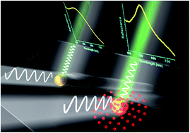
We report a significant modulation of the plasmon peak of a bare gold nanoparticle system after treatment with Ag+ or [AuCl4]− ions. The shift is highly selective to the presence of Ag+/[AuCl4]− metal ions and is not observed when other ions (Cu2+, Cd2+, Ce2+, Hg2+, Na+ and K+) are tested. The value of the red shift depends on the concentration of ions. As compared to Ag+ ions, the shift is larger in the presence of [AuCl4]− ions. We attribute the red-shift in plasmon to the formation of atomic clusters of Ag or Au and a change in relaxation times of hot electrons at the gold nanoparticle surface after treatment with Ag+ or [AuCl4]− ions. The red-shift is also sensitive to capping on gold nanoparticles, and silver ions do not shift the LSPR when gold nanoparticles are capped with citrate. The study of the effect of ions on the plasmon of AuNPs is significant from the viewpoint of designing highly selective AuNP based gold and silver ion sensors. The role of charge transfer and changes in electron dynamics, which lead to shifting of the localized surface plasmon resonance in AuNPs, has been exemplified as well. The versatility of the sensing approach has been tested in both solution-based and substrate-based configurations.
1 Introduction
The plasmon resonance is the most prominent aspect of gold nanoparticles (AuNPs) and has been thoroughly investigated for applications in nanotechnology. Various factors affect the localized surface plasmon resonance (LSPR), most notably the size and shape of AuNPs, dielectric properties of surrounding media and proximity of Au nanostructures to each other.1–3 The effects of the abovementioned factors have been studied and quantified theoretically as well as experimentally. Another factor which can significantly alter the LSPR is electron concentration in AuNPs.3,4 There are few studies which show red/blue shifts of the plasmon when the electronic concentration is altered through various processes such as electron irradiation, charge transfer, etc.5,6 If a AuNP sol contains active groups which can interact with gold, the shift in the plasmon is large due to attachment of solvent molecules to the gold surface and associated charge transfer; if solvent molecules do not bond covalently or ionically to the Au surface, then the shift is solely due to the dielectric constant of the solvent and is relatively small in magnitude (<5 nm).1,5,7
The dielectric properties of solvent can be varied by incorporating (dissolving) various moieties such as organic ligands (like molecules with thiol and amine groups) which have good affinity towards the gold surface.5,8–10 Use of organic molecules has been a predominant feature of many studies which focus on charge transfer, particularly due to capping of AuNPs by such ligands which prevents aggregation of particles.11,12 If inorganic ions are used to study their interaction with AuNPs, aggregation of particles makes the determination of red-shift due to charge transfer or possible reduction of ions (a reaction at the AuNPs surface) difficult.13–16 Thus, to determine red-shift of LSPR of uncapped AuNPs due to direct interaction with an ionic environment, we must first circumvent the problem of red-shift caused due to aggregation of AuNPs in an ionic environment. We use gold nanoparticles attached to silica nanospheres/glass to prevent aggregation of AuNPs inside the solution. Not only does the study serve as a model to sense ions in a simplified way but also demonstrates a facile approach towards fine modulation of LSPR in AuNPs while keeping the morphology intact.
2 Results and discussion
The silica nanospheres with AuNP decoration were subjected to various ionic environments and their optical properties in ultraviolet-visible region were tested (Fig. 1). We tried various alkaline ions and transition metal ions. It is found that the plasmon peak of supported AuNP particles does not shift unless the solution consists of silver or auric ions.
 |
| Fig. 1 (a), (b) and (c) represent TEM images of SiO2@AuNP (3 nm) systems covered with 3 nm AuNPs before any ionic treatment, after treating with AgNO3, and after treating with AuCl3, respectively. (d), (e) and (f) show the AuNP size distribution for small AuNPs attached over silica as seen in in (a), (b) and (c), respectively. The images confirm no change in morphology of AuNPs after treatment with ions (scale bar is 20 nm for TEM images; for particle size distribution diameters of more than 100 particles were measured). | |
2.1 Selectivity of shift in LSPR towards ionic species
Various ion treatments result in varying effect on plasmon band of SA3 (abbreviation for silica@AuNP particles with 3 nm as diameter of AuNPs) particles. Typically, the LSPR of SA3 particles appears at 510 nm and after testing various samples prepared following the same synthesis procedure we obtained standard deviation of ±2 nm in LSPR. The 2 nm standard deviation in plasmon peak is due to standard deviation in AuNP diameter and slight variation in density of AuNPs attached over the silica surface across various samples.17 While the ions such as Cu2+, Cd2+, Ce3+, Hg2+, Na+ and K+ (hereafter we will refer these ions collectively as CCCHNK ions) have negligible effect on plasmon band (and LSPR position) of AuNPs, Ag+ and [AuCl4]− are able to red-shift the plasmon peak by tens of nanometers (Fig. 2). With AgNO3 ions, the plasmon maximum of SA3 shifts from 510 nm to 517 nm. In presence of [AuCl4]− ions, the plasmon feature of SA3 is wide and is obscured by the presence of background absorption spectrum from [AuCl4]− ions itself and no clear plasmon maximum is observed; hence, to estimate the plasmon maxima and red-shift we eliminated background from both ‘SA3 in water’ and ‘SA3 in AuCl3’ spectra and the red-shift is found to be 35 nm. The plasmon peak variation (red-shift/blue-shift) of SA3 with CCCHNK ions is less than 2 nm and the width/extinction of LSPR peaks is either very similar (for Cu2+, Cd2+, Ce2+, Hg2+) to SA3 or decreases due to etching nature of some ionic solutions (KOH and NaOH).18 For all ions except the [AuCl4]− ions, the concentration of solution was 10 mM while for [AuCl4]−, 1 mM of solution concentration was utilized. If we use [AuCl4]− solution with concentration greater than 1 mM, then the LSPR peak is obscure due to large absorption by [AuCl4]− complex ions. When we centrifuge the ions out, the red-shift of LSPR (with respect to non-treated SA system) still exists but the value of shift decreases relative to the case when ionic solution was present alongside SA (Fig. 2b). For centrifuged ion out samples, prominent plasmon maxima is observed at 515 nm (for SA3 treated with AgNO3) and 524 nm (for SA3 treated with AuCl3) implying a red shift of 5 nm and 14 nm, respectively. The decrease in red-shift after centrifugation and redispersion process is probably due to change in dielectric constant of surroundings of SA3 particles after removal of excess ions since an ionic solution of AgNO3 (or AuCl3) will have its own contribution (due to electronic transitions) to extinction spectrum.19,20 We also tested other ionic salts of Ag and Au (i.e. Ag2SO4 and AuBr3) and found the value of red-shift in LSPR of SA3 to be nearly same to case of AgNO3 and AuCl3 (Fig. S6 in ESI†).
 |
| Fig. 2 Experimental UV-Vis spectra (normalized) results illustrate (a) plasmon peaks for SiO2@AuNPs (3 nm) particles in different ionic media, and (b) shifts in LSPR of SA3 particles in [AuCl4]− and Ag+ environments (dotted curve lines depict non-centrifuged sample cases). | |
In search of a more easy and efficient way to conduct detection and physical studies, we designed and tested AuNP-glass slide in thin film form (Fig. 3), for studying the effect of ionic treatment on LSPR of AuNPs in both transmittance and reflectance modes. After subjecting AuNP thin film on glass substrate with AgNO3 (or AuCl3) solution followed by rinsing with water, UV-Vis spectra shows red-shift with respect to non-treated case. It is observed that LSPR peaks are more prominent in reflectance spectra as compared to ones obtained in transmittance mode (Fig. 4a–c). There have been reports regarding LSPR transducers showing better refractive index sensitivity in reflection mode.21 We do not observe any LSPR of AuNP (3 nm) monolayer over glass substrate (referred to as Au3 hereafter) in transmittance measurement mode. A weak LSPR band appears at 545 nm after treatment of Au3 substrate with AgNO3 (10 mM) solution. When Au3 slide is treated with AuCl3 (0.5 mM) solution, a stronger plasmon (compared to Au3 and AgNO3 treated Au3 slide case) appears at 558 nm. The transmission spectra have been plotted after factoring in the contribution to transmission from glass slide (i.e. actual transmittance of sample is measured transmittance of sample divided by measured transmittance of glass slide).22 In reflectance measurement mode, plasmon peak of Au3 appears at 515 nm which shifts to 524 nm (or 542 nm) after treatment with 10 mM AgNO3 (or 0.5 mM AuCl3) solution. The standard deviation in all peak position measurements is ±2 nm. Thus, the red-shift of LSPR of AuNPs due to [AuCl4]− ions (red shift: 27 nm) is much greater compared to case of Ag+ ions (red shift: 9 nm).
 |
| Fig. 3 (a), (b) and (c) show SEM images of AuNP (3 nm) thin film on glass substrate before any ionic treatment, after treating with AgNO3, and after treating with AuCl3, respectively (scale bar: 100 nm). | |
 |
| Fig. 4 UV-Vis spectra of AuNP (3 nm) covered glass slides shows (a) appearance of plasmon band after ionic treatment in transmittance (obtained after dividing by transmittance of bare glass), (b) concentration dependent red-shift in LSPR for various concentrations of AuCl3, (c) concentration dependent red-shift in LSPR for various concentrations of AgNO3. The plot (d) shows the concentration dependent red-shift in LSPR of AuNP (7 nm) covered glass slides for various concentrations of AuCl3. Reflectance percent has been plotted in (b)–(d). | |
It must be noted that we carried out detailed studies with glass supported AuNPs only, the reasons being:
• The LSPR peaks are more prominent in reflectance spectra.
• Working with AuNP film on glass substrate is relatively hassle free since after ionic treatment slides can be quickly rinsed and subjected to UV-Vis characterization after drying.
On the contrary, silica@AuNP system has limitations of possibility of measurements in transmission mode only and a laborious procedure of removing ionic solution from SA particles via repeated centrifugation cycles. Repeated centrifugation cycles demand careful control on concentration of SA particles since each centrifugation results in loss of particles which affects extinction spectrum of sample.
2.2 Variation in LSPR shift with concentration of ions
We studied the dependence of shift of plasmon on concentration of ions (Ag+ or [AuCl4]−) to obtain the lowest possible concentration order which can be reliably detected. It is found that [AuCl4]− concentration as low as 10−9 M red-shifts the LSPR of Au3 by 8 nm (from 515 nm to 523 nm) while for Ag+ ion treatment the detection limit is of the order of 10−5 M (Fig. 4). Below the stated concentration limits, the difference in spectra of Au3 and ion treated Au3 slide is not appreciable and red-shifts are less than 2 nm (standard deviation limit for LSPR peak positions makes such detections unreliable). The red-shift of LSPR is a function of concentration of ions (in solution which Au3 slides are treated with) and larger concentrations induce larger red-shift. However, the red-shift does not increase indefinitely with increase in ion concentration but saturates after a limit. For example, in case of AgNO3 treatment of Au3 the maximum red-shift is 10 nm and the concentrations of AgNO3 > 50 mM do not change the LSPR position anymore (see Fig. S3†).
2.3 Interaction of charged species with AuNP assemblies
AgNO3 dissociates to Ag+ and NO3− in water while AuCl3 when dissolved in water gives negatively charged [AuClx(OH)4−x]− species where ‘x’ varies with the pH of the solution.23 The AuNPs prepared using a halide salt of gold are typically negatively charged due to strong adsorption of halide species over the AuNP surface.24–26 Both Ag+ and hydroxylated [AuCl4]− species are conducive to adsorption on Au surface; Ag+ due to ionic attraction and [AuCl4]− due to Au–Au coordination.27,28 The treatment of AuNPs with Ag+ leaves the surface of AuNPs positively charged while adsorption of [AuCl4]− over AuNPs leaves them negatively charged. The adsorption of ions and subsequent change of charge on particles is verified by measuring zeta potential of particles: −0.17 mV (for SA3), +0.40 mV (AgNO3 treated SA3) and −0.17 mV (AuCl3 treated SA3). X-ray photoelectron spectroscopy (XPS) measurements (Fig. 5) are carried out to ascertain valence state of Ag after interaction with AuNPs. The XPS shows that Ag is present (in AgNO3 treated Au3 system) as Ag0 (peaks at 368 and 374 eV correspond to Ag0) i.e. in chargeless or neutral form.29 The binding energy (Au4f) of Au is blue-shifted by 0.2 eV after treatment with Ag+ ions which indicates interaction of Au surface with Ag (Fig. S4†).30,31 It is known that Au surface of small gold nanoparticles (diameter < 10 nm) can reduce Ag+ ions to Ag0 (via anti-galvanic reaction) yielding atomic clusters of Ag attached to the AuNP surface.32–34 In such anti-galvanic reaction, the Ag does not replace Au but atomic clusters of Ag are present on the AuNP surface.35 The neutral charge of Ag on AuNP surface in conjunction with positive zeta potential of AgNO3 treated AuNPs indicates that Ag tends to replace chloride ions from the AuNP surface. The removal of chloride ions from AuNP surface makes AuNPs neutral as well results in a positive zeta potential of silica@AuNP particles due to free –NH2 terminations (which becomes NH3+ after protonation in an aqueous solution) on the silica surface. Anti-galvanic reactions and associated atomic cluster formation are known to depend on concentration of ion precursor, which matches well with variation of LSPR shift with concentration of ions in our study (Fig. 4).36 However, the anti-galvanic reaction on AuNP surface is still being investigated and as of now there is no clear explanation of process via which Ag+ reduces to Ag0. In addition to XPS measurements, EDX mapping (in STEM mode) carried out over SA3 particles also verifies the presence of silver over SA3 particle surface (Fig. S1†).
 |
| Fig. 5 XPS data shows the presence of gold in both (a) Au3 and (b) AgNO3 treated Au3 samples. Ag is not present in Au3 as seen in (c) while (d) shows the presence of Ag in AgNO3 treated Au3 sample. | |
We carried out control experiments to investigate whether aminosilane groups on APTES functionalized glass substrate itself is able to adsorb or reduce Ag+ and AuCl4− ions. The XPS studies on Ag+ and AuCl4− ion treated silanized glass substrates revealed presence of elemental Ag and Au (Fig. S5†). For Ag+ treated silanized glass substrate we observe peaks at 368 and 374 eV corresponding to the presence of Ag0. In case of AuCl4− ion treated silanized glass substrate, prominent peaks at 87.4 eV and 83.7 eV accompanied by satellite peaks at 88.4 eV and 84.8 eV are observed which corresponds to presence of elemental Au; the satellite peaks most probably correspond to Au bonded to –NH2 termination on silanized glass.37,38 Furthermore, the reflection spectra of Ag+ and AuCl4− ion treated silanized glass substrates show no LSPR peak (Fig. S5†). The XPS result in conjunction with non-observance of LSPR in reflectance spectra implies presence of Au (or Ag) in cluster form (<2 nm in size). TEM studies for analogous control experiments with silica particles also confirm the formation of ultrasmall Ag and Au clusters over –NH2 functionalized silica particles (Fig. S7†). The control studies thus strongly suggest the possibility of formation of atomic clusters by free amine termination on Au3 samples which may affect the surrounding dielectric constant of thin film and hence shift the LSPR.
2.4 Effect of nanoparticle size on ion sensing
It is a well-known fact that smaller nanoparticles have higher surface energies compared to larger sized particles and any surface dependent phenomenon will rely on surface to volume ratio of particle.39,40 Since LSPR, adsorption of ions and anti-galvanic reaction are surface related phenomena, we also probe the effect of size of AuNPs on magnitude of red-shift in LSPR due to the presence of [AuCl4]− ions.3,41,42 For treatment with AuCl3, our studies when repeated with 7 nm AuNP monolayer (Au7) over glass substrate show a shift of 20 nm (LSPR of Au7 shifts from 520 nm to 540 nm after treatment with 0.5 mM AuCl3) and the shift depends on concentration of ions (Fig. 4d). If we compare Au3 and Au7 studies, it is evident that for a treatment with same [AuCl4]− concentration, red-shift of plasmon maxima in case of Au3 (red shift = 27 nm) is larger as compared to Au7 particles (red-shift = 20 nm). Thus, smaller AuNPs are better for ion detection since they illustrate larger shift in LSPR for same concentration of ions. From a physical point of view, it implies that atomic clusters formed on AuNP surface are active participants in process where free electrons inside nanoparticles give rise to plasmonic features.42
2.5 Effect of citrate capping on AuNPs in ion sensing
If we use AuNPs capped with citrate moieties (prepared using Turkevich method) in place of bare AuNPs attached over silica, no red-shift of plasmon peak is observed after AgNO3 (20 mM) treatment of particles (Fig. 6a). For AuCl3 (0.5 mM) case as well, the red-shift (10 nm: from 538 nm to 548 nm) is smaller as compared to SA3 case (red-shift was 27 nm). The non-shifting or lesser magnitude of shift in LSPR for citrate capped AuNPs (SAu10cit) suggests that the red-shift depends on the direct interaction between Au surface and ions in the solution. Most probably, the interaction between Au surface and ions is inhibited by capping agent on AuNPs surface and the red-shift is less in comparison to bare AuNP case. The hindrance explanation is further substantiated by results obtained after treatment of SAu10cit particles with a blue-shifting (electron donating) agent like NaBH4; LSPR blue-shifts only by 3 nm after treatment with NaBH4. Thus, if any capping molecules are present over the surface of AuNPs, any possible charge transfer or interaction between AuNPs and ions is largely hindered and red shift in plasmon is restricted. Various researchers have proposed blue shift in plasmon peak due to charge transfer from anions (like BH4−, I−) to AuNP but similar shift in plasmon peak due to cations or metal ion complexes is not that well reported.3,43
 |
| Fig. 6 Experimental UV-Vis spectra shows (a) plasmon peaks after normalization for SiO2@AuNPs (10 nm, citrate capped) particles in different ionic media as stated on the figure, and (b) stabilization of red-shift in LSPR of SA3 particles in Ag+ environment after 6 minutes (for better clarity graphs have been plotted after y-offset). | |
2.6 Eliminating probability of red-shift in LSPR due to aggregation
To confirm that red-shift in plasmon is only due to ionic environment and not due to any aggregation phenomena catalyzed by Ag+/[AuCl4]− ions, we centrifuge out AgNO3 solution from mixture containing particles for TEM imaging. The TEM images show no change in morphology of AuNPs attached over the silica particles (Fig. 1). As evident from statistical distribution, the particle size variation across the samples is less than 1 nm which will not give large red-shifts (>10 nm) in extinction spectra of samples (Fig. 1d–f).44 Variation of AuNP particle size (attached over silica nanospheres) from 3 to 5 nm typically increases LSPR extinction but does not red-shift the LSPR wavelength.15,45 Similarly for AuNP-glass system, the SEM images indicate that the AuNP film/monolayer over glass is physically not affected by ionic treatment or there is no obvious change in particle morphology (Fig. 3a–c). The SEM image shows that no aggregation or size change of AuNPs takes place after the treatment of glass slides with AgNO3 (10 mM) or AuCl3 (1 mM) solutions. Since resolution of SEM is limited, SEM imaging cannot determine presence of small atomic clusters which possibly nucleate on silanized glass substrate itself after ionic treatment.
2.7 Time dependent red-shift and the effect of concentration of silica@AuNPs on LSPR
The change in LSPR is fast and red-shift in LSPR stabilizes within 6 minutes. The gradual shift of LSPR points to fast reaction kinetics for attachment and anti-galvanic reduction of ions around AuNPs (Fig. 6b). Even though little is known about kinetics of anti-galvanic reaction between Au and Ag+ ions, there are literature reports which suggest rapid reduction of Ag+ ions over Au surface (from 30 seconds to 5 minutes).34,46 The rapid change of LSPR observed in our study is probably due to two processes responsible for shift in LSPR viz. anti-galvanic reaction at AuNP surface and formation of Ag or Au clusters on functionalized silica surface. It is possible that the simultaneous reduction of Ag+ (or AuCl4−) ions at AuNP surface as well aminosilane terminated silica surface lead to faster formation of Ag (or Au) clusters and thus a rapid change in LSPR. The concentration of particles in sol does not affect the red shift significantly. The plasmon peaks show similar shift for low as well as high concentration of SA3 particles in the solution after AgNO3 is added (Fig. S2†). When we use 0.6 mg ml−1 of concentration (c) of particles, the plasmon peak is shifted by 9 nm after addition of AgNO3 and as we move down to lower concentrations up to 0.07 mg ml−1, shift in LSPR is typically 7 nm (constant for 0.6 < c ≤ 0.07 mg ml−1). We could not go below 0.07 mg ml−1 of particle concentration as the LSPR signal to noise ratio decreases at concentrations lower than that and thus shift in LSPR cannot be quantified reliably (Fig. S2†).
Though the understanding of blue-shift in plasmon peak due to adsorption of anions like I− over bare plasmonic nanoparticles is clear, there are not many reports on red-shift of LSPR due to cations. The gradual shift of LSPR with time implies that red-shift is not due to change in refractive index of the solution. Moreover, the refractive index for aqueous solutions of AgNO3 and AuCl3 is not drastically different as compared to pristine water. Hence, adsorption of cations/ions either over the AuNPs or in surrounding silica seems to be sole explanation of red-shift of plasmon. AgNO3 dissociates into Ag+ and NO3− ions in solution and Ag+ ions will be attracted to negatively charged AuNP surface. The Ag+ ions attached to the surface of ultrasmall AuNPs very likely reduce to Ag atomic clusters thus changing the electron dynamics of the system and red-shifting the LSPR (as explained in next section). More the concentration of AgNO3, higher the red-shift till attachment of Ag+ over AuNPs saturates; the negligible changes in LSPR for AgNO3 conc. > 25 mM substantiate this explanation.
2.8 Mechanism responsible for the red-shift of LSPR
There can be several reasons for red-shift in LSPR such as higher refraction, change in particle size/shape, inter-particle coupling or aggregation.7,47–49 However, other reasons might also be the variations in electron concentration in AuNPs (due to changes at surface) or change in lifetimes of hot electrons.50 The attachment of entities (Ag and Au atomic clusters) causes a red-shift in LSPR of AuNPs. The transfer of electrons to adsorbate will cause a scarcity of electrons in AuNPs and with lesser number of electrons available, the LSPR will red-shift in comparison to the case when no adsorbates are there on the surface. However, the magnitude of changes in LSPR seen after ion treatment of AuNPs indicates that LSPR shift cannot be wholly due to expected change in electron concentration.3 We can calculate the change in electron concentration in AuNPs using51,52 |
 | (1) |
where, Δλ is the change in LSPR wavelength λ being shifted peak wavelength, λp (=130 nm) is bulk plasma wavelength for gold, ε∞ (=12.2) is high-frequency dielectric constant for gold, ΔN is change in nanoparticle electron concentration, L (=1 for spherical shape) is shape factor for nanoparticle, and εm is dielectric constant of surrounding media (1 for air and 1.77 for water).
From eqn (1) it can be calculated that a red-shift of LSPR by 30 nm implies a 13% lower concentration of free electrons for ion-treated AuNPs. However, the possible presence of Au or Ag atomic clusters over ion treated AuNP is unlikely to trigger mass withdrawal of electrons from AuNPs. Other than the number of electrons, the plasmon wavelength also depends on the scattering of free electrons at metal–dielectric interface.49,53 In case of ion treated AuNPs, the plasmonic electrons will not be directly scattered by the AuNP-dielectric interface; rather, they will be scattered at AuNP-atomic cluster interface. It has been known that lifetimes of electrons in AuNPs can be altered by attaching different moieties over AuNP surface.8,54,55 The attachment of chemical moieties can lead to overall reduction in electron scattering and a stronger plasmon.8,54 For small nanoparticles, electron scattering is large in comparison to larger sized nanoparticles which results in lower extinction values for plasmonic signal.3,49,56 When suitable chemical entities which can participate in electron transfer with AuNPs (and thus can alter lifetimes of electrons) are adsorbed on AuNPs surface, it is expected that we get a red-shifted and strong plasmon. We strongly believe that change in electron lifetime is the phenomenon which is responsible for red-shift and a strong plasmon which is observed for bare AuNPs treated with Ag+ (or [AuCl4]−) ions. Since calculation of red half-width at half maximum (Γ1/2) can give a good idea about dephasing of plasmon in nanoparticle ensemble, we calculated Γ1/2 for SA3 nanoparticles from their normalized absorption spectra (Fig. 7). It is found that Γ1/2 is smaller for Ag+ and AuCl4− treated SA3 colloids with its value being 0.33 eV, 0.25 eV and 0.26 eV for SA3, SA3 (Ag+ treated) and SA3 (AuCl4− treated), respectively. The smaller values of Γ1/2 for ion-treated systems indicate that in comparison to untreated AuNPs, plasmon dephases slowly in Ag+ and AuCl4− treated AuNPs.
 |
| Fig. 7 Red half-width at half maximum (Γ1/2) calculated using normalized extinction spectra (UV-visible spectra after normalization) of silica@AuNPs (AuNP diameter = 3 nm) after treatment with AgNO3 and AuCl3. | |
To measure the lifetime of electrons in AuNPs (supported over glass substrate) we carried out time resolved absorption spectroscopy measurements before and after ionic treatment of AuNP thin film on glass substrate (Fig. 8). It is noticed that decay times for hot electrons increase after the treatment of AuNP/glass substrates with AgNO3 or AuCl3 solutions. For untreated and ion treated AuNPs on glass slide, the differential absorption versus time plot yields two decay times after biexponential fitting. The hot electron relaxation has two components and decay times (τe–ph, τph–ph) are (3.86 ps, 112 ps), (3.98 ps, 179 ps) and (2.78 ps, 246 ps) for AuNPs, AgNO3 treated AuNPs and AuCl3 treated AuNPs, respectively. The hot electrons relax through electron phonon interactions in AuNP lattice and the lattice further relaxes through phonon–phonon relaxation. The electron–phonon relaxation dynamics primarily determines the lifetime and thus the linewidth of plasmon band.53 However, even though we observe greater τe–ph for AgNO3 treated AuNPs, the τe–ph is significantly smaller for AuCl3 treated AuNPs. If only lifetime is to be taken into account, line-width would be greater and plasmon band would not red-shift in case of AuCl3 treated AuNPs. The significant red-shift as well as smaller linewidth (in spite of smaller τe–ph) for AuCl3 treated AuNPs implies that AuCl3 treatment induces changes in refractive index around AuNPs (Fig. 9). The greater τph–ph also points to different mode of lattice relaxation in case of AuCl3 treated AuNPs which is possible if AuCl3 treatment makes significant changes to lattice for small AuNPs (<10 nm).57,58 It is well known that AuNPs smaller than 10 nm are more sensitive towards external agents and electron as well as phonon dynamics in them can be significantly altered to modify their plasmonic characteristics.49,56 In conjunction with XPS studies and possibility of atomic cluster formation at AuNPs, the transient spectroscopy suggests that formation of atomic clusters alters the electron dynamics in ion treated AuNP thin films. Furthermore, control studies suggest formation of atomic clusters (of Ag and Au) on substrate itself which implies change in effective dielectric constant of AuNP thin films. We thus infer that treatment of AuNPs with AgNO3 or AuCl3 besides altering the relaxation dynamics of hot electrons in AuNPs also changes effective dielectric constant of AuNP thin film. The changes in relaxation dynamics of hot electrons and dielectric constant of thin film are thus responsible for red-shift of LSPR in AuNP ensemble after.
 |
| Fig. 8 Experimental transient absorption spectroscopy plots (differential absorption vs. time) for (a) AuNP thin film over glass, (b) AgNO3 treated AuNP thin film, (c) AuCl3 treated AuNP thin film. | |
 |
| Fig. 9 Schematic showing monolayer of AuNPs over glass with Ag or Au clusters attached on the surface of glass as well as AuNPs. The ions induce distortions at AuNP surface which changes the electron–phonon and phonon–phonon relaxation. | |
3 Conclusions
We have shown significant manipulation and redshift of LSPR characteristics of ultrasmall bare AuNPs using Ag+/AuCl4− ion treatment. The red-shifts up to 10 nm and 30 nm are possible using millimolar concentrations of Ag+ and AuCl4− ions, respectively. The reflection geometry with AuNP monolayer coated over glass slide proves to be better at sensing of ions (down to 100 nM or 10 ppb) and with certain modifications can be explored further to detect other ions. The probable mechanism responsible for red-shift due to ion treatment is formation of Ag or Au nanoclusters over AuNP surface which changes the hot electron dynamics in AuNPs. The adsorption of gold and silver ions supposedly perturbs the surface of AuNPs as well as substrate which is reflected as variations in LSPR.
Abbreviations
AuNPs | Gold nanoparticles |
SA | Silica@AuNPs particles |
SA3 | Silica@AuNPs (3 nm) particles |
SAu10cit | Silica@AuNPs (10 nm citrate capped AuNPs) |
Au3 | AuNP (3 nm) monolayer over glass substrate |
Au7 | AuNP (7 nm) monolayer over glass substrate |
Acknowledgements
We thank CRNTS (IIT Bombay) and SAIF (IIT Bombay) for providing FEG-TEM and FEG-SEM facilities. We also thank Prof. Anindya Datta (Department of Chemistry, IIT Bombay) for providing us access to his time resolved absorption spectroscopy set-up. We gratefully acknowledge the Industrial Research and Consultancy Center (IRCC) of IIT Bombay, Council of Scientific and Industrial Research (CSIR, New Delhi) and National Center for Photovoltaic Research and Education (NCPRE-project funded by MNRE, the Government of India) for the financial support of this work.
References
- K. L. Kelly, E. Coronado, L. L. Zhao and G. C. Schatz, J. Phys. Chem. B, 2003, 107, 668–677 CrossRef CAS.
- C. Tabor, D. Van Haute and M. A. El-Sayed, ACS Nano, 2009, 3, 3670–3678 CrossRef CAS PubMed.
- P. Mulvaney, Langmuir, 1996, 12, 788–800 CrossRef CAS.
- C. Goldmann, R. Lazzari, X. Paquez, C. Boissière, F. Ribot, C. Sanchez, C. Chanéac and D. Portehault, ACS Nano, 2015, 9, 7572–7582 CrossRef CAS PubMed.
- A. Henglein, Chem. Mater., 1998, 10, 444–450 CrossRef CAS.
- A. Henglein and C. Brancewicz, Chem. Mater., 1997, 9, 2164–2167 CrossRef CAS.
- A. C. Templeton, J. J. Pietron, R. W. Murray and P. Mulvaney, J. Phys. Chem. B, 2000, 104, 564–570 CrossRef CAS.
- K. O. Aruda, M. Tagliazucchi, C. M. Sweeney, D. C. Hannah, G. C. Schatz and E. A. Weiss, Proc. Natl. Acad. Sci. U. S. A., 2013, 110, 4212–4217 CrossRef CAS PubMed.
- Y.-R. Kim, R. K. Mahajan, J. S. Kim and H. Kim, ACS Appl. Mater. Interfaces, 2010, 2, 292–295 CAS.
- P. K. Jain and M. A. El-Sayed, Nano Lett., 2008, 8, 4347–4352 CrossRef CAS PubMed.
- G. Sener, L. Uzun and A. Denizli, ACS Appl. Mater. Interfaces, 2014, 6, 18395–18400 CAS.
- G. Sener, L. Uzun and A. Denizli, Anal. Chem., 2014, 86, 514–520 CrossRef CAS PubMed.
- G. Aragay, J. Pons and A. Merkoçi, Chem. Rev., 2011, 111, 3433–3458 CrossRef CAS PubMed.
- Y. Liu, C.-y. Liu, L.-b. Chen and Z.-y. Zhang, J. Colloid Interface Sci., 2003, 257, 188–194 CrossRef CAS PubMed.
- H. Tyagi, J. Mohapatra, A. Kushwaha and M. Aslam, ACS Appl. Mater. Interfaces, 2013, 5, 12268–12274 CAS.
- Z. Zhang, H. Li, F. Zhang, Y. Wu, Z. Guo, L. Zhou and J. Li, Langmuir, 2014, 30, 2648–2659 CrossRef CAS PubMed.
- T. C. Preston and R. Signorell, ACS Nano, 2009, 3, 3696–3706 CrossRef CAS PubMed.
- Q. Zhang, T. Zhang, J. Ge and Y. Yin, Nano Lett., 2008, 8, 2867–2871 CrossRef CAS PubMed.
- A. K. Gangopadhayay and A. Chakravorty, J. Chem. Phys., 1961, 35, 2206–2209 CrossRef.
- C. Durou, J.-C. Giraudou and C. Moutou, J. Chem. Eng. Data, 1973, 18, 289–290 CrossRef CAS.
- O. Kedem, A. Vaskevich and I. Rubinstein, J. Phys. Chem. Lett., 2011, 2, 1223–1226 CrossRef CAS PubMed.
- A. K. Kulkarni, K. H. Schulz, T. S. Lim and M. Khan, Thin Solid Films, 1999, 345, 273–277 CrossRef.
- S. Wang, K. Qian, X. Bi and W. Huang, J. Phys. Chem. C, 2009, 113, 6505–6510 CAS.
- G. N. Kastanas and B. E. Koel, Appl. Surf. Sci., 1993, 64, 235–249 CrossRef CAS.
- O. M. Magnussen, Chem. Rev., 2002, 102, 679–726 CrossRef CAS PubMed.
- W. Gao, T. A. Baker, L. Zhou, D. S. Pinnaduwage, E. Kaxiras and C. M. Friend, J. Am. Chem. Soc., 2008, 130, 3560–3565 CrossRef CAS PubMed.
- S. Biggs, P. Mulvaney, C. F. Zukoski and F. Grieser, J. Am. Chem. Soc., 1994, 116, 9150–9157 CrossRef CAS.
- M. Grzelczak, J. Perez-Juste, P. Mulvaney and L. M. Liz-Marzan, Chem. Soc. Rev., 2008, 37, 1783–1791 RSC.
- L. Ma, Y. Huang, M. Hou, Z. Xie and Z. Zhang, Sci. Rep., 2015, 5, 15442 CrossRef CAS PubMed.
- M.-J. Kim, H.-J. Na, K. C. Lee, E. A. Yoo and M. Lee, J. Mater. Chem., 2003, 13, 1789–1792 RSC.
- J. Radnik, C. Mohr and P. Claus, Phys. Chem. Chem. Phys., 2003, 5, 172–177 RSC.
- C. Yao, J. Chen, M.-B. Li, L. Liu, J. Yang and Z. Wu, Nano Lett., 2015, 15, 1281–1287 CrossRef CAS PubMed.
- J. Sun, H. Wu and Y. Jin, Nanoscale, 2014, 6, 5449–5457 RSC.
- B. J. Plowman, M. R. Field, S. K. Bhargava and A. P. O'Mullane, ChemElectroChem, 2014, 1, 76–82 CrossRef CAS.
- Z. Wu, Angew. Chem., Int. Ed., 2012, 51, 2934–2938 CrossRef CAS PubMed.
- S. Tian, C. Yao, L. Liao, N. Xia and Z. Wu, Chem. Commun., 2015, 51, 11773–11776 RSC.
- L.-C. Wang, Y. Zhong, H. Jin, D. Widmann, J. Weissmüller and R. J. Behm, Beilstein J. Nanotechnol., 2013, 4, 111–128 CrossRef PubMed.
- T. Yamashita and P. Hayes, Appl. Surf. Sci., 2008, 254, 2441–2449 CrossRef CAS.
- R. C. Tolman, J. Chem. Phys., 1949, 17, 333–337 CrossRef CAS.
- K. K. Nanda, A. Maisels, F. E. Kruis, H. Fissan and S. Stappert, Phys. Rev. Lett., 2003, 91, 106102 CrossRef CAS PubMed.
- V. Juvé, M. F. Cardinal, A. Lombardi, A. Crut, P. Maioli, J. Pérez-Juste, L. M. Liz-Marzán, N. Del Fatti and F. Vallée, Nano Lett., 2013, 13, 2234–2240 CrossRef PubMed.
- J.-P. Choi, C. A. Fields-Zinna, R. L. Stiles, R. Balasubramanian, A. D. Douglas, M. C. Crowe and R. W. Murray, J. Phys. Chem. C, 2010, 114, 15890–15896 CAS.
- Y. Choi, Y. Park, T. Kang and L. P. Lee, Nat. Nanotechnol., 2009, 4, 742–746 CrossRef CAS PubMed.
- V. Myroshnychenko, J. Rodríguez-Fernández, I. Pastoriza-Santos, A. M. Funston, C. Novo, P. Mulvaney, L. M. Liz-Marzán and F. J. G. de Abajo, Chem. Soc. Rev., 2008, 37, 1792–1805 RSC.
- M. N. Martin, J. I. Basham, P. Chando and S.-K. Eah, Langmuir, 2010, 26, 7410–7417 CrossRef CAS PubMed.
- H. Kang, B.-G. Kim, H. B. Na and S. Hwang, J. Phys. Chem. C, 2015, 119, 25974–25982 CAS.
- K. Saha, S. S. Agasti, C. Kim, X. Li and V. M. Rotello, Chem. Rev., 2012, 112, 2739–2779 CrossRef CAS PubMed.
- P.
K. Jain and M. A. El-Sayed, Chem. Phys. Lett., 2010, 487, 153–164 CrossRef CAS.
- S. Link and M. A. El-Sayed, J. Phys. Chem. B, 1999, 103, 4212–4217 CrossRef CAS.
- M. Zapata Herrera, J. Aizpurua, A. K. Kazansky and A. G. Borisov, Langmuir, 2016, 32, 2829–2840 CrossRef CAS PubMed.
- C. Novo, A. M. Funston and P. Mulvaney, Nat. Nanotechnol., 2008, 3, 598–602 CrossRef CAS PubMed.
- P. Mulvaney, J. Pérez-Juste, M. Giersig, L. M. Liz-Marzán and C. Pecharromán, Plasmonics, 2006, 1, 61–66 CrossRef CAS.
- G. V. Hartland, Chem. Rev., 2011, 111, 3858–3887 CrossRef CAS PubMed.
- K. O. Aruda, M. Tagliazucchi, C. M. Sweeney, D. C. Hannah and E. A. Weiss, Phys. Chem. Chem. Phys., 2013, 15, 7441–7449 RSC.
- D. Sil, K. D. Gilroy, A. Niaux, A. Boulesbaa, S. Neretina and E. Borguet, ACS Nano, 2014, 8, 7755–7762 CrossRef CAS PubMed.
- S. Berciaud, L. Cognet, P. Tamarat and B. Lounis, Nano Lett., 2005, 5, 515–518 CrossRef CAS PubMed.
- W. Huang, W. Qian, M. A. El-Sayed, Y. Ding and Z. L. Wang, J. Phys. Chem. C, 2007, 111, 10751–10757 CAS.
- C. A. Thibodeaux, V. Kulkarni, W.-S. Chang, O. Neumann, Y. Cao, B. Brinson, C. Ayala-Orozco, C.-W. Chen, E. Morosan, S. Link, P. Nordlander and N. J. Halas, J. Phys. Chem. B, 2014, 118, 14056–14061 CrossRef CAS PubMed.
Footnote |
† Electronic supplementary information (ESI) available. See DOI: 10.1039/c6ra23403h |
|
This journal is © The Royal Society of Chemistry 2016 |
Click here to see how this site uses Cookies. View our privacy policy here.