Total protein concentration quantification using nanobeads with a new highly luminescent terbium(III) complex

Piotr J. Cywiński *ab, Marek Pietraszkiewicz *b, Michał Maciejczyk b, Krzysztof Górski b, Tommy Hammann a, Konstanze Liermann ac, Bernd-Reiner Paulke d and Hans-Gerd Löhmannsröben c
aFunctional Materials and Devices, Fraunhofer Institute for Applied Polymer Research, Geiselbergstr. 69, 14476 Potsdam-Golm, Germany
bInstitute of Physical Chemistry, Polish Academy of Sciences, Kasprzaka 44-52, 01-224 Warsaw, Poland. E-mail: pcywinski@ichf.edu.pl; mpietraszkiewicz@ichf.edu.pl; Fax: +49-331-568-3000; Fax: +48-22-3433333; Tel: +49-331-568-3332 Tel: +48-22-3433416
cPhysical Chemistry, Institute of Chemistry, University of Potsdam, Karl-Liebknecht-Str. 24-25, 14476 Potsdam-Golm, Germany
dMicroencapsulation and Particle Applications, Fraunhofer Institute for Applied Polymer Research, Geiselbergstr. 69, 14476 Potsdam-Golm, Germany

Received 25th September 2016 , Accepted 2nd December 2016

First published on 6th December 2016


Abstract

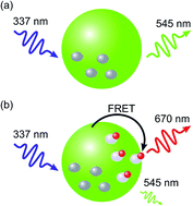
Total protein concentration (TPC) is a key parameter in many biochemical experiments and its quantification is often necessary for isolation, separation, and analysis of proteins. A sensitive and rapid nanobead-based TPC quantification assay based on Förster Resonance Energy Transfer (FRET) has been developed. A new, highly luminescent Tb(III) complex has been synthesised and applied as donor in this FRET assay with an organic dye (Cy5) as acceptor. FRET-induced changes in luminescence have been investigated both at donor and acceptor emission wavelength using time-resolved luminescence spectroscopy with time-gated detection. In the assay, the Tb(III) complex and fine-tuned polyglycidyl methacrylate (PGMA) nanobeads ensure that an improvement in sensitivity and background reduction is achieved. Using 40 nm large PGMA nanobeads loaded with the Tb(III) complex, it is possible to determine TPC down to 50 ng mL−1 in just 10 minutes. Through specific assay components the sensitivity has been improved when compared to existing nanobead-based assays and to currently known commercial methods. Additionally, the assay is relatively insensitive to the presence of contaminants, such as non-ionic detergents commonly found in biological samples. Due to no need for any centrifugal steps, this mix-and-measure bioassay can easily be implemented into routine TPC quantification protocols in biochemical laboratories.


1. Introduction

Protein quantification is an important task realized daily in medical and biological laboratories as well as in biopharmaceutical and food industries.1 In medical applications, the concentration of albumin and globulin is mostly determined, while lactoglobulins are relevant for high protein content foods and natural ingredients. In humans, elevated protein concentrations can be associated with health disorders such as leukaemia or dehydration.2 Similarly, low protein concentration can be a symptom for liver diseases.3 The ratio of fructose levels to TPC have also been found to be useful to estimate the concentration of glycoproteins that are involved in drug absorption. Reliable and fast tests to determine total protein concentration are therefore sought to improve the overall healthcare quality.

The most commonly used TPC determination methods are either based on direct measuring protein absorbance, absorbance of species reacting with proteins4 or on measuring fluorescence of fluorophores added to protein solution.5 The absorbance methods, however, are limited in their sensitivity with detection limits in mg mL−1 concentration range, low dynamic range and can produce significantly varying readouts for different proteins. The direct absorbance method considers measuring absorption characteristic for tyrosine and tryptophan amino acids, which absorb light at 280 nm. This method could be reliable, but one needs to notice that every protein has a different amount of these amino acids. Moreover, many other chemicals such as buffer salts, alcohols or nucleic acids, which also absorb at 280 nm, can give false positive signal in this case. In the bicinchoninic acid (BCA) assay, involving the chelating of copper ions with BCA, the presence of other copper chelating compounds as for example ammonia can interfere the analytical signal. Since BCA assay is based on measuring absorbance, it is also greatly influenced by the presence of tyrosine, tryptophan, and cysteine amino acids. Other absorbance based quantification methods are either incompatible with common chemicals used in biochemical investigations such as EDTA, Tris or carbohydrates (Folin–Lowry method), or require large amount of sample and tedious and time-consuming preparation involving heated sulfuric acid, steam distillation, and back-titration with sodium hydroxide (Kjeldahl method). Similarly, to bead-based assays, the Bradford assay is also based on electrostatic interactions. In this assay, the Coomassie brilliant blue dye changes its colour from red to blue after the interaction with positively charged proteins. The assay, however, can only detect proteins larger than 3 kDa and it is sensitive to detergents such as SDS and Triton X-100. Fluorescence methods are more sensitive than absorbance methods, but usually they require relatively long incubation times (around one hour) or toxic reagents (e.g. cyanides). Both absorbance and fluorescence methods involve heating steps and are sensitive to contaminants such as free parts of biomaterial, salts and organic compounds. The methods based on specific interactions (e.g. antibody–antigen or DNA pairing) are impractical due to the variety of proteins being involved in the protein composition. Therefore, the particle-based non-specific adsorption methods are a promising alternative to commercial TPC determination methods, particularly when combined with such a sensitive technique as Förster Resonance Energy Transfer (FRET).6

For the last ten years, detection systems based on FRET from lanthanide complexes to organic dyes or to quantum dots have drawn attention due to high sensitivity and the potential for multiplex detection.7 In such systems the use of lanthanide complexes, either Eu(III) or Tb(III), assures long luminescence decay times and well-defined and sharp emission peaks. These properties facilitate time-gated time-resolved luminescence signal acquisition. Due to the long decay time, the background signal from undesired effects, such as biosample autofluorescence, short-living fluorescence of fluorophores as well as the scattered light, is reduced significantly. FRET systems based on the lanthanide complexes have been used for numerous applications such as multiplexed diagnostics,8,9 molecular ruler,10 biotin-streptavidin assays11 as well as in tests for the determination of various biomarkers12 or proteins.13

FRET assays based on nanoparticles loaded with Eu(III) complex have been developed for the determination of protein isosbestic point14 and cell counting.15 However, to the best of our knowledge assays with Tb(III) complex loaded nanoparticles have not been reported up to date. As outlined in Förster theory, in every FRET system the donor photoluminescence quantum yield (ΦF) is crucial factor for efficient energy transfer. In this view, particularly important is the Förster radius (R0), a FRET characteristic value that determines the donor–acceptor distance at which FRET efficiency is equal to 50%. In accordance with the Förster theory, R0 is dependent on donor luminescence quantum yield (ΦL) and spectral overlap between donor emission and acceptor absorption. Thus, the donor properties significantly influence R0 and consequently the performance of the whole sensing system. The finding of a new stable lanthanide complexes with high luminescence quantum yields is also highly desirable for FRET-based detection. This fact tempted us to develop novel Tb(III) complex with high luminescence quantum yield.

In the present work, we report the synthesis of a new Tb(III) complex with ΦL reaching 97% in solid state, 70% in organic solution (DCM) and 50% in water-dispersible polyglycidyl methacrylate (PGMA) nanoparticles. This complex was also successfully applied in bioassay to determine the TPC in the presence of Bovine Serum Albumin (BSA) as a model protein.

2. Materials and methods

Synthesis of the (tpip)3Tb–TAP complex

The tetraphenylimidodiphosphinate (tpip) and its terbium complex (tpip)3Tb were prepared in accordance with a method known in the literature. All analytical characterisation of novel compounds was performed at the Institute of Organic Chemistry of the Polish Academy of Sciences. The preparation of the ternary complex (tpip)3Tb-TAP followed the procedure, already published.2

The (tpip)3Tb complex (0.1 mmol, 140.8 mg) and 1,4,8,9-tetraazatriphenylene (TAP) (0.1 mmol, 23.2 mg) were placed in a round-bottom flask (10 mL) with stirring bar, and charged with 5 mL of toluene. Stirring at reflux was maintained, till all materials were dissolved completely. Upon standing colourless crystals deposited, which were filtered off and dried under reduced pressure. The second crop was obtained from the mother liquor by diffusion with hexane, layered on toluene solution. Overall yield: 155.8 mg, 95%. Elem. anal. calc. for C86H68N7O6P6Tb; C 62.972, H 4.178, N 5.977, found C 62.95, H 4.17, N 5.96.

Synthesis of PGMA nanoparticles

0.655 g SDS (sodium dodecylsulphate), 0.2 g Carbowax (PEG 6.000) und 0.1372 g Borax (sodium tetraborate) were dissolved in 176 mL ultra-pure water (Merck Millipore). This solution was filled into a 250 mL double-walled glass reactor (HWS Mainz) with reflux condenser and anchor stirrer. The reactor content was slowly stirred at ambient temperature. 0.108 g PPS (potassium peroxidisulphate) were dissolved in 20 mL ultra-pure water (Millipore). The solution was filled into a dropping funnel. 4 g EPMA (2,3-epoxypropyl methacrylate) and 0.2 g MAA (methacrylic acid) were added to the reactor content. Stirring was adjusted to 400 min−1. The polymerisation fleet was continuously purged with nitrogen. (In the bubble counter on the reflux condenser, a nitrogen bubble was registered approx. each second.) After 20 min, the emulsion in the reactor was heated up to 60 °C. 10 min after reaching the polymerisation temperature, the reaction was started by adding the PPS solution from the dropping funnel (fast). After 8 h, the polymerisation was stopped by cooling down and aeration.

Solid content of the polymer dispersion was determined by means of a moisture analyser (Sartorius). Particle size was measured by dynamic light scattering (Brookhaven BI-90). Latex purification was carried out by dialysis against deionised water over 3 days (Visking dialysis tubing, SERVA), followed by ultrafiltration (0.4 L-cell [Berghof], 0.5–0.8 bar overpressure, 25 nm track-etched membrane [Waters]). Zeta potential as a function of pH was determined by means of a combined system, consisting of Zetasizer S (Malvern Instruments) and auto-titrator DL21 (Mettler Toledo).

Staining of nanoparticles using (tpip)3Tb–TAP

100 μL of a 5 mM (tpip)3-Tb–TAP solution in THF were added to 600 μL of a 0.5% solution of nanoparticles in water. The mixture was gently stirred for at least 2 hours. Then, the swollen nanoparticles were centrifuged three times at 10[thin space (1/6-em)]000 rpm for 20 min. Each time the supernatant was discarded and the precipitated nanoparticles were re-dispersed in double distilled water by ultrasonification. Then, further purification was realised with three filtrations through a 50 kDa centrifugational microfilter at 10[thin space (1/6-em)]000 rpm for 5 min each time. Finally, fully purified nanoparticle solution was diluted 10.000 times and used for FRET assay.

Steady-state luminescence measurements

The steady-state luminescence measurements were carried out on a FluoroLog-3 spectrofluorometer (Horiba Jobin Yvon, Bensheim, Germany). Luminescence was excited at 337 nm and collected in the range of 400–700 nm in the usual rectangular configuration in a 200 μL cubic quartz cuvette (Hellma, Mühlheim, Germany) placed in a cuvette holder, whose temperature was maintained at 25 ± 0.2 °C. Both excitation and emission slits were set to 5 nm bandpass. Luminescence quantum yield of nanoparticles labelled with (tpip)3Tb–(TAP) complex was measured on a C9920-02 Absolute PL Quantum Yield Measurement System (Hamamatsu, Herrsching am Ammersee, Germany) with an integrate sphere detection unit, a xenon lamp as the monochromatic excitation light source and unlabelled nanoparticles as a reference.

Time-resolved luminescence measurements

The time-gated time-resolved measurements were carried out on a Kryptor luminescence reader (Cezanne, Nimes, France) equipped with a nitrogen laser (λem = 337.1 nm) as an excitation source. Luminescence was collected using one photomultiplier positioned in the donor spectral channel (488 ± 10 nm) and another in the acceptor channel (665 ± 13 nm). The signals were collected in the range of 2 μs to 8 ms with a resolution of 2 μs. The measurements were executed at constant donor nanoparticles and acceptor protein concentrations. The BSA concentration was varied from 10 ng mL−1 to 100.000 ng mL−1. The (tpip)3Tb–(TAP) and Cy5 luminescence signals were also measured separately to estimate the potential background signals.

3. Results and discussion

System design

Förster radius (R0) is the distance between two dyes (donor and acceptor), wherein the efficiency of non-radiative energy transfer via Förster resonance mechanism between these two dyes is 50%. The Förster radius is defined as follows:
 
image file: c6ra23207h-t1.tif(1)
where R0 is Förster radius, Φ0 is luminescence quantum yield of a donor in the absence of an acceptor, κ2 is the orientation factor, which is the angle between the emission dipole of a donor and absorption dipole of an acceptor (for virtually freely rotating dyes the value is equal to 2/3), n is refraction index of the surrounding medium, NA is Avogadro constant, and J(λ) is the spectral overlap between donor emission and acceptor absorption spectrum.

A closer look on eqn (1) let us notice that there are two crucial factors influencing the Förster radius, namely, quantum yield of a donor in the absence of an acceptor and spectral donor–acceptor overlap. All other factors are constant.

Noticeably, the Förster radius is proportional to the inverse sixth power of the donor luminescence quantum yield (Φ). In the case when Φ would change from 0.1 to 0.9 the Förster radius rises by around 30%, but for a realistic scenario considers increasing Φ from 0.2 to 0.5 what increases Förster radius by 10%, which means in practice the change in R0 of around 0.5 nm. Since lanthanide complexes exhibit emission with narrow and well-defined peaks, the spectral donor–acceptor overlap depends stronger on the acceptor absorptivity than on the shape of the donor emission spectrum. In this case, either the organic dyes with molar extinction coefficients ε around 250[thin space (1/6-em)]000 M−1 cm−1 can be applied or quantum dots with ε reaching 1 × 10⁶ M−1 cm−1 can also be considered.

In typical TPC nanobead-based assays usually polystyrene nanoparticles with the size of around 100 nm are used due to commercial availability or relatively easy preparation. However, the synthesis of highly size-monodispersed polymer nanoparticles with sizes below 40 nm is not a trivial task and it does not work for every polymer. Therefore, as the polymer to prepare nanoparticles in our case we used polyglycidyl methacrylate PGMA, which is known to form nanoparticles below 40 nm, is optically transparent material at the macroscale, while at the nanoscale its transparency can reduce scattering.

In our approach, the nanoparticles are stained with lanthanide complexes over the whole volume, but only the complexes being close to the nanoparticle surface (at distances not exceeding 2R0) can efficiently take part in the FRET process. Unfortunately, all other complexes remaining within the nanoparticle interior also produce luminescence signal that is not being quenched in the FRET process, therefore, it is the component increasing background signal and associated “donor leak”. Using nanoparticles with smaller diameter, the number of ineffective complexes is reduced as well as the background signal. Reducing the size also increases the surface-to-volume ratio that provides lager surface to bind proteins that can increase the dynamic range in charge association based nanosystems. In our case the reduction of nanoparticle diameter by the factor of 2.5 i.e. from 100 nm down to 40 nm is equal to volume reduction by the factor of 15.

(tpip)3Tb–TAP complex

In order to provide a terbium complex with high Φ value, the (tpip)3Tb–TAP complex has been designed and synthesised according to the procedure presented in Scheme 1. Tetraphenylimidodiphosphinate Tb(III) complex (tpip3Tb) 1, prepared according to the literature,16 was subjected for additional complexation with 1,4,8,9-tetraazatriphenylene 2 (ref. 17) in toluene at elevated temperature to form coordinatively saturated complex 3. This approach has also worked well with (tpip)3Eu complexes with auxiliary ligands, which dramatically increased the luminescence quantum yield, acting as additional antenna.18 The (tpip)3Tb and (tpip)3Eu complexes display rather moderate Φ values (<10%), due to high triplet state energy matching poorly with the emissive levels of Tb and Eu cations. However, the addition of the tetraazatriphenylene antenna yields a significant increase in Φ up to 97% (Fig. 1).
image file: c6ra23207h-s1.tif
Scheme 1 Preparation of the luminescent Tb(III) complex (tpip)3Tb–TAP (3).

image file: c6ra23207h-f1.tif
Fig. 1 (a) Luminescence (in cps (counts per second)) emission spectra taken to follow the formation of (tpip)3Tb–(TAP) complex at different concentrations of TAP (0–40 μM). (b) Normalized luminescence intensity (20 values) as a function of TAP concentration. A full complexation (saturation of the luminescence signal) has been achieved at a TAP concentration of ∼20 μM.

Taking advantage of this fact, we developed a nanobead-based sensing system for total protein concentration. Its working principle is schematically shown in Fig. 2. The system comprises negatively charged PGMA nanobeads (40 nm diameter) loaded with (tpip)3Tb–TAP complexes that act as FRET donors, and proteins labelled with organic dyes being FRET acceptors. The sensing proceeds in accordance with the following procedure. In the first step, the donor nanoparticles are mixed with analyte proteins.


image file: c6ra23207h-f2.tif
Fig. 2 The working principle of the nanobead-based sensing system using luminescent Tb3+ complex (tpip)3Tb–TAP to determine total protein concentration.

At pH value below their isoelectric points the proteins become positively charged and adsorb firmly to the negatively charged nanoparticles. Afterwards, the dye-labelled proteins are added. Consequently, FRET is observed from the (tpip)3Tb–TAP to a dye (here Cy5). Depending on the photoluminescence changes observed both in donor and acceptor emissions a total concentration can be determined with high accuracy. Noticeably, for the whole concentration series a precise result can be obtained within 10 min.

A calibration curve for the [(tpip)3Tb–TAP]–Cy5 FRET pair with BSA as a model protein is presented in Fig. 3. As can be seen an eight-fold increase could be observed in the dynamic range of 50–3000 ng mL−1 BSA.


image file: c6ra23207h-f3.tif
Fig. 3 The calibration curve determined for BSA concentrations from 10 ng mL−1 to 0.1 mg mL−1 measured with the developed TR-FRET system. A logistic function was fitted to the data.

Smaller nanoparticles facilitate lower background due to reduced light scattering and assure that most (tpip)3Tb–TAP complexes take part in the FRET process. The limit of detection (LOD) has been estimated to be ca. 50 ng mL−1, which is over 200 times more sensitive than methods conventionally used for TPC determination19 and around 2–5 times more sensitive than other nanoparticle-based systems.13 Therefore, the intrachannel donor “emission leak” into the acceptor channel is also significantly reduced. In nanoparticle-based assays, nanoparticles of around 100 nm are usually used as donors.13,20 Even though the nanoparticles are loaded with complexes over the whole volume, only the complexes placed in the outer shell can efficiently participate to the energy transfer. According to the Förster theory, the critical distance for FRET to occur is equal to 2R0. For typical lanthanide complex-dye FRET pair 2R0 is in the range of 9–12 nm.9 In the case of our system the critical distance for FRET was found to be 2R0 = (11.2 ± 0.2) nm (the calculation (in ESI) has been executed based on constants presented in the literature11,13). Therefore, in our experiments we used 40 nm large nanoparticles to provide significant number of complexes that could contribute in FRET. Additionally, through the application of nanoparticles with the diameter of 40 nm, the surface-to-volume ratio increased significantly providing larger surface for proteins to bind, which extended the dynamic range up to 2 μg mL−1.

4. Conclusions

A new Tb(III) complex has been synthesised and characterised both in solid state and in solution. The complex provides high and stable luminescence in comparison to other known systems containing tetraazatriphenylene. Based on this new complex, we developed a simple, rapid, sensitive, and separation-free nanoparticle-based assay that can be used to determine TPC in high-throughput systems. Our sensing system proven to be over 200 times more sensitive than commercial methods currently available in the market for total protein concentration. Due to the application of a new efficient, highly luminescent Tb(III) complex and smaller nanoparticles our system has two times lower limit of detection (LOD) when compared to other nanoparticle based TR-FRET systems. The assay is highly sensitive to proteins, it can be performed at room temperature and the signal remains stable for more than 5 hours. Common contaminants, such as buffer salts, solvents, or DNA are well tolerated in the assay. The assay can be accurate in the presence of reducing reagents, but not in the presence of a large amount of detergents. If known contaminants are present in the protein solution the assay protocol modifications must be applied to achieve unambitious results.

Acknowledgements

MP, MM and KG are grateful for financial support from the Science Centre, Poland, grant No. OPUS9 2015/17/B/ST5/01038.

References

  1. J. E. Noble, A. E. Knight, A. J. Reason, A. Di Matola and M. J. A. Bailey, A comparison of protein quantitation assays for biopharmaceutical applications, Mol. Biotechnol., 2007, 37(2), 99–111 CrossRef CAS PubMed; R. Owusu-Apenten, Colorimetric analysis of protein sulfhydryl groups in milk: applications and processing effects, Crit. Rev. Food Sci. Nutr., 2005, 45(1), 1–23 CrossRef PubMed.
  2. F. Almanjomi, D. Armstrong, O. Abla, A. Naqvi, S. Weitzman and J. Hitzler, Lumbar spinal fluid collections in children treated with intrathecal chemotherapy: elevated CSF protein as a diagnostic clue, Pediatr. Blood Cancer, 2008, 51(2), 295–298 Search PubMed.
  3. J. A. Joles, N. WillekesKoolschijn and H. A. Koomans, Hypoalbuminemia causes high blood viscosity by increasing red cell lysophosphatidylcholine, Kidney Int., 1997, 52(3), 761–770 CrossRef CAS PubMed.
  4. M. M. Bradford, Rapid and Sensitive Method for Quantitation of Microgram Quantities of Protein Utilizing Principle of Protein–Dye Binding, Anal. Biochem., 1976, 72(1–2), 248–254 CrossRef CAS PubMed; O. H. Lowry, N. J. Rosebrough, A. L. Farr and R. J. Randall, Protein measurement with the Folin phenol reagent, J. Biol. Chem., 1951, 193(1), 265–275 Search PubMed; P. K. Smith, R. I. Krohn, G. T. Hermanson, A. K. Mallia, F. H. Gartner, M. D. Provenzano, E. K. Fujimoto, N. M. Goeke, B. J. Olson and D. C. Klenk, Measurement of Protein Using Bicinchoninic Acid, Anal. Biochem., 1985, 150(1), 76–85 CrossRef PubMed.
  5. A. Lorenzen and S. W. Kennedy, A Fluorescence-based protein assay for use with a microplate reader, Anal. Biochem., 1993, 214(1), 346–348 CrossRef CAS PubMed; L. J. Jones, R. P. Haugland and V. L. Singer, Development and characterization of the nanoorange (R) protein quantitation assay: a fluorescence-based assay of proteins in solution, BioTechniques, 2003, 34(4), 850–854 Search PubMed; W. W. You, R. P. Haugland, D. K. Ryan and N. P. Haugland, 3-(4-Carboxybenzoyl)quinoline-2-carboxaldehyde, a reagent with broad dynamic range for the assay of proteins and lipoproteins in solution, Anal. Biochem., 1997, 244(2), 277–282 CrossRef PubMed.
  6. S. K. Fischer, A. Joyce, M. Spengler, T. Y. Yang, Y. Zhuang, M. S. Fjording and A. Mikulskis, Emerging Technologies to Increase Ligand Binding Assay Sensitivity, AAPS J., 2015, 17(1), 93–101 CrossRef CAS PubMed.
  7. N. Hildebrandt, K. D. Wegner and W. R. Algar, Luminescent terbium complexes: Superior Forster resonance energy transfer donors for flexible and sensitive multiplexed biosensing, Coord. Chem. Rev., 2014, 273, 125–138 CrossRef.
  8. D. Geissler, L. J. Charbonniere, R. F. Ziessel, N. G. Butlin, H.-G. Löhmannsröben and N. Hildebrandt, Quantum Dot Biosensors for Ultrasensitive Multiplexed Diagnostics, Angew. Chem., Int. Ed., 2010, 49(8), 1396–1401 CrossRef CAS PubMed.
  9. D. Geissler, S. Stufler, H.-G. Löhmannsröben and N. Hildebrandt, Six-Color Time-Resolved Forster Resonance Energy Transfer for Ultrasensitive Multiplexed Biosensing, J. Am. Chem. Soc., 2013, 135(3), 1102–1109 CrossRef CAS PubMed.
  10. F. Morgner, S. Stufler, D. Geissler, I. L. Medintz, W. R. Algar, K. Susumu, M. H. Stewart, J. B. Blanco-Canosa, P. E. Dawson and N. Hildebrandt, Terbium to Quantum Dot FRET Bioconjugates for Clinical Diagnostics: Influence of Human Plasma on Optical and Assembly Properties, Sensors, 2011, 11(10), 9667–9684 CrossRef CAS PubMed.
  11. P. J. Cywiński, T. Hammann, D. Hühn, W. J. Parak, N. Hildebrandt and H.-G. Löhmannsröben, Europium-quantum dot nanobioconjugates as luminescent probes for time-gated biosensing, J. Biomed. Opt., 2014, 19(10), 101506 CrossRef CAS PubMed; P. J. Cywinski, K. Nchimi Nono, L. J. Charbonnière, T. Hammann and H.-G. Löhmannsröben, Photophysical evaluation of a new functional terbium complex in FRET-based time-resolved homogenous fluoroassays, Phys. Chem. Chem. Phys., 2014, 16, 6060–6067 RSC.
  12. K. D. Wegner, Z. Jin, S. Linden, T. L. Jennings and N. Hildebrandt, Quantum-Dot-Based Forster Resonance Energy Transfer Immunoassay for Sensitive Clinical Diagnostics of Low-Volume Serum Samples, ACS Nano, 2013, 7(8), 7411–7419 CrossRef CAS PubMed; H. Zeng, J. Wu, M. T. Bedford, G. Sbardella, F. M. Hoffmann, K. Bi and W. Xu, A TR-FRET-Based Functional Assay for Screening Activators of CARM1, ChemBioChem, 2013, 14(7), 827–835 CrossRef PubMed; K. Nchimi-Nono, K. D. Wegner, S. Linden, A. Lecointre, L. Ehret-Sabatier, S. Shakir, N. Hildebrandt and L. J. Charbonniere, Activated phosphonated trifunctional chelates for highly sensitive lanthanide-based FRET immunoassays applied to total prostate specific antigen detection, Org. Biomol. Chem., 2013, 11(38), 6493–6501 Search PubMed.
  13. H. Härmä, S. Pihlasalo, P. J. Cywinski, P. Mikkonen, T. Hammann, H.-G. Löhmannsröben and P. Hanninen, Protein Quantification Using Resonance Energy Transfer between Donor Nanoparticles and Acceptor Quantum Dots, Anal. Chem., 2013, 85(5), 2921–2926 CrossRef PubMed.
  14. S. Pihlasalo, L. Auranen, P. Hanninen and H. Harma, Method for Estimation of Protein Isoelectric Point, Anal. Chem., 2012, 84(19), 8253–8258 CrossRef CAS PubMed.
  15. S. Pihlasalo, A. Engbert, E. Martikkala, P. Ylander, P. Hanninen and H. Harma, Nonspecific Particle-Based Method with Two-Photon Excitation Detection for Sensitive Protein Quantification and Cell Counting, Anal. Chem., 2013, 85(5), 2689–2696 CrossRef CAS PubMed.
  16. S. W. Magennis, S. Parsons, A. Corval, J. D. Woollins and Z. Pikramenou, Imidodiphosphinate ligands as antenna units in luminescent lanthanide complexes, Chem. Commun., 1999, 1, 61–62 RSC.
  17. E. Amouyal, A. Homsi, J. C. Chambron and J. P. Sauvage, Synthesis and study of a mixed-ligand ruthenium(II) complex in its ground and excited-states – bis(2,2′-bipyridine)(dipyrido 3,2-a,2′,3′-c-phenazine-n4n5)ruthenium(II), J. Chem. Soc., Dalton Trans., 1990,(6), 1841–1845 RSC.
  18. M. Pietraszkiewicz, O. Pietraszkiewicz, J. Karpiuk, E. Karpiuk, T. Borowiak and G. Dutkiewicz, The first ternary europium tetraphenylimidodiphosphinate complex: X-ray structure and photoluminescent properties, Inorg. Chim. Acta, 2012, 387, 426–430 CrossRef CAS.
  19. T. Zor and Z. Seliger, Linearization of the Bradford protein assay increases its sensitivity: Theoretical and experimental studies, Anal. Biochem., 1996, 236(2), 302–308 CrossRef CAS PubMed.
  20. S. Pihlasalo, J. Kirjavainen, P. Hanninen and H. Harma, High Sensitivity Luminescence Nanoparticle Assay for the Detection of Protein Aggregation, Anal. Chem., 2011, 83(4), 1163–1166 CrossRef CAS PubMed.

Footnote

Electronic supplementary information (ESI) available. See DOI: 10.1039/c6ra23207h

This journal is © The Royal Society of Chemistry 2016