White light emission from quantum dot and a UV-visible emitting Pd-complex on its surface

Madhulekha Gogoi* and Arun Chattopadhyay
Department of Chemistry and Centre for Nanotechnology, Indian Institute of Technology Guwahati, Guwahati-781039, Assam, India. E-mail: madhulekha@gmail.com

Received 14th September 2016 , Accepted 25th October 2016

First published on 25th October 2016


Abstract

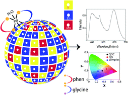
Near white light emission was achieved following surface functionalization of Mn-doped ZnS quantum dots (Qdots) by [Pd(glycine)(phenanthroline)]Cl complex with a proposed bonding of the complex through the dangling sulphide ion of the Qdot. Reaction between the complex ion and Qdot lead to the formation of quantum dot complex. Independent emissions in the UV-vis region from the complex and orange from the doped quantum dot lead to such color. Also, the material exhibited a dual decay mechanism comprising of fluorescence and phosphorescence pathways. The strategy described in the present report, with two independent emission channels, is highly adaptable for different choices of complementary color emitting coordination complexes and Qdots.


Introduction

White light emitting materials have been the subject of extensive research in recent years, owing to their enormous potential for use in lighting and display devices.1–5 At present, the most popular white light emitting devices (WLEDs) are based on blue light emitting diodes (LEDs) along with a yellow emitting phosphor6 and compact fluorescent light (CFL) consisting of mercury excited semiconductor phosphors to achieve composite white photoluminescence.7 However, the reabsorption between multiple phosphors leading to decreased luminous efficiency of WLED and environmental toxicity issues related to mercury in CFL8 and lead solder used on LED circuit board9 have been of great concern. These have given rise to the search for novel white light emitting materials, where emission efficiency is complemented with the environmental friendliness of the constituent materials. In this regard, semiconductor quantum dots (Qdots) hold several advantages over other conventional materials, due to their size-dependent tunable emission characteristics, narrow emission spectra, high quantum yield and ease of production via colloidal methods of synthesis.10 A general approach of having simultaneous multiple color emission is to physically mix the materials of the constituent colors.11 There are also efforts to fabricate composite (by mechanical mixing) of the constituent materials.12 However, these methods and materials – although popular – have the inherent limitation of reabsorption and energy transfer in case of ordinary mixing of Qdots to achieve white light emission.

In order to overcome the limitations as described above a viable option could be in the form of multicomponent emitting species being present in a singular entity and having objects of nanoscale dimensions. A conventional approach is to incorporate dopant ions as additional emitting species. For example, Mn2+ and or Cu2+-doped ZnS, ZnSe, PbS, PbSe, CdS or CdSe Qdots are quite popular with tunable, multiple and wide ranging emission color (including white light emission).13–15 However, issues like ‘self-purification’,16 ‘manipulation of surface state emission’17 and ‘intrinsic toxicity due to Hg and Pb’8,9 call for further development of Qdot based multi-component emitting species. A recent idea that has come up is based on systematic functionalization of the surface of Qdot with inorganic complex(s).18 This is based on the concept that the surface cations of a Qdot are chemically active.19–24 The whole species consisting of Qdot and surface complex has been defined as quantum dot complex (QDC). The complexation reaction could be achieved in two ways. In the first method, the surface cations of the Qdots are reacted with selected organic ligand(s) in order to form the complex(s). Secondly, the same inorganic complex synthesized separately could be made to react with the surface anion to form the same final complex on the surface. In both the cases the bonding between the complex and Qdot is achieved through the anion present on the surface. Importantly, more than one ligand could be simultaneously reacted with the surface cations (which also could be more than one species) in order to form multiple complexes on the same Qdot. The optical emission due to the surface complex – in combination with the emission from the dopant ion – has resulted in white light emission from single Qdot with high efficiency in terms of chromaticity coordinates (0.30, 0.33).18 The complexation reaction on the surface of a Qdot is expected to open up newer vistas through conferring rich and diverse optical properties to the species. Although initial works related to the surface complexation reaction of Qdots are already in the literature, the field needs to grow to its full potential. This may involve extending the emission wavelength range through formation of a variety of complexes. In this regard, emission in the near UV region and the whole visible range of wavelengths would offer new characteristics to the dots. Recently phosphorescent metal organic complexes have attracted considerable attention for their application in organic light emitting diode (OLED) because they can generate 100% internal quantum efficiency, resulting from both singlet and triplet formation.25 Hence, developing Qdot based white light emitting diode material possessing both fluorescence and phosphorescence decay pathways with the potential for achievement of 100% internal quantum efficiency is another thrust area in this field.

Herein we report the generation of a new QDC with emission in the range of 390–700 nm from a single species. The Qdot used was Mn2+-doped ZnS nanocrystal with trap-state emission in the host Qdot in the range of 454 nm and that due to dopant ion Mn2+ (4T16A1) emission at 597 nm. The complex ion of [Pd(Phen)(Gly)]+ was used as the third emitting species possessing a non-structural emission band either due to intraligand transition, vibronic structure-based emission or possibly aggregate induced emission. The formation of the QDC was achieved through the reaction of the complex with the Qdot at room temperature. Thus while white light emitting Qdots have been obtained through doping and complexation reaction, the achievement of near UV to orange emission leading to white light, from QDC by single wavelength excitation can be considered a first herein.

Experimental section

Materials and synthesis

Zinc acetate dihydrate (99%, Merck), manganese acetate tetrahydrate (Pure grade, Merck), sodium sulphide (58%, Merck), sodium hydroxide (Merck), palladium chloride (99%, Aldrich), glycine (99.5%, Sisco Research Laboratories Pvt. Ltd.), 1,10-phenanthroline (99.5%, Sisco Research Laboratories Pvt. Ltd.), tyrosine (99%, Sisco Research Laboratories Pvt. Ltd.), DL-tryptophan (99+%, Aldrich), ethanol (Merck), ethanol (HPLC), potassium bromide (Sigma Aldrich) were used as received without further purification. Milli-Q grade water was used in all experiments.

Synthesis of Mn doped ZnS quantum dots (Qdot)

Mn doped ZnS quantum dots were synthesized by following an established method.22 To synthesize the quantum dots, 5.0 mM zinc acetate dihydrate and 0.1 mM manganese acetate tetrahydrate were dissolved in 30 mL of water and put in stirring condition. The mixture was being heated. When temperature reached 70 °C, a 20 mL solution of 5.0 mM sodium sulphide was added and white turbidity appeared instantly. The whole reaction mixture was stirred at 100 °C for 3 h. After that, the colloidal dispersion was centrifuged at 25[thin space (1/6-em)]000 rpm for 15 min. The pellet was washed with water and ethanol and redispersed in water followed by centrifugation and the cycle was continued for one more time. The purified pellet was then dispersed in 100 mL water by sonication and was used for further experiments.

Synthesis of Pd(II) complex

The Pd(II) complex of the type [Pd(Phen)(AA)]+ (where AA is an anion of amino acid, in our case AA was glycinato) was prepared by following a reported procedure.26 Briefly, each of 4.0 mM 10 mL solution of 1,10-phenanthroline, 4.0 mM 10 mL solution of PdCl2 and 13.3 mM 10 mL solution of glycine was prepared in a 1[thin space (1/6-em)]:[thin space (1/6-em)]1 water[thin space (1/6-em)]:[thin space (1/6-em)]ethanol medium using sonication for 15 min. Then all three solutions were mixed together and kept on stirring for 12 h. The medium turned light yellow, which was centrifuged at 25[thin space (1/6-em)]000 rpm for 30 min to precipitate the complex. It was dried in oven at 60 °C for 12 h to obtain the yellow colored powder of the complex. Further characterization was performed with this powder of the complex.

Synthesis of quantum dot complex (QDC)

The quantum dot complex (QDC) was prepared by simple addition of [Pd(Phen)(Gly)]Cl complex to Mn doped ZnS Qdot dispersion in water. Briefly, the pellet of the Qdot obtained after purification and centrifugation was dispersed in 30 mL water. To it a solution of the complex, obtained by dissolving 3 mg of powder in 30 mL water followed by sonication for 15 min, was added and the mixture was kept as such for 12 h. A bright yellow (light brownish) precipitate was obtained with clear supernatant on centrifugation at 25[thin space (1/6-em)]000 rpm for 15 min. The precipitate was washed with water and was redispersed in water following sonication and used for further studies.

Results and discussion

Blue emitting [Pd(Phen)(Gly)]Cl complex was synthesized by a chemical process using PdCl2, 1,10-phenanthroline and glycine and was collected as a yellowish powder.26 The details of the procedure is mentioned in the Experimental section. The complex was characterized through UV-vis absorption, fluorescence, Fourier transform infra-red (FTIR) and nuclear magnetic resonance (NMR) spectroscopic techniques. The absorption and fluorescence spectra of the complex are shown in Fig. 1(a). The observed absorption peaks at 218 nm and 272 nm are due to intraligand transitions viz. π to π* transition of 1,10-phenanthroline.27 The weak shoulder at 305 nm is due to n to π* transition within the phenanthroline ring.28 In this regard, the absorption spectra of only ligands were examined and the results are presented in Fig. S1(a), ESI. Glycine absorbs at 200–230 nm due to the excitation of p-orbital non-bonding electron to anti-bonding σ*-orbital and due to π to π* transition in the carboxylate group.29 Another glycine peak observed at 268 nm originated from interaction between individual amino acid molecules due to aggregate formation.30 The emission spectrum of the complex, shown in Fig. 1(a), exhibited a broad featureless characteristic basically centred in the blue range of the visible spectrum. The emission starting at 380 nm is attributed to intraligand transitions as observed from the emission spectra of ligand solutions [Fig. S1(b)] and excitation spectra of ligands and complex [Fig. S1(c) and (d), ESI]. But the formation of such unstructured broad emission band with sharp features at 468 nm, needs to be addressed as the emission peaks due to d–d transitions in d8 system of Pd(II) are reported to be too weak to be detected because these are both spin and symmetry forbidden.31 We have considered four possible origins for the unconventionally sharp emission band with maximum at 468 nm. (1) Luminescence may be due to protonated species of phen ligand in excited state, which can be detected by observation of shifts in the emission maximum with pH variation.32 (2) It may be due to excimer formation between aromatic molecules viz. phen. This can be monitored by the appearance of monomer and excimer luminescence simultaneously.33 (3) The emission could be due to aggregation induced emission (AIE) of the complex molecules. Earlier, such AIE has been observed in case of Au(I)–thiolate complexes.34,35 This possibility can be checked by performing solid state fluorescence spectroscopy and by studying the luminescence property of the complex solution after addition of a poor solvent to cause aggregation.36 (4) Emission could also be due to vibronic structure of coordinating ligand. To see the possibility of the first hypothesis, pH-dependent luminescence spectra of the complex were recorded and the results are shown in Fig. S2(a), ESI. As observed, with pH variation, neither shifting nor evolution of the new band was there, which discarded the hypothesis. However, the dispersibility of complex in aqueous medium decreased with increase in pH which led to precipitation and to overall loss of the luminescence intensity. In this regard, it is important to note that the pH value of the solution of complex in water as such was measured to be 3.2 while pH variation was done with dilute HCl and NaOH solutions. Excimer formation hypothesis can also be discarded since this structureless emission band did not occur in the spectrum of only phenanthroline solution (Fig. S1(b), ESI). Moreover, there was no observable quenching of the phen (or glycine) emission at 380 nm with the evolution of new emission bands in the spectrum of complex (Fig. 1(a)).
image file: c6ra22985a-f1.tif
Fig. 1 (a) UV-vis absorption and emission spectra of the complex, (b) FTIR spectra of glycine, phenanthroline and the complex.

In order to probe whether the emission band at 468 nm was associated with AIE, we have studied the emission spectrum of the same amount of complex dispersion in tetrahydrofuran/hexane solvent mixture with increasing fraction of the poor solvent hexane (Fig. S2(b), ESI). There was no significant change in the spectrum. However, luminescence study of the complex solution in water, with continuous dilution (Fig. S2(c), ESI), revealed that the overall intensity of the emission spectra had increased with increasing concentration. Hence the emission behavior of the complex was opposite to the aggregation caused quenching (ACQ) phenomenon28 in which luminescence is quenched in concentrated solution, as a whole. Another control experiment had been carried out to probe the effect of solvent on emission behavior of the complex. It is observed from the results presented in Fig. S3, ESI, that the structureless features in the spectrum disappeared upon dilution. Instead only the emission arising from the intraligand transitions or AIE of glycine molecules was observed prominently in the diluted samples. Looking into the above scenario, the contribution of AIE towards the origin of 468 nm emission band cannot be discarded fully. This band disappeared in the diluted dispersions of complex in solvents other than water, where dispersibility of complex is less than that in water. If AIE was responsible then this can be correlated to the pi-stacking of phenanthroline ligands of neighbouring complex molecules leading to AIE.37–40 Aggregation causes restriction of intramolecular rotation (RIR) or vibration (RIV) decreasing the probability of non-radiative decay.41,42 Hence, solubility in both phases (aqueous and organic) due to the presence of phenanthroline and glycine ligands in the complex may lead to π-stacking of phenanthroline molecules in concentrated solutions, giving rise to AIE in all of the above solvents. In this regard, solid state fluorescence spectrum of the complex is presented in Fig. S3(d), which is similar to that found in the solution state (Fig. 1(a)). However, the last possibility of vibronic structure as the origin of the emission band at 468 nm can also contribute to the overall spectrum as reported in the literature.43,44

The formation of the complex of the type [Pd(Phen)(Gly)]+ was further substantiated from the FTIR spectral analysis. The spectra corresponding to glycine, phenanthroline and the complex are shown in Fig. 1(b). Further details of the analysis are presented in Fig. S4 and Table S1 of the ESI. The presence of quadruple bands for ring frequencies in the spectrum of the complex in the range 1640–1562 cm−1 corresponds to phenanthroline ring.45,46 For the glycinato ion, N–H stretching and wagging at 3428 and 920 cm−1, respectively; hydrogen bonded symmetric and antisymmetric stretching and bending of NH3+ at 3063, 3081 and 1517 cm−1, respectively, were observed in the spectrum of the complex.47,48 In addition, C–H deformation bands at 1454 and 1341 cm−1,39 in-plane and out-of-plane H-motion corresponding to phenanthroline in the range 1200 and 850 cm−1 were present in the spectrum demonstrating its proposed formula.37 Since the COO symmetric and antisymmetric vibrations are close to NH3+ and NH2 deformations, it is difficult to assign these bands. Besides, the C[double bond, length as m-dash]C stretching frequency of phenanthroline at 1626 cm−1 in the complex indicated coordination with the metal.49 However, because of the presence of phenanthroline and glycine, aromatic and aliphatic signatures of C–Nstretch were observed in the FTIR spectrum of the complex at 1341 and 1110 cm−1, respectively.50

Surface functional group characterization results of Qdot and QDC obtained through FTIR spectroscopy are presented in Fig. S5 and Table S2, ESI. In the FTIR spectrum of Qdot, the presence of COO symmetric, antisymmetric and C–H vibrational bands pointed towards the chemisorption of acetate ions from Zn and Mn precursors on Qdot surface to act as the capping agent. Previously, in case of ZnO nanowire formation, similar capping effect of acetate ions from precursor was observed and was assumed to be the factor responsible for constancy in nanowire diameter.51 In addition to this, O–H bending and stretching bands were present in the Qdot spectrum at 1620 and 3385 cm−1.52 The complexation of Qdot with Pd(II) complex to form QDC was demonstrated by the presence of broad band at 1600 cm−1 corresponding to quadruple for ring frequencies, out of plane motion of ring H of phenanthroline moiety at 720 and 844 cm−1, metal–N stretch at 472 cm−1,53 and C–H symmetric and antisymmetric stretch of CH2 group at 2853 and 2920 cm−1 in the QDC spectra. The results indicated the formation of the complex on the surface of the Qdot in the QDC. From the comparative FTIR spectra analysis above, among complex, Qdot and QDC it is observed that the complex spectra consists of various stretches from its constituents. In the Qdot spectra, acetate ions stretches are observed which may act as capping agent whereas in the QDC spectra, the stretches corresponding to the complex are observed.

The 1H-NMR spectra of the complex and QDC were recorded in D2O (Fig. S6 and S7, ESI). The proton peaks due to 1,10-phenanthroline in the complex were observed in the range from 8.76 to 7.85 ppm, which exhibited downfield shifts as compared to the pristine phenanthroline protons. This may be due to coordination of the molecule (ligand) to Pd(II).26 On the other hand, the coordinated glycine in the complex showed a singlet at 3.79 ppm, which was again downfield shifted in comparison to zwitterionic glycine. The results indicated bidentate coordination of glycine to Pd(II) through –NH2 and –COO groups.26 However, in the NMR spectra of QDC, the singlet for methylene proton has been observed at 3.67 ppm which is very close to that of zwitterionic glycine (3.59 ppm). This indicates towards monodentate coordination (through only COO– group) of glycine with Pd(II) ion in QDC while NH2 group remains free. This is because in the monodentate coordination, the methylene proton of glycine experiences similar chemical environment as in the free ligand.54 The detailed mechanism of QDC formation has been described in subsequent sections.

The microstructural and compositional analyses of the developed Qdot and QDC were performed by X-ray diffraction (XRD), high-resolution transmission electron microscopy (HRTEM), selected area electron diffraction (SAED), energy dispersive X-ray (EDS) and electron spin resonance (ESR) spectroscopic techniques. From the comparative XRD plot corresponding to Qdot and QDC in Fig. 2(a), the presence of cubic ZnS phase with diffraction peaks at 2θ values 29°, 48° and 57° corresponding to (111), (220) and (311) planes, respectively, was observed (JCPDS 05-0566). The HRTEM images of both Qdot and QDC in Fig. 2(c) and (d) exhibited single crystalline nature of the crystals with lattice fringes of 0.3 nm, corresponding to the spacing of (111) planes respectively, of cubic ZnS. There was no discernible change in the lattice spacing of (111) facet (0.3 nm) from the standard value of 0.31 nm, which can be corroborated to the substitution of few Zn2+ ions by Mn2+ ions having similar ionic radii.55,56 SAED analysis of Qdot and QDC shows the presence of (111) and (220) planes of cubic ZnS, which agree well with the conclusions drawn from XRD and HRTEM analyses (Fig. S8(a) and (b), ESI). From particle size distribution plot (as calculated from the TEM images), the average particle diameter of the Qdot (3.2 nm) seemed to have remained unaltered after complexation (Fig. S8(e) and (f), ESI). Analysis through ESR spectroscopy revealed the presence of Mn2+ ions in the crystals of Qdot as is evidenced by the occurrence of hyperfine splitting in the spectrum in Fig. 2(b).57 An ESR spectrum with a sextet due to I = 5/2 for Mn2+ ions is the consequence of hyperfine splitting.51 The weight average ratio between metal ion concentrations in the Qdots from EDS result was found to be same as the atomic ratio between metal ions from atomic absorption spectroscopy (AAS) result. The details of the composition are tabulated in Table S3, ESI.


image file: c6ra22985a-f2.tif
Fig. 2 (a) XRD plot of Qdot and QDC; (b) ESR spectrum of Qdot; HRTEM images of (c) Qdot and (d) QDC. The insets in (c) and (d) show the IFFT and FFT images of individual particles.

The complexation process on Qdot surface was further investigated by the changes in the optical properties. As is evident from Fig. 3(a), the band-edge absorption of the Qdot was observed at 300 nm. On the other hand, the absorption spectrum of QDC exhibited peaks of both Qdot (300 nm) and the complex, which appeared at 220 nm and at slightly blue shifted than that of the complex (270 nm). This was further examined for QDC formation through addition of different volumes of the complex solution (0.1 μM with respect to Pd2+ ion) to the same volume of Qdot dispersion and by recording the absorption spectra. It was observed (Fig. S9, ESI) that on increasing the concentration of the complex the overall absorption also increased. The coexistence of both the absorption peaks is clearly evident in the spectra of the QDCs obtained by adding higher concentrations of the complex (Fig. S9(b), ESI). Additionally, the samples were centrifuged and redispersed in water and absorption spectra were recorded. It was observed that both the spectra of the sample dispersions before and after centrifugation superimposed with each other (Fig. 3(b)). This indicated that the complex was attached to the surface of the Qdot.


image file: c6ra22985a-f3.tif
Fig. 3 Comparative UV-vis absorption spectra of (a) Qdot, QDC and complex and (b) QDC before and after centrifugation (superimposed on each other), complex and supernatant. The QDC herein was prepared from a set of samples, which were subjected to centrifugation and redispersion.

Further, treatment of Qdot dispersion with glycine (13.3 mM) and phenanthroline (4.4 mM) solutions, separately, did not change the UV-vis absorption spectrum of the Qdot (refer Fig. S10 and Table S4, ESI). This indicated that simple addition of ligands to the Qdot solution at room temperature did not result in the complexation on the surface. The absorption peaks of the ligands already described in Fig. 1(a) were observed in the spectra of the Qdot dispersions added with respective ligands and their supernatants but not in that of the redispersed pellets after centrifugation.

The complexation process was further investigated by fluorescence spectroscopy technique. Pristine Qdot exhibited dual emissions at around 454 nm and 597 nm when excited with 300 nm light (Fig. 4(a)). The sharp peak at 597 nm is due to 4T16A1 transition in Mn2+, while the broad peak at 454 nm is due to the host Qdot.18 In case of bulk ZnS band edge emission occurs in the UV range at around 330 nm. Therefore, on Qdot formation with reduction in size and quantum confinement effect, the emission should have been blue shifted. However, the aqueous Mn doped ZnS Qdots, exhibited a broad emission peak centred around 454 nm (blue region) along with the dopant emission peak at 597 nm. The reason for this shift is attributed to the trap-state emission, which has smaller energy (thus longer wavelength emission) than the band-edge emission in ZnS. There are two different types of trap states reported in case of transition metal doped ZnS quantum dots: shallow trap states originating basically due to sulfur vacancy and deep trap states due to Zn2+ vacancies or S2− dangling bonds.58–60 On the other hand, the only complex showed featureless emission spectrum (in the region from 400 nm to 557 nm) upon excitation with 270 nm light (Fig. 4(a)). These arise from intraligand transitions, MLCT, vibronic structure and AIE as explained earlier (refer Fig. 1(a)). However as can be observed in Fig. 4(a), emission spectrum of the QDC (with excitation 300 nm) consisted of blue shifted emission due to complex on the surface of Qdot at 390 nm and emission of the dopant (Mn2+) at 597 nm. To further investigate emission owing to QDC formation, different volumes of the solution of the complex (0.1 μM in terms of Pd2+ concentration) were added to the Qdot dispersion and subsequently fluorescence spectroscopy measurements were performed with 300 nm excitation. The results shown in Fig. S11(a), ESI revealed that with increasing volume addition of the complex, the intensity of the host emission peak decreased, accompanied by the evolution of a blue-shifted peak due to the emission from complex on the surface of the Qdot and complete quenching of the host emission after addition of certain volume of complex solution. For e.g. the emission due to the pristine Qdot was observed at 454 nm while that following the addition of 100 μL of complex solution (0.1 μM) it was observed at 390 nm. Quenching in 454 nm emission in QDC is due to photoinduced electron transfer from the Qdots to the surface complex facilitated by close proximity.61 The possibility of energy transfer has been ruled out as there was no overlap between the absorption and emission maxima of the complex and Qdots respectively. The quenching in host and dopant emissions and evolution of new blue shifted emission indicated towards complexation between Qdot and added complex to form QDC. It is evident by the observation of gradual increase in the blue shifted emission of QDC with increasing volume of complex addition after certain volume (60 μL to 100 μL in this case). Besides, with increasing volume of the added complex solution, the structureless features in the spectra appeared indicating AIE. The dopant emission seems to have been decreased in intensity with complex addition but remained unshifted in terms of peak position. It was found that the pH of the medium remained same after each addition of the complex solution, thus discounting the role of pH in the change in the emission characteristics. The excitation wavelength independence of the emission spectrum of the QDC (Fig. S11(b), ESI) discounted Raman scattering as the origin of the observed spectrum. Additionally, the excitation spectra of both Qdot and QDC exhibited peaks at around 320 nm when excited with their corresponding emission wavelengths i.e. 450 nm and 597 nm for Qdot and 400 nm and 597 nm for QDC, respectively (refer Fig. 4(b) and (c)).


image file: c6ra22985a-f4.tif
Fig. 4 (a) Fluorescence spectra of Qdot, QDC and complex (excitation 300 nm) and (b) and (c) excitation spectra of Qdot and QDC respectively (excitation wavelengths are indicated in legend).

The mechanism for complexation on Qdot surface is depicted schematically in Fig. 5. The surface ions of the Qdot played a vital role in the complexation process. To understand the complexation process fully, X-ray photoelectron spectroscopy (XPS) of the QDC has been studied and the results are included in Fig. S12, ESI. Based on the XPS analysis, a mechanism is being proposed for the observed emissions. According to the proposition, the surface S2− ions of Qdots with dangling bonds are assumed to be coordinating to the Pd(II) ion through the removal of one of the coordinating atoms of glycinate ligand while the glycinate coordination becomes monodentate. In this regard, it is observed from Pd3d XPS spectra (Fig. S12(c), ESI) that only the peak corresponding to Pd(II) state is present at 336.8 eV.62,63 Hence the formation of octahedral complex i.e. Pd(IV) state by coordination through S2− ions of Qdots at sixth coordination site, can be nullified. On the other hand, presence of N1s XPS peak (Fig. S12(e), ESI) at 400.2 eV corresponds to the zwitterionic NH3+ which indicates the presence of free amino group.64 Also, the analysis of NMR spectra of complex and QDC (Fig. S6 and S7, ESI) pointed towards similar inferences of free amino group in glycine ligand in QDC. All of the above observations corroborate to the assumption that S2− ions of Qdots replace the N-mediated coordination of glycinate with Pd(II) ions forming the QDC. The “affinity” between transition metal and sulfur has been remained as an important concern regarding the formation of coordination complexes involving both bridging and terminal sulfide ligands.65–67 Additionally, the observed blue shift in the emission of the complex on QDC formation may be caused by the influence of different charge transfer processes between newly coordinating S2− ligand and existing moieties in the complex, viz. [S(p) to π*] ligand to ligand charge transfer, and mixed [Pd(d)/S(p) to π*] transitions.68 This assumption is further validated by monitoring UV-vis absorption and fluorescence spectrum of the complex added with different volumes of Na2S solution (Fig. S13(a) and (b), ESI). It was observed that the color of the complex solution turned yellowish on adding sulfide solution, which was observed during QDC formation too (Fig. S13(c), ESI). However, blue shifting in both the absorption and photoluminescence peaks is clearly evident in case of complex solution added with sulfide (Fig. S13(a) and (b), ESI). These observations corroborate to coordination between Qdot and complex mediated through surface S2− ions.


image file: c6ra22985a-f5.tif
Fig. 5 Schematic diagram of the complex, Mn doped ZnS Qdot and QDC. Structures of the molecules were drawn with Chem 3D Ultra 3.0 and Adobe Illustrator.

Further investigations have been done through time resolved photoluminescence (TRPL) study. Regarding the lifetime measurement, it is to be mentioned that the measurement in μs and ns regimes were carried out in two different instruments. The average lifetime (in each case) has been calculated using the formula described in (eqn (1)).23 The details of lifetime are given in Table S5, ESI.

 
image file: c6ra22985a-t1.tif(1)
where, αi and τi are the pre-exponential factors and excited-state luminescence decay time associated with the i-th component, respectively.

The decay curve of Mn doped ZnS Qdot (Fig. 6(a)) has been fitted with biexponential function. The fastest lifetime on order of ∼2 ns is associated with shallow trap emission.69 Whereas the slow decaying component on order of ∼28 ns corresponds to the deep trap emission state.69 However, excitation of QDC resulted into a biexponential decay with lifetime of 1.5 ns and 9 ns respectively (Fig. 6(b)). The lifetime associated with deep trap state emission in case of Mn doped ZnS Qdots decreased significantly on complexation (from 28 ns to 9 ns). The decrease in lifetime is associated with quenching caused by charge transfer between Qdots and the complex due to close proximity (the possibility of energy transfer has been ruled out by the absence of spectral overlap) governed by the equation of lifetime,70 presented below (eqn (2)).

 
image file: c6ra22985a-t2.tif(2)
where, τ is the lifetime, k1 is radiative decay constant, k2 is non-radiative decay constant, kq is bimolecular quenching constant and [Q] is the quencher concentration. Complex formation on Qdot surface led to static quenching, which decreased lifetime as it is an additional process depopulating the excited state.71


image file: c6ra22985a-f6.tif
Fig. 6 Time resolved photoluminescence (TRPL) plot of (a) Qdot and (b) QDC with excitation set at 290 nm. (c) Cyclic voltammetry curve of complex, (d) energy level diagram of ZnS, Mn2+ and complex. Phosphorescence decay plots of (e) complex and QDC (excitation 270 nm) and (f) Qdot (regarding Mn2+ dopant emission) with excitation set at 320 nm.

To understand the charge transfer process between Qdot and complex, the HOMO and LUMO energy levels of the complex has been determined from the cyclic voltammetric (CV) results applying Bredas et al. equation (eqn (3)).72 The CV plot of the complex is shown in Fig. 6(c). Whereas the energy levels of ZnS Qdots has been taken as standard values.73 The energy level diagram of the two systems has been included in Fig. 6(d). The values of Eg (HOMO LUMO gap) have been estimated from the respective Tauc's plots (Fig. S14(a) and (b), ESI).

 
image file: c6ra22985a-t3.tif(3)

The phosphorescence decay measurements of the complex and the complex in QDC (Fig. 6(e)) and Mn2+ emission in Qdot (Fig. 6(f)) have been carried out and the fitting parameters are included in Table S6, ESI. The average lifetimes have been calculated using eqn (1). The relatively longer lifetime in μs range correspond to phosphorescence due to spin forbidden π to π* intraligand transition, the corresponding absorption peak of which is observed in the spectra in Fig. 1(a).74 It is observed from the table that phosphorescence decay of complex becomes faster i.e. average lifetime decreases from 5 μs to 1.7 μs on QDC formation. In this case, it is unambiguous whether charge transfer from Qdot occurs exactly to the same energy level of complex from where phosphorescence emission is occurring or to substate just above that energy level. Hence, it cannot be correlated here that decrease in lifetime is associated with quenching. Meanwhile, the Mn2+ related emission is fitted by biexponential fittings. The comparatively longer lifetime of 785 μs is due to spin forbidden 4T1 to 6A1 transition while the shorter lifetime of 6 μs is due to hybridisation between s–p electrons of ZnS host and d electrons of Mn2+ ions.75

Quantum yield (QY) measurement of Qdot and QDC has been carried out by using the comparative method of Williams et al.76 and tryptophan as the standard dye. The details of the measurement is described in ESI. From the measurement, the photoluminescence QY values of Mn doped ZnS Qdot and QDC are obtained to be 0.35% and 0.2% respectively. The synthesis technique applied for Qdot synthesis comprises of an aqueous method without using any external capping agent. However, from the FTIR spectroscopy results of Qdot (Fig. S5), the capping effect of acetate ions of precursors has been observed. It is previously reported that the capping effect of acetate ions increases with temperature (100–180 °C)77 whereas our synthesis technique requires refluxing at 100 °C. The consequence of all these circumstances is the evolution of defects in the nanocrystals. Now the defect sites facilitate a number of non-radiative pathways for the photoinduced excitation relaxation which ultimately decreases the QY of the Qdots. However, in case of Mn doped ZnS Qdots, synthesized even by aqueous techniques, QY can be increased by increasing the dopant concentration. Alternatively, surface passivation of Mn doped ZnS Qdots has been accomplished by growing a shell either of another ZnS layer or CdS layer to completely eliminate the defect related blue emissions.78 But for the purpose of obtaining a white light emission from the final product i.e. QDC, the dopant concentration has been optimized during Qdot synthesis so that the blue and orange emissions emerge out with equivalent intensities. Moreover, on complexation the QY value seems to be decreased by a very small percentage. Such reduction in QY can be understood by the observed quenching in photoluminescence of quantum dots due to charge transfer. In this regard, it is worthwhile to mention that static quenching by complex formation decreases the QY as it depopulates the number of fluorophores.79 However, in case of QDC formation through complexation, new fluorescence results at the expense of the original ZnS emission, limiting the decrease in QY to a negligible percentage.

In this regard, it is noteworthy that QDC exhibited both room temperature fluorescence and phosphorescence when excited with two different wavelengths.

The so-developed nanosystem consisting of Mn doped ZnS quantum dot, on the surface of which formed a coordination complex of Pd(II) via S2− group, exhibiting near white light emitting characteristic. The chromaticity color coordinates have been found to be (0.35, 0.29) for QDC based on the CIE (Commission International de L'Eclairage) and as shown in Fig. 7(a), these are close to that of perfect white light emission (0.33, 0.33).80 Whereas only Qdot as well as complex showed CIE of (0.46, 0.35) and (0.24, 0.28), respectively, which are far away from pure white light emission. This was also visualized by recording digital photographs of Qdot, QDC and complex dispersed in water inside a fluorimeter excited with their respective excitation maximum, as shown in Fig. 7(b).


image file: c6ra22985a-f7.tif
Fig. 7 (a) CIE chromaticity diagram of (i) QDC (excitation at 300 nm), (ii) Qdot (excitation at 300 nm) and (iii) the complex (excitation at 270 nm). (b) Digital photographs of Qdot, QDC and complex in solution phase recorded inside a fluorimeter excited by their respective excitation maximum wavelengths.

Conclusion

In conclusion, a new QDC, synthesized by complexation with a [Pd(glycine)(phenanthroline)]+ complex ion on Mn doped ZnS quantum dot surface mediated through S2− coordination, has been reported. Such a QDC exhibited blue-shifted complex emission and dopant emission (ranging from 390 nm to 700 nm) with equivalent intensity. As a consequence, near white light emission characteristics were displayed based on the combination of colors ranging from near UV to orange following a single wavelength excitation. The CIE coordinates were calculated to be (0.35, 0.29), which is close to that of perfect white light (0.33, 0.33). Additionally, the QDC possessed both fluorescence and phosphorescence properties owing to the presence of complex and semiconductor Qdot. Such a material emitting white light as a combination of near UV, blue-green and orange emissions and possessing two decay paths may emerge out to be efficient in engineering white light emitting devices based on a new strategy.

Acknowledgements

MG acknowledges financial support from DST Nanomission Post Doctoral Fellowship (JNC/AO/A-0610/14-1588 dated 09.06.2014). MG also thanks CIF, IITG for providing characterization facilities; Dr S. Gogoi, Assistant Professor, Gauhati University, for phosphorescence decay measurement in his lab and Dr S. Bhandari for assistance.

Notes and references

  1. E. Fanniza, C. Urso, V. Pinto, A. Cardone, R. Ragni, N. Depalo, M. L. Curri, A. Agostiano, G. M. Farinola and M. Striccoli, J. Mater. Chem. C, 2014, 2, 5286 RSC .
  2. R. Liang, D. Yan, R. Tian, X. Yu, W. Shi, C. Li, M. Wei, D. G. Evans and X. Duan, Chem. Mater., 2014, 26, 2595 CrossRef CAS .
  3. H. S. Chen, S. J. J. Wang, C. J. Lo and J. Y. Chi, Appl. Phys. Lett., 2005, 86, 131905 CrossRef .
  4. S. J. Yang, J. H. Oh, S. Kim, H. Yang and Y. R. Do, J. Mater. Chem. C, 2015, 3, 3582 RSC .
  5. X. Wang, X. Yan, W. Li and K. Sun, Adv. Mater., 2012, 24, 2742 CrossRef CAS PubMed .
  6. S. Nakamura and G. Fasol, The Blue Laser Diode, Springer, Berlin, 1997 Search PubMed .
  7. J. D. Furman, A. Y. Warner, S. J. Teat, A. A. Mikhailovsky and A. K. Cheetham, Chem. Mater., 2010, 22, 2255 CrossRef CAS .
  8. E. Engelhaupt, Environ. Sci. Technol., 2008, 42, 8176 CrossRef CAS .
  9. S. R. Lim, D. Kang, O. A. Ogunseitan and J. M. Schoenung, Environ. Sci. Technol., 2011, 45, 320 CrossRef CAS PubMed .
  10. G. K. Grandhi and R. Viswanatha, J. Phys. Chem. Lett., 2013, 4, 409 CrossRef CAS PubMed .
  11. M. G. Bawendi, J. Heine, K. F. Jensen, J. N. Miller and R. L. Moon, Quantum Dot White and Colored Light Emitting Diodes, US Pat., 6,501,091 B1, Dec 31, 2002 .
  12. B. Y. Li, A. Rizzo, R. Cingolani and G. Gigli, Adv. Mater., 2006, 18, 2545 CrossRef .
  13. B. Zhou, Y. Yao, M. Gao, K. Sun, J. Zhang and W. Li, Nanoscale, 2015, 7, 17231 RSC .
  14. X. Yuan, R. Ma, W. Zhang, J. Hua, X. Meng, X. Zhong, J. Zhang, J. Zhao and H. Li, ACS Appl. Mater. Interfaces, 2015, 7, 8659 CAS .
  15. X. Yuan, J. Hua, R. Zeng, D. Zhu, W. Ji, P. Jing, X. Meng, J. Zhao and H. Li, Nanotechnology, 2014, 25, 435202 CrossRef PubMed .
  16. M. Gogoi, P. Deb and A. Kostka, Phys. Status Solidi A, 2012, 209, 1543 CrossRef CAS .
  17. S. K. Panda, S. G. Hickey, H. V. Demir and A. Eychmüller, Angew. Chem., Int. Ed., 2011, 50, 4432 CrossRef CAS PubMed .
  18. S. Pramanik, S. Bhandari, S. Roy and A. Chattopadhyay, J. Phys. Chem. Lett., 2015, 6, 1270 CrossRef CAS PubMed .
  19. R. Begum and A. Chattopadhyay, J. Phys. Chem. Lett., 2014, 5, 126 CrossRef CAS PubMed .
  20. R. Begum and A. Chattopadhyay, Langmuir, 2012, 28, 9722 CrossRef CAS PubMed .
  21. R. Begum, S. Bhandari and A. Chattopadhyay, Langmuir, 2012, 28, 9722 CrossRef CAS PubMed .
  22. S. Bhandari, S. Roy, S. Pramanik and A. Chattopadhyay, Langmuir, 2015, 31, 551 CrossRef CAS PubMed .
  23. S. Bhandari, S. Roy and A. Chattopadhyay, RSC Adv., 2014, 4, 24217 RSC .
  24. S. Bhandari, S. Roy and A. Chattopadhyay, Langmuir, 2014, 30, 10760 CrossRef CAS PubMed .
  25. V. W. W. Yam and K. M. C. Wong, Chem. Commun., 2011, 47, 11579 RSC .
  26. R. Mital, T. S. Srivastava, H. K. Parekh and M. P. Chitnis, J. Inorg. Biochem., 1991, 41, 93 CrossRef CAS PubMed .
  27. E. Sakuda, Y. Ando, A. Ito and N. Kitamura, Inorg. Chem., 2011, 50, 1603 CrossRef CAS PubMed .
  28. P. Mazumdar, D. Das, G. P. Sahoo, G. Salgado-Moŕan and A. Misra, Phys. Chem. Chem. Phys., 2014, 16, 6283 RSC .
  29. J. S. McConnell, R. M. McConnell and L. R. Hossner, Proc. Arkansas Acad. Sci., 1993, 47, 73 CAS .
  30. L. Homchaudhuri and R. Swaminathan, Chem. Lett., 2001, 30, 844 CrossRef .
  31. W. Güntner, G. Gliemann, H. Kunkely, C. Reber and J. I. Zink, Inorg. Chem., 1990, 29, 5238 CrossRef .
  32. A. Listorti, A. D. Esposti, R. S. K. Kishore, V. Kalasani, M. Schmittel and N. Armaroli, J. Phys. Chem. A, 2007, 111, 7707 CrossRef CAS PubMed .
  33. H. Kunkely and A. Vogler, J. Am. Chem. Soc., 1990, 112, 5625 CrossRef CAS .
  34. Z. Luo, X. Yuan, Y. Yu, Q. Zhang, D. T. Leong, J. Y. Lee and J. Xie, J. Am. Chem. Soc., 2012, 134, 16662 CrossRef CAS PubMed .
  35. T. Zhao, N. Goswami, J. Li, Q. Yao, Y. Zhang, J. Wang, D. Zhao and J. Xie, Small, 2016 DOI:10.1002/smll.201601420 .
  36. S. S. Pasha, P. Alam, S. Dash, G. Kaur, D. Banerjee, R. Chowdhury, N. Rath, A. Roy Choudhury and I. R. Laskar, RSC Adv., 2014, 4, 540549 RSC .
  37. W. Yang and T. Nakano, Chem. Commun., 2015, 51, 17269 RSC .
  38. G. G. Sadikov, A. S. Antsyshkina, M. N. Rodnikova and I. A. Solonina, Crystallogr. Rep., 2009, 54, 48 CrossRef CAS .
  39. A. Bencini, E. Berni, A. Bianchi, P. Fornasari, C. Giorgi, J. C. Lima, C. Lodeiro, M. J. Melo, J. S. de Melo, A. J. Parola, F. Pina, J. Pina and P. Valtancoli, Dalton Trans., 2004, 2180 RSC .
  40. N. Goswami, Q. Yao, Z. Luo, J. Li, T. Chen and J. Xie, J. Phys. Chem. Lett., 2016, 7, 962 CrossRef CAS PubMed .
  41. X. L. Xin, M. Chen, Y. Ai, F. Yang, X. L. Li and F. Li, Inorg. Chem., 2014, 53, 2922 CrossRef CAS PubMed .
  42. T. Nishiuchi, k. Tanaka, Y. Kuwatani, J. Sung, T. Nishinaga, D. Kim and M. Iyoda, Chem.–Eur. J., 2013, 19, 4110 CrossRef CAS PubMed .
  43. K. M. C. Wong, X. Zhu, L. L. Hung, N. Zhu, V. W. W. Yam and H. S. Kwok, Chem. Commun., 2005, 2906 RSC .
  44. K. Kimura and S. Iwasaki, Jpn. J. Appl. Phys., 1999, 38, 609 CrossRef CAS .
  45. A. A. Schilt and R. C. Taylor, J. Inorg. Nucl. Chem., 1959, 9, 2 CrossRef .
  46. M. Reiher, G. Brehm and S. Schneider, J. Phys. Chem. A, 2004, 108, 734 CrossRef CAS .
  47. G. Fischer, X. Cao, N. Cox and M. Francis, Chem. Phys., 2005, 313, 39 CrossRef CAS .
  48. M. H. Ahmad, J. A. Byrne, J. A. D. McLaughlin, A. Elhissi and W. Ahmed, Appl. Surf. Sci., 2013, 273, 507 CrossRef .
  49. V. X. Jin and J. D. Ranford, Inorg. Chim. Acta, 2000, 304, 38 CrossRef CAS .
  50. http://www.che.utah.edu/%7Ering/Instrumental%20Analysis%20CHE5503/IR%20Information/Peak%20Interpretation%20Lists.pdf.
  51. M. C. Akgun, Y. E. Kalay and H. E. Unalan, J. Mater. Res., 2012, 27, 1445 CrossRef CAS .
  52. M. Gogoi, P. Deb, G. Vasan, P. Keil, A. Kostka and A. Erbe, Appl. Surf. Sci., 2012, 258, 9685 CrossRef CAS .
  53. K. Nakamoto and P. J. McCarthy, Spectroscopy and Structure of Metal Chelate Compound, John Wiley, New York, 1968 Search PubMed .
  54. J. J. Criado, M. C. Hemera, M. F. Palomero, M. Medarde, E. Rodriguez and J. J. G. Marin, J. Lipid Res., 1997, 38, 1022 CAS .
  55. G. Srinivasan and J. Kumar, J. Cryst. Growth, 2008, 310, 1841 CrossRef CAS .
  56. Y. S. Wang, P. J. Thomas and P. O'Brien, J. Phys. Chem. B, 2006, 110, 21412 CrossRef CAS PubMed .
  57. N. Murase, R. Jagannathan, Y. Kanematsu, M. Watanabe, A. Kurita, K. Hirata, T. Yazawa and C. Method, J. Phys. Chem. B, 1999, 103, 754 CrossRef CAS .
  58. K. Saikia, P. Deb, B. Mondal and E. Kalita, Mater. Res. Express, 2014, 1, 015014 CrossRef .
  59. H. Li, W. Y. Shih and W. H. Shih, Nanotechnology, 2007, 18, 205604 CrossRef .
  60. K. Sooklal, B. S. Cullum, S. M. Angel and C. J. Murphy, J. Phys. Chem., 1996, 100, 4551 CrossRef CAS .
  61. S. A. Gallagher, S. Comby, M. Wojdyla, T. Gunnlaugsson, J. M. Kelly, Y. K. Gun'ko, I. P. Clark, G. M. Gretham, M. Towrei and S. J. Quinn, Inorg. Chem., 2013, 52, 4133 CrossRef CAS PubMed .
  62. L. S. Kibis, A. I. Stadnichenko, S. V. Koscheev, V. I. Zaikovskii and A. I. Boronin, J. Phys. Chem. C, 2012, 116, 19342 CAS .
  63. L. S. Kibis, A. I. Titkov, A. I. Standnichenko, S. V. Koscheev and A. I. Boronin, Appl. Surf. Sci., 2009, 255, 9248 CrossRef CAS .
  64. M. Z. Atassi and E. Appella, Methods in Protein Structure Analysis, Springer Science and Business Media, 2013, p. 256 Search PubMed .
  65. H. Vahrenkamp, Angew. Chem., Int. Ed., 1975, 14, 322 CrossRef .
  66. R. Benavente, P. Espinet, J. M. Martín-Álvarez, J. A. Miguel and G. Aullón, Inorg. Chem., 2007, 46, 2035 CrossRef CAS PubMed .
  67. N. Goswami, A. Giri, S. Kar, M. S. Bootharaju, R. John, P. L. Xavier, T. Pradeep and S. K. Pal, Small, 2012, 8, 3175 CrossRef CAS PubMed .
  68. B. C. Tzeng, S. C. Chan, M. C. W. Chan, C. M. Che, K. K. Cheung and S. M. Peng, Inorg. Chem., 2001, 40, 6699 CrossRef CAS PubMed .
  69. S. Gul, J. K. Cooper, C. Corrado, B. Vollbrecht, F. Bridges, J. Guo and J. Z. Zhang, J. Phys. Chem. C, 2011, 115, 20864 CAS .
  70. Fluorescence lifetimes and dynamic quenching, http://www2.stetson.edu/%7Ewgrubbs/datadriven/quenching/quenchingwtg.html, accessed June 2016.
  71. Y. Axmann, PhD thesis, Ecole Polytechnique Fédérale de Lausanne, EPFL-STI-IMX-LTP, 2004 .
  72. L. Leonat, G. Sbârcea and I. V. Brânzoi, UPB Sci. Bull. B, 2013, 75, 111 CAS .
  73. Y. Xu and M. A. A. Schoonen, Am. Mineral., 2000, 85, 543–556 CrossRef CAS .
  74. http://www.uniregensburg.de/Fakultaeten/nat_Fak_IV/Physikalische_Chemie/Yersin/yesp04.htm.
  75. C. Gan, Y. Zhang, D. Battaglia, X. Peng and M. Xiao, Appl. Phys. Lett., 2008, 92, 241111 CrossRef .
  76. A. T. R. Williams, S. A. Winfield and J. N. Miller, Analyst, 1983, 108, 1067 RSC .
  77. M. C. Akgun, Y. E. Kalay and H. E. Unalan, J. Mater. Res., 2012, 27, 1445 CrossRef CAS .
  78. H. Labiadh, T. B. Chaabane, D. Piatkowski, S. Mackowski, J. Lalevée, J. Ghanbaja, F. Aldeek and R. Schneider, Mater. Chem. Phys., 2013, 140, 674 CrossRef CAS .
  79. http://www.jhinst.cas.cz/%7Efluorescence/support/Lectures/UFCH_fluor04.pps.
  80. C. Y. Sun, X. L. Wang, X. Zhang, C. Qin, P. Li, Z. M. Su, D. X. Zhu, G. G. Shan, K. Z. Shao, h. Wu and J. Li, Nat. Commun., 2013, 4, 2717 Search PubMed .

Footnote

Electronic supplementary information (ESI) available: A detailed description of characterization techniques, control experiments to understand the emission characteristics of [Pd(Gly)(Phen)]Cl complex and QDC as well as spectroscopic and microstructural properties of the two are reported. See DOI: 10.1039/c6ra22985a

This journal is © The Royal Society of Chemistry 2016