DOI:
10.1039/C6RA22841K
(Paper)
RSC Adv., 2016,
6, 102814-102820
Hierarchically nanostructured MnO2 electrodes for pseudocapacitor application†
Received
13th September 2016
, Accepted 22nd October 2016
First published on 24th October 2016
Abstract
The supercapacitive properties of hierarchically nanostructured manganese oxide (MnO2) electrodes were investigated. The hierarchical MnO2 nanostructures consisting of nanowires in a small length scale and inverse-opal nanostructures in a large length scale were successfully fabricated using a combination of colloidal nanosphere lithography and cyclic voltammetric deposition. A maximum specific capacitance, Csp, of 714 F g−1 at the scan rate of 10 mV s−1 was obtained, which was significantly larger than 556 F g−1 observed for the MnO2 electrode with simple nanowire structures. The enhancement of the Csp value is attributed to the lower charge-transfer and diffusive resistance of the hierarchical nanostructures, which was estimated by electrochemical impedance measurements. The other capacitive properties were also improved considerably. The Csp retention for the hierarchical nanostructure electrode after 5000 charge–discharge cycles was 75.4%, and the voltammetric response at high scan rates was 1.8 times larger than that for the simple nanowire electrode.
Introduction
Supercapacitors are attracting growing attention for their wide range of potential applications in portable electronic equipment, hybrid vehicles and cellular devices due to their extraordinarily high energy density and capacitance.1–3 In particular, pseudocapacitors which use fast surface faradic charge-transfer reactions recently have attracted considerable interest because they exert superior capacitive performances compared to other types of supercapacitors, such as electrochemical double layer capacitors (EDLCs).3,4 Typical electrode materials used for pseudocapacitors are transition metal oxides and conductive polymers.4–6 Since Lee et al. first reported on the application of manganese oxide (MnO2) to supercapacitors,7 MnO2 has been widely studied as a promising electrode material due to its high theoretical capacitance, environmentally friendly characteristics and low price. For the charge storage of MnO2, two mechanisms were proposed, both involving a redox reaction between the III and IV oxidation states of Mn. One is based on the intercalation and deintercalation of electrolyte cations (C+) in the bulk (eqn (1)),8 and the other involves the surface adsorption of the cations on MnO2 (eqn (2)).9| | |
MnO2 + C+ + e− ⇄ MnOOC
| (1) |
| | |
(MnO2)surface + C+ + e− ⇄ (MnO2−C+)surface
| (2) |
where C+ = Na+, K+, Li+. However, these charge storage processes occur only near the electrode surface and therefore the surface area is a major factor in governing the device performance.10 In this context, various types of MnO2 nanostructures have been proposed as an electrode for supercapacitors.11–20 For example, MnO2 nanoparticles (NPs) electrochemically deposited onto Ni foil showed the significantly improved specific capacitance of 355 F g−1 compared to values below 200 F g−1 for conventional MnO2 electrodes.12 MnO2 nanowire structures were fabricated by cyclic13 and cathodic14 electrodeposition, and the specific capacitances reached 350 F g−1 at 50 mV s−1 and 353 F g−1 at 2 mV s−1, respectively. MnO2 nanotubes have also been prepared by mixing KMnO4 and HCl in aqueous solution followed by a hydrothermal treatment, which resulted in the specific capacitance of 220 F g−1 at 5 mV s−1.15 Although the introduction of various nanostructures enhanced the specific capacitance of MnO2 electrodes to some extent, it is still far below its theoretical value of 1370 F g−1,10 which is probably attributed to incomplete usage of the enhanced surface area and poor electronic/ionic conductivity.1 In order to improve the conductivity, there also have been attempts to couple nanostructured MnO2 with carbon-based nanomaterial such as carbon nanotubes (CNTs)21,22 and graphene.23,24 However, the improvement was limited and the specific capacitances ranging from 310 F g−1 to 600 F g−1 were reported.
In this work, we successfully fabricated hierarchically nanostructured MnO2 electrodes which can exploit the synergetic joining of two different length scales. The hierarchical nanostructures were formed by electrodeposition of MnO2 nanowires with dimension of a few nanometers onto substrates nanopatterned by colloidal nanosphere lithography with dimensions of several hundred nanometers. The nanostructure with larger length scale improves the electronic/ionic conductivity of MnO2 by reducing the average thickness of the MnO2 layer at equal deposition weight. The nanowire structure with smaller length scale provides large surface area and short diffusion paths for ions. The specific capacitance of this hierarchically nanostructured MnO2 electrode reached 714 F g−1 at the scan rate of 10 mV s−1 without using any additive carbon materials.
Experimental
Fabrication of hierarchically and simply nanostructured MnO2 electrodes
First, two-dimensionally (2D) close-packed polystyrene (PS) nanospheres with diameter of 580 nm (Interfacial Dynamics Co.) were transferred onto thoroughly cleaned fluorine-doped tin oxide (FTO)-coated glass substrate (FTO thickness of 1 μm, resistance of 7 Ω □−1) by a scooping transfer technique25 for fabricating periodically arrayed nanostructures. The electrodeposition of MnO2 was performed in a three-electrode system in which MnO2-deposited FTO substrate was used as working electrode, a platinum plate as counter electrode and Ag/AgCl (in saturated KCl aqueous solution) as reference electrode. The cyclic voltammetric technique was used to deposit MnO2 nanowires on the PS-coated FTO substrates by cycling the potential between 0.4 and 1.3 V in 5 mM aqueous manganese sulfate monohydrate (MnSO4·H2O, ≥ 99%, Sigma-Aldrich) solution at the scan rate of 30 mV s−1 with 0.1 M sodium sulfate (Na2SO4, ≥ 99%, Sigma-Aldrich) as supporting electrolyte and 0.01 wt% of sodium dodecyl sulfate (SDS, ≥ 99%, Sigma-Aldrich) as wetting agent. The PS nanospheres were then removed by immersing the electrodes into tetrahydrofuran (THF, ≥ 99%, Daejung Chemical and Metals Co. Ltd.) solution under stirring for 12 h. For comparison, simple nanostructures of MnO2 films were also fabricated using the same electrodeposition procedure on bare FTO-coated glass substrates without nanopatterning. After deposition, the electrodes were washed with ethanol and distilled water, followed by annealing at 60 °C for 2 h to remove residues and enhance their capacitive performance. The weight of the deposited MnO2 was determined by a quartz crystal microbalance (QCM, Stanford Research Systems QCM200).
Characterization
The crystalline structure and electron binding energies of the electrodeposited MnO2 were confirmed by X-ray diffraction (XRD, Philips PW1827) and X-ray photoelectron spectroscopy (XPS, K-alpha, Thermo UK), respectively. The results (Fig. S1†) are in agreement with previously reported MnO2 nanowires.26 The structure and surface morphology of the MnO2 electrodes were characterized by field emission scanning electron microscopy (FE-SEM, JEOL JSM-7410F, JEOL Ltd.) and high-resolution transmission electron microscopy (HRTEM, JEM-2100F, JEOL Ltd.). The electrochemical properties were evaluated in 1 M aqueous Na2SO4 solution at room temperature using a cyclic voltammeter (ZIVE SP2, WonATech). The cyclic voltammetry (CV) was measured over the potential range from 0.0 to 1.0 V at scan rates of 10–200 mV s−1, and the electrochemical impedance spectroscopy (EIS) was measured over the frequency range from 10−1 to 105 Hz at open-circuit potential with an amplitude of 10 mV.
Results and discussion
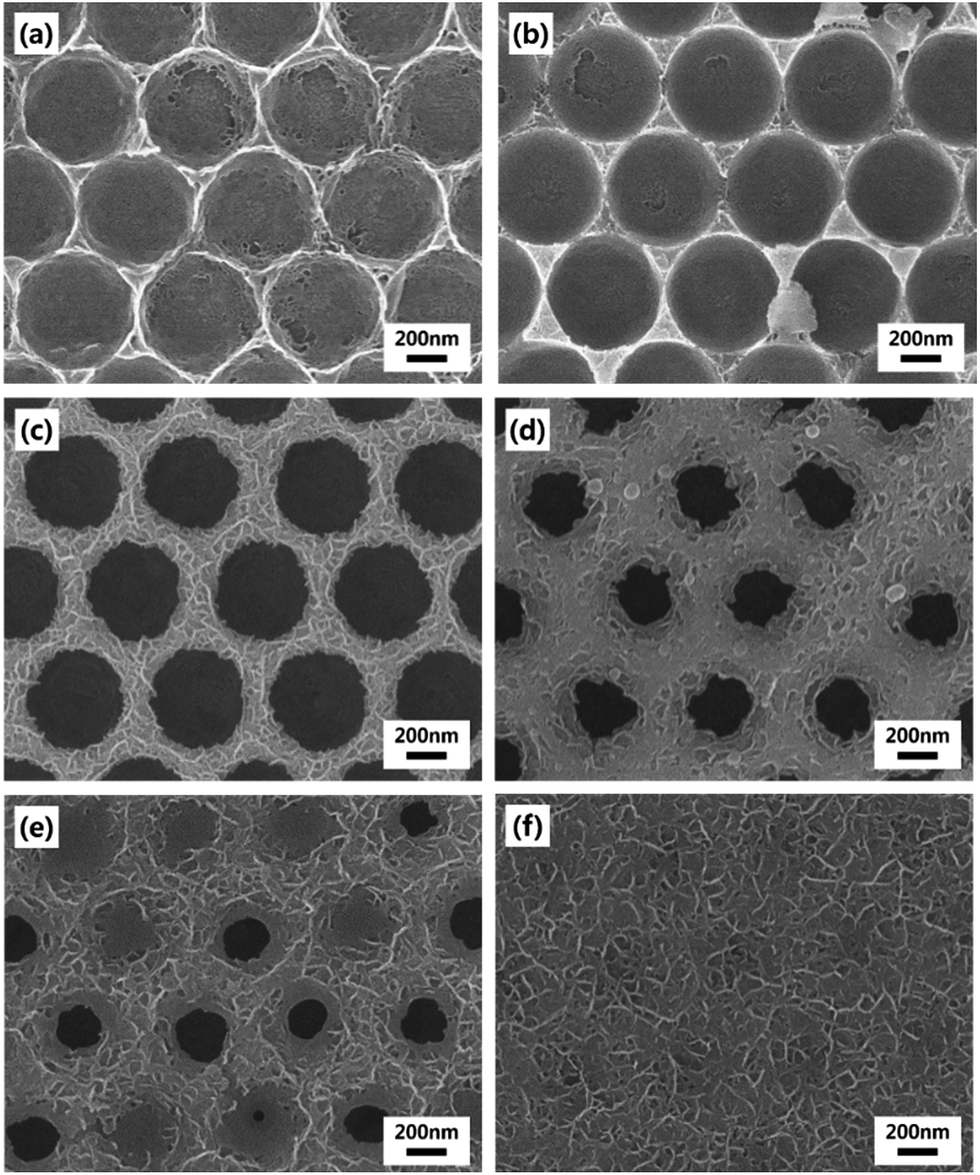
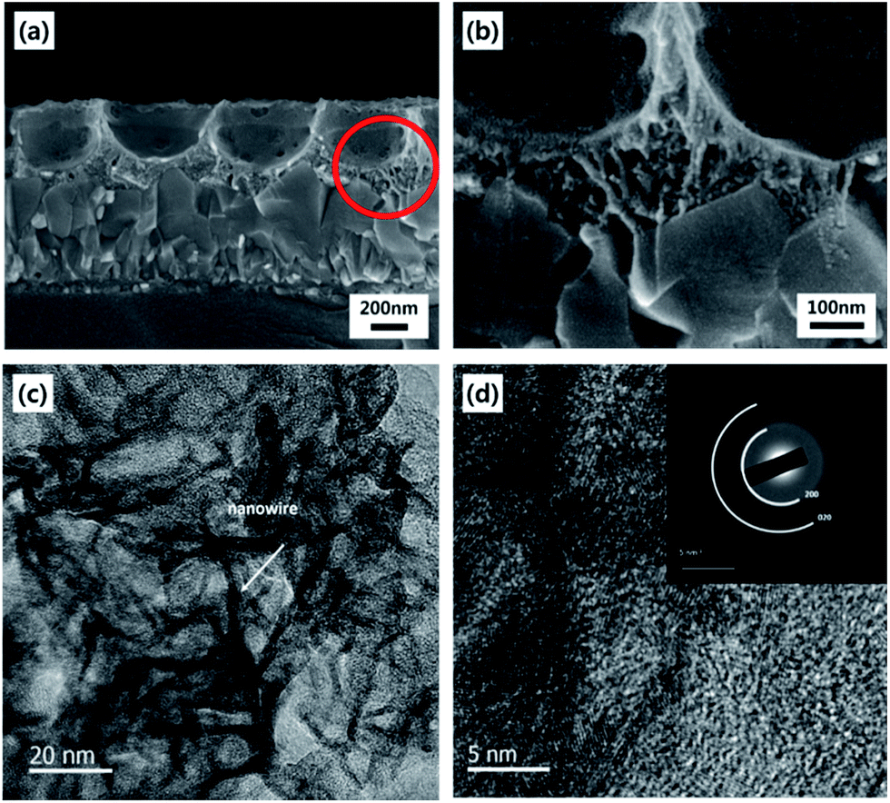
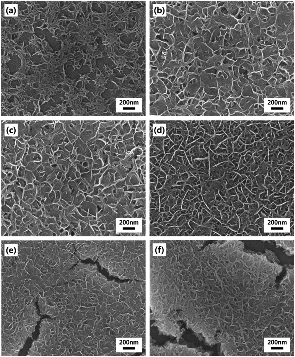
The basic strategy to fabricate hierarchical MnO2 nanostructures is illustrated in Fig. 1. First, close-packed 2D array of PS nanospheres was prepared on FTO-coated glass substrates. Then, a thin layer of MnO2 nanowires was deposited on the PS nanosphere-covered surface using the cyclic voltammetric deposition method. Finally, the PS nanospheres were extracted by the THF treatment. The FE-SEM images below the schematic illustration in Fig. 1 show that the periodically arrayed nanostructure was successfully fabricated on a large area of the substrate surface. The morphological evolution of the surfaces as a function of the number of MnO2 deposition cycles is shown in Fig. 2. MnO2 nanowires with a few nanometers in width formed on the inverse-opal nanostructures with dimension of 580 nm between the nearest-neighbour holes are clearly observed. The area of the surface covered with MnO2 nanowires gradually increased as the number of deposition cycles increased. The MnO2 layer seemed to reach the half-height of the PS nanospheres at around 15 deposition cycles (Fig. 2c). After more than 20 cycles (Fig. 2d–f), it was observed that the PS-removed spaces were gradually covered with MnO2 nanowire layers. The formation of MnO2 nanowires was more closely observed by cross-sectional FE-SEM (Fig. 3a and b) and TEM/HRTEM (Fig. 3c and d) images. The lattice fringes in Fig. 3d and SAED pattern (inset of Fig. 3d) reveal the formation of birnessite-type MnO2 polycrystalline nanowires.26 The deposit weight of the MnO2 layer as measured by QCM was almost proportional to the number of electrodeposition cycles (Fig. S2†), reaching approximately 300 μg after the 25 cycles. For comparison, the MnO2 nanowires were also electrodeposited on bare FTO-coated glass substrates, which will be denoted as simple nanostructures in contrast to the hierarchical nanostructures. The surface morphologies of these simple nanostructures after various numbers of deposition cycles were also characterized by FE-SEM as shown in Fig. 4. At 15 cycles (Fig. 4c), it is observed that almost the whole surface was covered with MnO2 nanowires. The FE-SEM image of the whisker-like nanowires became clearer at 20 cycles of deposition (Fig. 4d). After 25 cycles, the nanowires became shorter and more slender, as shown in Fig. 4e and f. In addition, some cracks on the electrode surface which are probably caused by detachment of MnO2 layers from the substrate were observed. The number and size of these cracks increased as the electrodeposition proceeded.
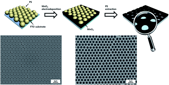 |
| | Fig. 1 (Upper part) Schematic illustration of the fabrication of hierarchical MnO2 nanostructures and (lower part) surface FE-SEM images taken after PS transfer on the FTO-coated glass substrate (left) and after MnO2 layer growth at 15 electrodeposition cycles, followed by PS extraction (right). | |
 |
| | Fig. 2 FE-SEM images of hierarchical MnO2 nanostructures electrodeposited on the PS nanosphere-covered FTO substrates, followed by PS extraction. The number of electrodeposition cycles is (a) 5, (b) 10, (c) 15, (d) 20, (e) 25 and (f) 30. | |
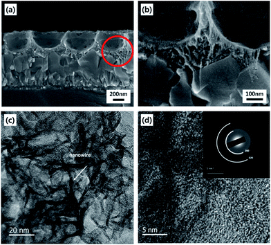 |
| | Fig. 3 Cross-sectional FE-SEM images of (a) low-magnification and (b) high-magnification view of hierarchically nanostructured MnO2 electrodes. (c) TEM and (d) HRTEM images of the MnO2 of hierarchical electrodes, and SAED pattern (inset of d). The number of MnO2 electrodeposition cycles was 15 for all samples. | |
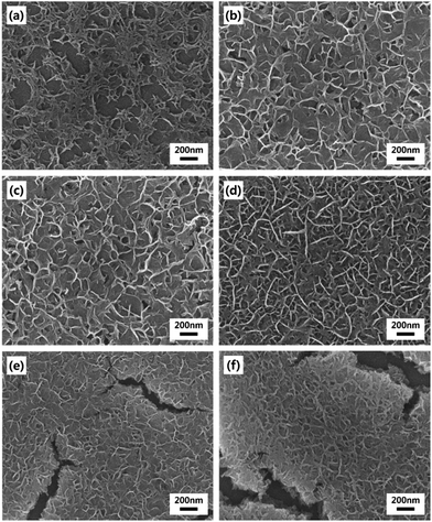 |
| | Fig. 4 FE-SEM images of simple MnO2 nanostructures electrodeposited on bare FTO substrates. The number of electrodeposition cycles is (a) 5, (b) 10, (c) 15, (d) 20, (e) 25 and (f) 30. | |
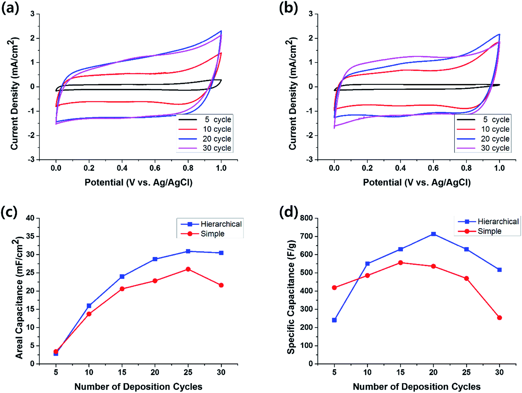
The electrochemical performance of half-cell supercapacitors based on the hierarchical and simple MnO2 nanostructures was investigated by cyclic voltammetry (CV) measured in 1 M aqueous Na2SO4 solution within the potential range from 0.0 to 1.0 V. The typical cyclic voltammograms measured at the scan rate of 100 mV s−1 for the electrodes with hierarchical and simple nanostructures are shown in Fig. 5a and b, respectively. The almost symmetric CV curves indicate the reversibility of the redox transition of MnO2. In the case of simple nanostructure electrodes, the areal capacitance was easily saturated as the MnO2 deposition preceded, or even decreased after 25 deposition cycles (red circles in Fig. 5c). This is probably because the bottom part MnO2 molecules of the thick film become electrochemically difficult to be accessible to the electrolyte. The cracks observed in Fig. 4 may also contribute to the decrease of the capacitive performance for thick electrodes. In contrast, for the cells with hierarchical MnO2 nanostructures, the increment of the areal capacitance with the number of deposition cycles (blue squares in Fig. 5c) was larger than that of the cells with simple nanostructures. At small number of deposition cycles, the areal capacitance of the cells with hierarchical nanostructure is similar to that of the cells with simple nanostructure. For example, after 5 deposition cycles, the areal capacitance for the cells with hierarchical and simple nanostructures is 2.8 mF cm−2 and 3.4 mF cm−2, respectively. However, the areal capacitance of the cells with hierarchical nanostructures increased rapidly as the deposition proceeded and exceeded that of the simple nanostructure cells after 10 deposition cycles. Notably, the hierarchical nanostructures retarded the saturation of the areal capacitance, as compared to the simple nanostructures, indicating that the benefits of nanostructures, i.e. enhancement in electrolytic accessibility, are more effectively retained for the MnO2 electrode grown on the arrayed pattern.
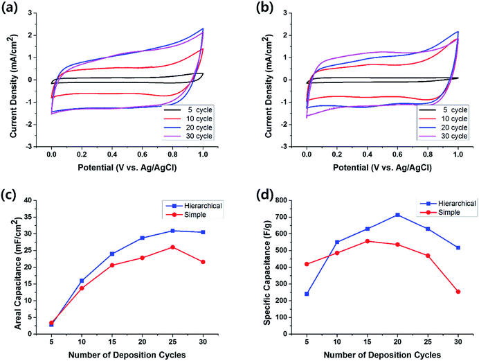 |
| | Fig. 5 Representative cyclic voltammograms of the (a) hierarchical and (b) simple MnO2 nanostructures at various numbers of MnO2 electrodeposition cycles. Plots of (c) areal and (d) specific capacitances measured at a scan rate of 10 mV s−1 for the hierarchical ( ) and simple ( ) and simple ( ) MnO2 nanostructure electrodes as a function of the number of MnO2 electrodeposition cycles. ) MnO2 nanostructure electrodes as a function of the number of MnO2 electrodeposition cycles. | |
The specific capacitance, Csp, was also calculated by the following equation
| |
 | (3) |
where
I (A) is the average current,
m (g) is the mass of deposited MnO
2, and d
V/d
t (mV s
−1) is the scan rate.
27 While the specific capacitance of the cells with simple nanostructures (red circles in
Fig. 5d) reached 556 F g
−1 at 15 deposition cycles and then decreased to 254 F g
−1 at 30 deposition cycles, that of the cells with hierarchical nanostructures (blue squares in
Fig. 5d) increased until 20 deposition cycles and reached its maximum value of 714 F g
−1 at the scan rate of 10 mV s
−1. This is the largest value of MnO
2 electrodes fabricated without supportive material, such as CNTs and/or graphene, to the best of our knowledge. This improvement in
Csp can be attributed to the effective charge transport in the hierarchically nanostructured electrodes along with considerably increased surface area. However, the benefits of the hierarchical structures were reduced after more than 20 deposition cycles, which is probably because the PS-removed holes began to be blocked, as shown in the FE-SEM images of
Fig. 2, and hence the electrolytic accessibility became decreased.
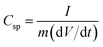
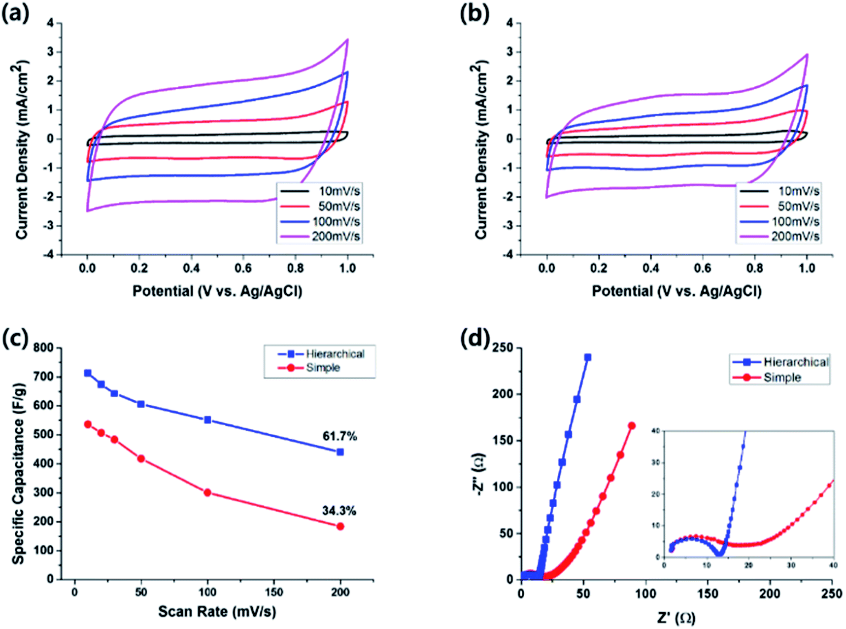
Fig. 6 shows the CV curves and specific capacitances at various scan rates ranging from 10 mV s
−1 to 200 mV s
−1. For the electrode with hierarchical nanostructures (
Fig. 6a and blue squares in
Fig. 6c),
Csp gradually decreased from 714 F g
−1 at the slowest scan rate of 10 mV s
−1 to 440 F g
−1 at 200 mV s
−1. In contrast, for the electrode with a simple nanostructure (
Fig. 6b and red circles in
Fig. 6c), the
Csp values varied from 536 F g
−1 at 10 mV s
−1 to 184 F g
−1 at 200 mV s
−1. The relatively larger retention of
Csp for the hierarchical nanostructure, namely 61.7%, as compared to 34.3% for the simple nanostructure implies that the access for ions to surface active sites is more efficient in case of the hierarchically nanostructured electrode. Nyquist plots of the hierarchical and simple nanostructure electrodes obtained in the frequency range from 10
−1 to 10
5 Hz where
Z′ and
Z′′ are the real and imaginary part of the impedance, respectively, are shown in
Fig. 6d. The smaller diameter of the semicircle in the high-frequency region for the hierarchical nanostructure electrode indicates that the electrode possesses smaller charge-transfer resistance,
Rct.
28–30 The slope of the line in the low-frequency region is related to the diffusive resistance,
Rd, and the steeper line of the hierarchical nanostructure electrode reflects better capacitive behavior.
30–32 Improved capacitive properties of the hierarchical nanostructure electrode,
i.e. large capacitance and rapid response, can therefore be explained by these reduced resistances and consequently enhanced charge transfer as well as increased contact between the electrode and electrolyte. The Bode |
Z| and Bode angle plots also supported the better capacitive behavior of the hierarchically nanostructured electrode as shown in Fig. S3.
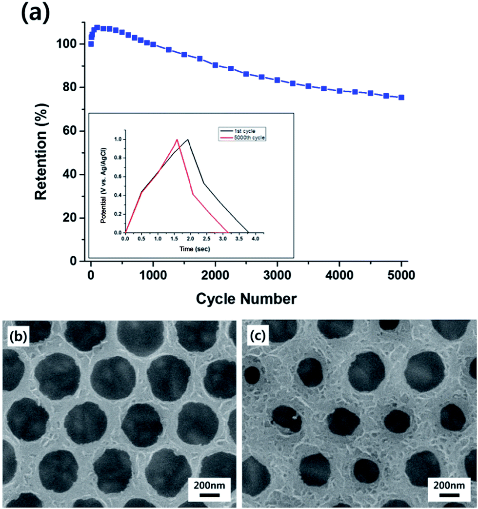
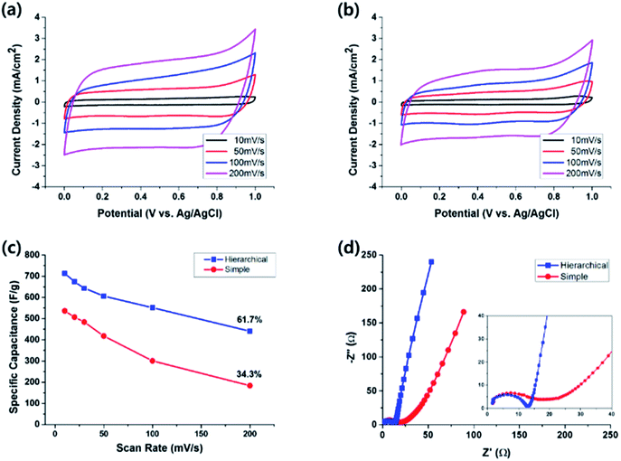
† The phase angle of the hierarchical electrode is −77°, closer to the value of an ideal capacitor, −90°, compared to −62° for the simple nanostructure electrode. The cycle life of the hierarchical nanostructure electrode was also examined by continuous galvanostatic charge–discharge cycles at the current density of 10 mA cm
−2 (
Fig. 7a). As shown in the inset of the figure, the shape of the curve after 5000 charge–discharge cycles little deviated from that after the first cycle indicating that the capacitive behavior retained even after 5000 cycles. The capacitance increased slightly to 107.6% in the first 100 cycles. Similar enhancements have been previously reported and are probably due to the activation of electrochemical cycling.
21,33 The retention of the specific capacitance was 75.4% after 5000 cycles, indicating that the MnO
2 nanowires will not easily detach from the current collector substrate.
Fig. 7b and c show that the MnO
2 nanowires on the electrode surface became less apparent after 5000 charge–discharge cycles, compared to the initial nanostructures (
Fig. 2c and d). The gradual decrease of the areal capacitance may be attributed to this morphological deterioration of the nanostructures over the charge–discharge cycles, although various factors such as reduction of surface Mn ions are also related.
34
 |
| | Fig. 6 Representative cyclic voltammograms of the (a) hierarchical and (b) simple MnO2 nanostructures at various scan rates. The number of MnO2 electrodeposition cycles was 20 for both samples. (c) Specific capacitances as a function of the scan rate and (d) Nyquist plots for the hierarchical ( ) and simple ( ) and simple ( ) MnO2 nanostructures. ) MnO2 nanostructures. | |
 |
| | Fig. 7 (a) Csp retention for the hierarchical MnO2 nanostructures as a function of the number of galvanostatic charge–discharge cycles at the current density of 10 mA cm−2. (Inset) Galvanostatic curves after 1 and 5000 charge–discharge cycles at a constant current density of 10 mA cm−2. FE-SEM images of hierarchically nanostructured MnO2 electrodes at (b) 15 and (c) 20 electrodeposition cycles after 5000 charge–discharge cycles. | |
Conclusions
The MnO2 electrode with hierarchical nanostructures was successfully fabricated by electrodepositing MnO2 nanowires onto FTO substrates covered with periodically arrayed PS nanospheres, followed by PS extraction. The hierarchical structure consisted of inverse-opal nanostructures with dimension of a few hundred nanometers, e.g. 580 nm in this work, and nanowires with width of a few nanometers. The specific capacitance of the hierarchically nanostructured MnO2 electrode reached 714 F g−1 at the scan rate of 10 mV s−1, which is significantly larger than the maximum Csp value for the MnO2 electrode with simple nanowire structures of 556 F g−1. This enhancement of the specific capacitance without using any supportive material, such as CNTs and/or graphene, could be attributed to the synergetic effect of the two different length scales, i.e. reduced MnO2 thickness and consequently improved electronic/ionic conductivity owing to the large length-scale of the inverse-opal nanostructures, and increased surface area and consequently shortened diffusion paths for ions owing to the small length-scale of the nanowire structures. The specific capacitance of the hierarchical nanostructures retained 61.7% at the rapid scan rate of 200 mV s−1, which is significantly larger than the retention of 34.3% observed for the simple nanostructures at the same scan rate. In addition to the increased surface area, the EIS measurements confirmed that the arrayed nanopatterns lowered the charge transfer and diffusive resistance of the electrode, which also contributed to the improvement of the capacitive properties, i.e. large capacitance and rapid response. Moreover, the Csp retention of the hierarchically nanostructured electrode after 5000 charge–discharge cycles was 75.4%. All the results presented here demonstrate the benefits of the hierarchical MnO2 nanostructures for the application to pseudocapacitor electrodes.
Acknowledgements
This work was supported by National Research Foundation of Korea (NRF) Grant (No. 2016R1A5A1012966, NRF-2013R1A1A2A10012336, and No. 2011-0030233) funded by the Korean Government.
Notes and references
- G. Wang, L. Zhang and J. Zhang, Chem. Soc. Rev., 2012, 41, 797 RSC.
- J. Yan, Q. Wang, T. Wei and Z. Fan, Adv. Energy Mater., 2014, 4, 1300816 CrossRef.
- L. Zhang and X. S. Zhao, Chem. Soc. Rev., 2009, 38, 2520 RSC.
- R. Liu, J. Duay and S. B. Lee, Chem. Commun., 2011, 47, 1384 RSC.
- M. Winter and R. J. Brodd, Chem. Rev., 2004, 104, 4245 CrossRef CAS PubMed.
- R. Liu, J. Duay, T. Lane and S. B. Lee, Phys. Chem. Chem. Phys., 2010, 12, 4309 RSC.
- H. Y. Lee, V. Manivannan and J. B. Goodenough, C. R. Acad. Sci., Ser. IIc: Chim., 1999, 2, 565 CrossRef CAS.
- S. C. Pang, M. A. Anderson and T. W. Chapman, J. Electrochem. Soc., 2000, 147, 444 CrossRef CAS.
- H. Y. Lee and J. B. Goodenough, J. Solid State Chem., 1999, 144, 220 CrossRef CAS.
- M. Toupin, T. Brousse and D. Belanger, Chem. Mater., 2004, 16, 3184 CrossRef CAS.
- S. Liu, S. Sun and X.-Z. You, Nanoscale, 2014, 6, 2037 RSC.
- S. Devaraj and N. Munichandraiah, J. Electrochem. Soc., 2007, 154, A901 CrossRef CAS.
- M.-S. Wu, Appl. Phys. Lett., 2005, 87, 153102 CrossRef.
- J. Wei, N. Nagarajan and I. Zhitomirsky, J. Mater. Process. Technol., 2007, 186, 356 CrossRef CAS.
- W. Xiao, H. Xia, J. Y. H. Fuh and L. Lu, J. Power Sources, 2009, 193, 935 CrossRef CAS.
- V. Subramanian, H. Zhu, R. Vajtai, P. M. Ajayan and B. Wei, J. Phys. Chem. B, 2005, 109, 20207 CrossRef CAS PubMed.
- S. W. Lee, J. Kim, P. T. Hammond and Y. Shao-Horn, ACS Nano, 2010, 4, 3889 CrossRef CAS PubMed.
- R. Liu, J. Duay and S. B. Lee, ACS Nano, 2010, 4, 4299 CrossRef CAS PubMed.
- H. Xia, J. Feng, H. Wang, M. O. Lai and L. Lu, J. Power Sources, 2010, 195, 4410 CrossRef CAS.
- Jaidev, R. I. Jafri, A. K. Mishra and S. Ramaprabhu, J. Mater. Chem., 2011, 21, 17601 RSC.
- X. Dong, W. Shen, J. Gu, L. Xiong, Y. Zhu, H. Li and J. Shi, J. Phys. Chem. B, 2006, 110, 6015 CrossRef CAS PubMed.
- K.-W. Nam, C.-W. Lee, X.-Q. Yang, B. W. Cho, W.-S. Yoon and K.-B. Kim, J. Power Sources, 2009, 188, 323 CrossRef CAS.
- J. Yan, Z. Fan, T. Wei, W. Qian, M. Zhang and F. Wei, Carbon, 2010, 48, 3825 CrossRef CAS.
- G. Yu, L. Hu, M. Vosgueritchian, H. Wang, X. Xie, J. R. McDonough, X. Cui, Y. Cui and Z. Bao, Nano Lett., 2011, 11, 2905 CrossRef CAS PubMed.
- J. R. Oh, J. H. Moon, S. Yoon, C. R. Park and Y. R. Do, J. Mater. Chem., 2011, 21, 14167 RSC.
- D. P. Dubal, D. Aradilla, G. Bidan, P. Gentile, T. J. S. Schubert, J. Wimberg, S. Sadki and P. Gomez-Romero, Sci. Rep., 2015, 5, 9771 CrossRef CAS PubMed.
- C. D. Lokhande, D. P. Dubal and O. S. Joo, Curr. Appl. Phys., 2011, 11, 255 CrossRef.
- L. Wang, H. Ji, F. Zhu, Z. Chen, Y. Yang, X. Jiang, J. Pinto and G. Yang, Nanoscale, 2013, 5, 7613 RSC.
- J.-W. Wang, Y. Chen and B.-Z. Chen, J. Electrochem. Soc., 2015, 162, A1654 CrossRef CAS.
- E. Barsoukov and J. R. Macdonald, Impedance Spectroscopy: Theory, Experiment, and Applications, Wiley Interscience, 2005 Search PubMed.
- Z. Lei, J. Zhang and X. S. Zhao, J. Mater. Chem., 2012, 22, 153 RSC.
- J. Zhi, O. Reiser and F. Huang, ACS Appl. Mater. Interfaces, 2016, 8, 8452 CAS.
- H. Jiang, L. Yang, C. Li, C. Yan, P. S. Lee and J. Ma, Energy Environ. Sci., 2011, 4, 1813 CAS.
- F. Ataherian and N.-L. Wu, J. Electrochem. Soc., 2011, 158, A422 CrossRef CAS.
Footnote |
| † Electronic supplementary information (ESI) available: Powder XRD pattern of electrodeposited MnO2 thin films, Bode |Z| and Bode angle plots of the hierarchical and simple MnO2 nanostructure electrodes, a photograph of a red LED lightened by three asymmetric supercapacitor units connected in series. See DOI: 10.1039/c6ra22841k |
|
| This journal is © The Royal Society of Chemistry 2016 |
Click here to see how this site uses Cookies. View our privacy policy here. 

 ) and simple (
 ) MnO2 nanostructure electrodes as a function of the number of MnO2 electrodeposition cycles.
 ) and simple (
 ) MnO2 nanostructures.