DOI:
10.1039/C6RA22827E
(Paper)
RSC Adv., 2016,
6, 111747-111753
Preparation of pH- and magnetism-responsive sodium alginate/Fe3O4@HNTs nanocomposite beads for controlled release of granulysin
Received
13th September 2016
, Accepted 18th November 2016
First published on 18th November 2016
Abstract
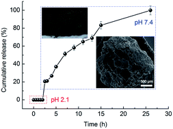
The burst release of drug molecules from natural biopolymer-based hydrogels is a common issue in the field of controlled drug delivery systems. pH- and magnetism-responsive sodium alginate/Fe3O4@HNTs nanocomposite beads for controlled release of granulysin were prepared. Fe3O4@HNTs were prepared by growth of Fe3O4 nanoparticles on the surface of HNTs via solvothermal reactions. Then, the SA/Fe3O4@HNTs-granulysin nanocomposite beads were prepared by sol–gel transition of the SA-granulysin aqueous solution with homogeneously dispersed Fe3O4@HNTs. The nanocomposite beads were characterized by Fourier transform infrared spectroscopy and scanning electron microscopy. In addition, the factors, e.g., the concentration of SA, weight ratio of Fe3O4@HNTs to SA and weight ratio of FeCl3·6H2O to HNTs, influencing granulysin loading and controlled release behavior were systematically studied. The entrapment efficiency was enhanced to 61.4% and the burst release of granulysin was overcome by introducing appropriate amounts of HNTs and the Fe3O4 nanoparticles. Granulysin was released at a steady rate in pH 7.4 PB owing to the synergistic effects of SA, HNTs and the Fe3O4 nanoparticles. The release kinetics of granulysin from the nanocomposite beads follows the Higuchi model. HNTs and the Fe3O4 nanoparticles formed additional crosslinkages in the nanocomposite beads, which changed the swelling behaviors of the beads, and then improved the controlled release behaviors of the model drug molecules.
1. Introduction
Hydrogels are composed of crosslinked hydrophilic polymers with a three dimensional network.1 Stimuli-responsive hydrogels exhibit a transition between swollen and de-swollen states in response to environmental changes such as pH, temperature, light or even magnetic field.1–4 In the past decades, stimuli-responsive hydrogels based on biopolymers have frequently been used as controlled drug delivery matrices owing to their excellent properties including environmental sensitivity, high swelling degree, non-toxicity, biocompatibility and biodegradability. Various biopolymers including sodium alginate (SA), chitosan and guar gum have been used for the preparation of stimuli-responsive hydrogels for controlled drug delivery.5–7 For example, the SA hydrogel is pH-sensitive, which swells in neutral- and basic aqueous media, but collapses in acidic aqueous solutions.8 Thus, the SA hydrogel is an ideal matrix for controlled drug delivery. However, when pristine biopolymers are used for the preparation of hydrogels, there are some significant limitations.2 First, the uncontrolled burst release of the drugs from the hydrogels is inevitable in most cases owing to the fact that the drug molecules are often loaded in the hydrogels by simple physical interactions. The other one is the weak mechanical properties of the hydrogels, which caused their fast disintegration in aqueous media. These problems have seriously hindered applications of biopolymer-based hydrogels for controlled drug delivery.
Inspired by shells of the marine animals, organic/inorganic bionanocomposites composed of the biopolymer matrices and inorganic materials are of great interests in various fields including tissue engineering and controlled drug delivery.9,10 Compared to the pristine biopolymers, the resulting bionanocomposites show excellent properties owing to the synergetic effects of biopolymers and inorganics.6,11,12 Various kinds of inorganics have been used to prepare bionanocomposites in order to improve the mechanical properties, overcome the burst release of drug molecules and enhance the drug loading efficiency of the pristine biopolymer matrices.13–15 In recent years, many studied showed that introduction of clay minerals into polymers is an effective approach to improve comprehensive properties of the polymeric materials.16,17
Different from the other natural alumino-silicate clay minerals, halloysite nanotubes (HNTs) have the same chemical composition as kaolinite but a hollow tubular structure like carbon nanotubes.18 The size of HNTs varies in the range of 10–15 nm in inner diameter and 1–15 μm in length. HNTs are promising natural nano-carriers for drugs owing to the unique morphology.19–21 The tubular structure of HNTs leads to retardation of drug release and is helpful to overcome the burst release of drugs. We have studied the interactions between HNTs and ofloxacin, and the effects of activation of HNTs on the release of ofloxacin, which are helpful for us to use HNTs rationally in the design of controlled drug delivery systems.22,23 We also have prepared magnetic 2-hydroxypropyltrimethyl ammonium chloride chitosan/Fe3O4/halloysite nanotubes microspheres for the controlled release of ofloxacin.17
Granulysin is a novel antimicrobial protein produced by human cytolytic T lymphocytes and natural killer cells.24 It is active against a broad range of microbes, including Gram-positive and Gram-negative bacteria, fungi, and parasites. The fact that it kills Mycobacterium tuberculosis is particularly important, since the current vaccine is of limited efficacy and antibiotic resistance is increasing. Although functionally related to other antibacterial peptides, defensins and magainins, granulysin is structurally distinct. In addition, the 9 kDa granulysin may also be used as a new anti-tumoral treatment.25
Here, we report preparation of pH- and magnetic-responsive SA/Fe3O4@HNTs-granulysin nanocomposite beads for controlled release of granulysin. Fe3O4@HNTs was prepared by growth of Fe3O4 nanoparticles on the surface of HNTs via solvothermal reactions. Then, the SA/Fe3O4@HNTs-granulysin nanocomposite beads were prepared by sol–gel transition of the SA-granulysin aqueous solution with homogeneously dispersed Fe3O4@HNTs. The nanocomposite beads were characterized by different techniques, and the factors influencing granulysin loading and controlled release behavior were systematically studied.
2. Experimental
2.1. Materials
HNTs were purchased from Sigma-Aldrich. SA with a kinetic viscosity of 200 ± 20 mPa s was obtained from the Shanghai Jingchun Reagent Co. Ltd. (Shanghai, China). The nucleotide sequence of 9 kD granulysin protein was cloned from human peripheral blood cDNA template by PCR. The prokaryotic expression plasmid of pEGM-4T-1-granulysin was constructed and transformed into Escherichia coli rosetta. The soluble granulysin-GST protein was expressed by 1 mM IPTG induction at 37 °C, purified by beaver beads GSH magnetic bead, and stored at −80 °C for further use. FeCl3·6H2O, ethylene glycol, polyethylene glycol (10
000 g mol−1) and sodium acetate were purchased from China National Medicines Co. Ltd. All the other chemical reagents used in this study were also purchased from China National Medicine Co. Ltd. and used as received. Deionized water was used for all the experiments.
2.2. Preparation of Fe3O4@HNTs
Fe3O4@HNTs was prepared by solvothermal reduction reactions between FeCl3 and ethylene glycol in the presence of HNTs.26 A proper amount of FeCl3·6H2O was dissolved in 40 mL of ethylene glycol to form a clear solution. 3.6 g of sodium acetate and 1.0 g of polyethylene glycol were added into the solution with constant stirring for 30 min. Then, 0.4 g of HNTs was ultrasonically dispersed into the above dispersion for 3 h. The mixture was sealed in a 50 mL Teflon lined stainless-steel autoclave, and then heated to and maintained at 200 °C for 48 h. After cooled to room temperature, the dark brown Fe3O4@HNTs was washed with deionized water and ethanol successively, and then dried at 60 °C.
2.3. Preparation of SA/Fe3O4@HNTs-granulysin nanocomposite beads
The SA/Fe3O4@HNTs-granulysin nanocomposite beads were prepared according to the following procedure. Typically, 0.1 g of Fe3O4@HNTs was dispersed in 5 mL of deionized water, and then ultrasonicated for 30 min to form a homogeneous suspension. 0.2 g of SA was dissolved in 10 mL of deionized water. A proper amount of granulysin was dissolved in 5 mL of deionized water. Then, the Fe3O4@HNTs suspension, the SA solution and the granulysin solution were mixed together. Subsequently, the suspension containing SA, Fe3O4@HNTs and granulysin was added dropwise into 200 mL of 7% Ca(NO3)2·4H2O aqueous solution at 4 °C with mild stirring (50 rpm). The nanocomposite beads were formed immediately and further crosslinked for 6 h at 4 °C to accomplish the reaction between SA and Ca2+. After that, the nanocomposite beads were filtered with a 100-mesh screen, and then dried at room temperature for 48 h. Factors influencing the entrapment efficiency (EE) and the cumulative release behavior were investigated. The factors are the concentration of SA (CSA), weight ratio of Fe3O4@HNTs to SA (mFe3O4@HNTs/mSA) and weight ratio of FeCl3·6H2O to HNTs (mFeCl3·6H2O/mHNTs) as shown in Table 1. The SA/HNTs beads were prepared according to the same procedure using HNTs instead of Fe3O4@HNTs.
Table 1 Composition, granulysin content and EE of the SA/Fe3O4@HNTs-granulysin nanocomposite beads
Samples |
CSA (wt%) |
mFe3O4@HNTs/mSA |
mFeCl3·6H2O/mHNTs |
EE (%) |
SA/Fe3O4@HNTs-granulysin-1 |
0.5 |
1 |
3.3 |
61.4 |
SA/Fe3O4@HNTs-granulysin-2 |
1 |
1 |
3.3 |
60.9 |
SA/Fe3O4@HNTs-granulysin-3 |
2 |
1 |
3.3 |
59.2 |
SA/Fe3O4@HNTs-granulysin-4 |
2 |
0 |
3.3 |
48.8 |
SA/Fe3O4@HNTs-granulysin-5 |
2 |
0.25 |
3.3 |
50.6 |
SA/Fe3O4@HNTs-granulysin-6 |
2 |
0.5 |
3.3 |
55.2 |
SA/Fe3O4@HNTs-granulysin-7 |
2 |
1 |
0 |
55.2 |
SA/Fe3O4@HNTs-granulysin-8 |
2 |
1 |
0.83 |
56.3 |
SA/Fe3O4@HNTs-granulysin-9 |
2 |
1 |
1.63 |
55.9 |
2.4. Measurement of swelling ratio
0.1 g of the sample was soaked in 250 mL of pH 7.4 phosphate buffer (PB) solution at 37 ± 0.5 °C. The PB solution was prepared by dissolving 4.80 g of NaOH and 20.40 g of KH2PO4 in 3000 mL of distilled water according to USP 30. After a pre-determined time interval, the swollen sample was separated from the unabsorbed fluid by filtering through a 100-mesh screen, blotted to remove excess fluid, and then weighed immediately. The swelling ratio of the samples at a given time (t), Qt, can be calculated using the following equation:where m0 and mt are the weights of the dry sample and the swollen sample, respectively. Qt is calculated as grams of water per gram of sample.
2.5. Measurement of EE for granulysin
0.1 g of the sample were soaked and shaken occasionally in 100 mL of pH 7.4 PB solution for 12 h, and then crushed by ultrasonication (100 W) for 30 min. The suspension was filtered, and the granulysin content was determined using a UV-Vis spectrophotometer at 202 nm. The EE of the beads was defined according to the following equation:where w1 is the actual drug content in the weighed quantity of the beads and w2 is the theoretical drug content in the corresponding weighed quantity of the beads.21 w2 was calculated as follows. In preparing the nanocomposite, x g of granulysin was used and y g of the nanocomposite was finally obtained. So, w2 = x/y × m. m is the weight of the granulysin-loaded sample for test.
2.6. In vitro cumulative release studies
Study of the in vitro granulysin cumulative release behavior of the samples was carried out in an intelligent dissolution apparatus (ZRS-8G, China). 0.2 g of the sample was immersed in 500 mL of pH 7.4 PB solution and stirred at 100 rpm at 37 ± 0.5 °C. At certain time intervals, 5.0 mL of the solution was withdrawn and replaced with the same amount of fresh PB solution. The solution was filtered through a 0.45 μm nylon membrane filter and analyzed by UV-Vis spectrophotometer at 202 nm. The cumulative granulysin released was determined using eqn (3): |
Cumulative granulysin released (%) = Rt/L × 100
| (3) |
where L and Rt represent the amount of granulysin loaded and the cumulative amount of granulysin released at time t, respectively.
All the tests, including measurement of the swelling ratio, determination of the EE and in vitro granulysin cumulative release studies, were carried out in triplicate, and the average values are presented in this study.
2.7. Characterization
Fourier transform infrared spectra (FT-IR) of the samples were taken as KBr pellets using a Thermo Nicolet NEXUS TM spectrophotometer. Morphology of the samples was investigated by field emission scanning electron microscopy (SEM, JSM-6701F, JEOL). Before SEM observation, all the samples were placed on round brass stubs and sputter coated with platinum under an argon atmosphere. A vibrating-sample magnetometer (VSM) (Lake Shore, 735 VSM, Model 7304, USA) was used to test the magnetic property of the samples.
3. Results and discussion
3.1. Preparation of Fe3O4@HNTs
The digital images of HNTs and Fe3O4@HNTs are shown in Fig. 1a and b. After solvothermal reduction reactions between FeCl3 and ethylene glycol in the presence of beige HNTs, the dark brown Fe3O4@HNTs was formed, indicating formation of Fe3O4 nanoparticles on the surface of HNTs. The Fe3O4 nanoparticles are 80–150 nm in diameter according to our previous study.27 Fe3O4@HNTs showed a standard paramagnetic characteristic curve with no hysteresis after removal of the magnetic field as shown in Fig. 1c. The saturation magnetization of Fe3O4@HNTs is 52.05 emu g−1 at 300 K. The Fe3O4@HNTs nanoparticles can be neatly manipulated using a magnet.
 |
| Fig. 1 Digital images of (a) HNTs and (b) Fe3O4@HNTs, and (c) the magnetic curve of Fe3O4@HNTs. | |
3.2. Preparation of SA/Fe3O4@HNTs-granulysin nanocomposite beads
The SA/Fe3O4@HNTs-granulysin nanocomposite beads were prepared by sol–gel transition of the SA-granulysin aqueous solution with homogeneously dispersed Fe3O4@HNTs nanoparticles. The sol–gel transition of the suspension occurred immediately after being dropped into the Ca(NO3)2 solution owing to the reaction between Ca2+ and SA. Consequently, the SA in the beads was separated from the Ca2+ in the external medium and could only react gradually with the interpenetrated Ca2+ ions. After a period of time, the SA/Fe3O4@HNTs-granulysin nanocomposite beads were successfully formed.
In order to study the influence of Fe3O4@HNTs on the microstructure of the beads, the SA-granulysin beads and SA/Fe3O4@HNTs-granulysin-3 nanocomposite beads were observed by SEM and are shown in Fig. 2. Serious contraction of the SA-granulysin beads was observed during room temperature drying. There are many wrinkles on the surface of the SA-granulysin beads even at high magnification as showing in Fig. 2a and b. After introduction of Fe3O4@HNTs, the contraction of the beads was partly inhibited as showing in Fig. 2c and d. This may be because the introduced Fe3O4@HNTs acted as inorganic skeleton of the beads, which formed more crosslinking points and strengthened the beads. Also, the surface of the SA/Fe3O4@HNTs-granulysin-3 beads became very dense, which may hinder the burst release of drug molecules from the pristine SA beads. The SA/Fe3O4@HNTs-granulysin-3 beads have a macroporous structure according to the cross-section. The size of the pores is in the range of 100–300 μm owing to evaporation of water in the drying process. The walls of the macropores are also very dense. The changes in the microstructure of the SA-granulysin beads are because of introduction of Fe3O4@HNTs, which is of helpful to regulate the controlled release behavior of granulysin.
 |
| Fig. 2 SEM micrographs of (a, b) SA-granulysin, (c, d) SA/Fe3O4@HNTs-granulysin-3 and (e, f) cross-section of SA/Fe3O4@HNTs-granulysin-3 beads. | |
The FT-IR spectra of Fe3O4@HNTs, SA, SA-granulysin and SA/Fe3O4@HNTs-granulysin-3 are shown in Fig. 3. In the FT-IR spectrum of Fe3O4@HNTs, the bands at 3695 and 3622 cm−1 are attributed to –OH groups on the internal surface of HNTs.28 The bands at 911 and 1033 cm−1 are attributed to the Al–O and Si–O stretching vibration, respectively.21 The bands at 1623 and 1418 cm−1 in the FT-IR spectrum of SA are attributed to asymmetric and symmetric stretching vibrations of the –COO− groups, respectively. After formation of the SA-granulysin beads, the band at 1623 cm−1 shifted to 1632 cm−1, and the band at 1418 cm−1 shifted to 1432 cm−1. Also, a new band appeared at 1375 cm−1. These changes are owing to the reaction between SA and Ca2+. The bands corresponding to granulysin cannot be detected by FT-IR owing to the low content of granulysin in the beads as shown in Table 1. After incorporating Fe3O4@HNTs in the SA-granulysin beads, the band at 1375 cm−1 further shifted to 1382 cm−1, indicating molecular level interaction between SA and Fe3O4@HNTs during formation of the SA/Fe3O4@HNTs-granulysin beads. In addition, the absorption band of Fe3O4@HNTs can be observed at 3690 cm−1, which is assigned to –OH groups of HNTs.
 |
| Fig. 3 FT-IR spectra of Fe3O4@HNTs, SA, SA-granulysin and SA/Fe3O4@HNTs-granulysin-3 beads. | |
3.3. Swelling behaviors
The swelling behaviors of the nanocomposites beads were investigated, as they are important properties of a hydrogel drug delivery vehicle. The swelling behaviors have great influence on the controlled release of the drug molecules. The quick disintegration of drug delivery vehicles is the main reason for the burst release of drug molecules, which may be improved by decreasing the dissolution rate of the vehicles. Thus, the swelling behaviors of the nanocomposites beads as a function of swelling time in the pH 7.4 PB solution are shown in Fig. 4.
 |
| Fig. 4 Variation of swelling ratio of SA/Fe3O4@HNTs-granulysin-3, SA/Fe3O4@HNTs-granulysin-4 and SA/Fe3O4@HNTs-granulysin-7 beads with swelling time in pH 7.4 PB solution. | |
For SA/Fe3O4@HNTs-granulysin-3, the swelling ratio increased quickly with time, which is attributed mainly to the electrostatic repulsion between the charged polymer chains and their hydrophilicity.29 The maximum swelling ratio of 6.0 g g−1 was achieved after kept in the PB solution for 4 h. Then, the swelling ratio decreased to about zero after 8 h, indicating completely disintegration of the nanocomposite beads. This is owing to the breakdown of the –COO⋯Ca⋯OOC– crosslinkages by PO43− ions in the PB solution. SA/Fe3O4@HNTs-granulysin-4 has an mFe3O4@HNTs/mSA of 0, which means there is no Fe3O4@HNTs in the beads. The maximum swelling ratio of the beads was achieved after 3 h, and the beads disintegrated after 6 h. The above changes in the swelling behaviors indicate that the time required to reach the maximum swelling ratio and the time required to complete disintegration of the beads are prolonged by the introduction of Fe3O4@HNTs. The Fe3O4@HNTs nanoparticles could contract and restrict the movability of the SA polymer chains, and then slowed down the swelling and disintegration of the beads. For SA/Fe3O4@HNTs-granulysin-7, the mFeCl3·6H2O/mHNTs is zero, indicating no Fe3O4 on the surface of HNTs. The beads have almost the same swelling behavior as SA/Fe3O4@HNTs-granulysin-3, except for a bit lower maximum swelling ratio. This means the changes in the swelling behavior of the nanocomposite beads are mainly because of HNTs.
3.4. EE of SA/Fe3O4@HNTs-granulysin beads for granulysin
The EE of the SA/Fe3O4@HNTs-granulysin beads for granulysin is listed in Table 1. The introduction of Fe3O4@HNTs has evidently enhanced the EE from 48.8% (SA/Fe3O4@HNTs-granulysin-4) to 57.2% (SA/Fe3O4@HNTs-granulysin-3). The EE increased gradually with the increase of the Fe3O4@HNTs content. This is because the introduced Fe3O4@HNTs acts as the crosslinker in the nanocomposite beads. The higher crosslinking density reduced leakage of granulysin during formation of the beads. Different from Fe3O4@HNTs, we did not found obvious influence of CSA and mFeCl3·6H2O/mHNTs on the EE.
3.5. Effect of CSA on the controlled release of granulysin
The effect of the CSA on the controlled release of granulysin from the SA/Fe3O4@HNTs-granulysin nanocomposite beads in pH 7.4 PB solution is shown in Fig. 5. For the beads prepared with a CSA of 0.5 wt%, the cumulative granulysin released increased quickly to 77.52% in 5 h, and almost all the granulysin was released in 7 h. A higher CSA obviously reduced the release rate of granulysin. For the beads prepared with a CSA of 1 wt%, only 55.7% of granulysin was released in 5 h, and about 13 h is needed for the complete release of granulysin. The release of granulysin was further slowed down by increasing the CSA to 2 wt%. This is because the crosslinking density of the nanocomposite beads increases with increasing the CSA as a result of the increased electrostatic attraction between SA and Ca2+ as well as the physical entanglement between the SA polymeric chains.30 Thus, a longer period of time is needed for the PO43− ions in the pH 7.4 PB solution to break the –COOCa1/2 crosslinkages in the nanocomposite beads prepared with a higher CSA.
 |
| Fig. 5 Effect of CSA on the controlled release of granulysin from the SA/Fe3O4@HNTs-granulysin nanocomposite beads in pH 7.4 PB solution. | |
3.6. Effect of mFe3O4@HNTs/mSA on the controlled release of granulysin
The effect of mFe3O4@HNTs/mSA on the release of granulysin from the SA/Fe3O4@HNTs-granulysin beads is shown in Fig. 6. For the beads prepared without Fe3O4@HNTs (mFe3O4@HNTs/mSA = 0), the cumulative release of granulysin increased to 76.08% in 6 h, and then to 98.09% in 10 h. The introduction of Fe3O4@HNTs obviously slowed down the release of granulysin from the nanocomposites beads. For the beads with an mFe3O4@HNTs/mSA of 0.25%, the cumulative release of granulysin is 57.67% in 6 h, and then increased to 75.85% in 10 h. A higher mFe3O4@HNTs/mSA further slowed down the release of granulysin. The release of granulysin is closely related to swelling behaviors of the nanocomposite beads. As mentioned above, the time required to achieve the maximum swelling ratio increased from 3 h to 4 h by the introduction of Fe3O4@HNTs (mFe3O4@HNTs/mSA = 1). During the swelling process, the PO43− in pH 7.4 PB solution penetrated into the beads with water and reacted with crosslinked calcium alginate. Consequently, the crosslinkages of the beads were partly destroyed, which resulted in dissolution of granulysin in pH 7.4 PB solution. Since the introduced Fe3O4@HNTs evidently prolonged the swelling time, it is reasonable for the evidently changed cumulative release behavior.
 |
| Fig. 6 Effect of mFe3O4@HNTs/mSA on the controlled release of granulysin from the SA/Fe3O4@HNTs-granulysin nanocomposite beads in pH 7.4 PB solution. | |
3.7. Effect of mFeCl3·6H2O/mHNTs on the controlled release of granulysin
The effect of mFeCl3·6H2O/mHNTs on the controlled release of granulysin in pH 7.4 PB solution is shown in Fig. 7. The release of granulysin from the nanocomposite beads is still very fast if HNTs were incorporated in the beads. The cumulative granulysin released from the beads is about 76.96% in 6 h. The introduction of Fe3O4 nanoparticles on the surface of HNTs obviously decreased the release of granulysin to 43.78% in 6 h with increasing the mFeCl3·6H2O/mHNTs from 0 to 3.33%. Thus, the Fe3O4 nanoparticles are also helpful to overcome the burst release problem of the model drug from the traditional SA-based hydrogel beads. This may be attributed to the following fact. As the mFeCl3·6H2O/mHNTs increased, more Fe3O4 nanoparticles were generated in the nanocomposite beads, which could form additional crosslinkages in the beads. Thus, the penetration of PO43− into the beads and the dissolution of SA and granulysin into the pH 7.4 PB solution were slowed down.
 |
| Fig. 7 Effect of mFeCl3·6H2O/mHNTs on the controlled release of granulysin from the SA/Fe3O4@HNTs-granulysin nanocomposite beads in pH 7.4 PB solution. | |
3.8. pH-controlled release of granulysin from the nanocomposite beads
After confirming the influences of various parameters on the release of granulysin, the pH-controlled release of granulysin from the nanocomposite beads were studied. The controlled release of granulysin from the SA/Fe3O4@HNTs-granulysin-3 nanocomposite beads in pH 2.1 PB solution (first 2 h) and pH 7.4 PB solution are shown in Fig. 8. Only the first two hour in pH 2.1 was studied because the beads will pass from stomach (pH ∼2.1) to intestinal tract (pH ∼7.4) in 2 h. No release of granulysin was detected in 2 h in pH 2.1 PB solution. This is owing to the pH sensitivity of the beads. In pH 2.1 PB solution, no obvious swelling of the beads was detected. So, the entrapped granulysin could not contact and dissolve in the PB solution. Once the nanocomposite beads were transferred into pH 7.4 PB solution, the release of granulysin started. Owing to the synergistic effects of SA, HNTs and the Fe3O4 nanoparticles, granulysin was released at a steady rate in pH 7.4 PB solution. The cumulative granulysin released increased gradually from zero to 51.27% after immersed in pH 7.4 PB solution for 5 h, and then increased to 83.35% in 13 h, and finally completely released in 24 h. Thus, the bust release of the model drug was effectively hindered by the collaboration of SA, HNTs and the Fe3O4 nanoparticles.
 |
| Fig. 8 Cumulative release of granulysin from the SA/Fe3O4@HNTs-granulysin-3 nanocomposite beads in pH 2.1 PB solution (first 2 h) and pH 7.4 PB solution. | |
In order to study the release behavior of granulysin from the magnetic nanocomposite beads in pH 7.4 PB solution, the in vitro release data of granulysin from the SA/Fe3O4@HNTs-granulysin-3 nanocomposite beads were fitted to various kinetic models including the zero-order equation, first-order equation and the Higuchi model as listed in Table 2. Ft is the drug release fraction at time “t”, k1 − k3 are the release constants of the respective equations, t is the release time. The correlation coefficient (r32) for the Higuchi model is 0.986, which is much higher than the zero-order and first-order equations. This means the release kinetics of granulysin from the SA/Fe3O4@HNTs-granulysin beads follows the Higuchi model. The release constant k3 of the Higuchi model is 0.214.
Table 2 Kinetic parameters for the release of granulysin from the SA/Fe3O4@HNTs-granulysin-3 nanocomposite beads in pH 7.4 PB solution
Zero-order |
First-order |
Higuchi model |
Ft = k1t |
Ft = 1 − e−k2t |
Ft = k3t1/2 |
k1 |
r12 |
k2 |
r22 |
k3 |
r32 |
0.0548 |
0.593 |
0.171 |
0.906 |
0.214 |
0.986 |
4. Conclusions
For the controlled delivery of granulysin, the SA/Fe3O4@HNTs-granulysin nanocomposite beads were successfully prepared by sol–gel transition of the SA-granulysin aqueous solution with homogeneously dispersed Fe3O4@HNTs. It was found that the CSA, mFe3O4@HNTs/mSA and mFeCl3·6H2O/mHNTs have great influences on the EE of granulysin and the controlled release behavior of granulysin. This is because the Fe3O4@HNTs nanoparticles could form additional crosslinkages in the nanocomposite beads. Consequently, the EE was enhanced and the burst release of granulysin was overcome by introducing appropriate amounts of HNTs and the Fe3O4 nanoparticles. Granulysin was released at a steady rate in pH 7.4 PB solution owing to the synergistic effects of SA, HNTs and the Fe3O4 nanoparticles. We hope this study could shed light on the design of novel biopolymer-based hydrogels for the controlled release of drug molecules.
Acknowledgements
We are grateful for financial support of the National Natural Science Foundation (81360344) and the “Hundred Talents Program” of the Chinese Academy of Sciences.
Notes and references
-
(a) Y. Zhao, J. Liu and Y. Hu, Adv. Mater., 2013, 25, 591 CrossRef PubMed;
(b) Y. Zhao, C. Hu and L. Song, Energy Environ. Sci., 2014, 7, 1913 RSC;
(c) C. Ma, Y. Shi, D. A. Pena, L. Peng and G. H. Yu, Angew. Chem., Int. Ed., 2015, 54, 7376–7380 CrossRef PubMed.
- J. Zhang, Q. Wang and A. Wang, Acta Biomater., 2010, 6, 445–454 CrossRef PubMed.
- L. Fan, J. P. Zhang and A. Q. Wang, J. Mater. Chem. B, 2013, 1, 6261–6270 RSC.
- J. Yoon, P. Bian, J. Kim, T. J. McCarthy and R. C. Hayward, Angew. Chem., Int. Ed., 2012, 51, 7146–7149 CrossRef PubMed.
- S. Chen, M. Liu, S. Jin and B. Wang, Int. J. Pharm., 2008, 349, 180–187 CrossRef PubMed.
- H. P. Brack, S. A. Tirmizi and W. M. Risen, Polymer, 1997, 38, 2351–2362 CrossRef.
- Y.-N. Dai, P. Li, J.-P. Zhang, A.-O. Wang and Q. Wei, J. Biomed. Mater. Res., Part B, 2008, 86, 493–500 CrossRef PubMed.
- J. P. Zhang, Q. Wang, X. L. Xie, X. Li and A. Q. Wang, J. Biomed. Mater. Res., Part B, 2010, 92, 205–214 CrossRef PubMed.
- C. Aguzzi, C. Viseras, P. Cerezo, I. Salcedo, R. Sanchez-Espejo and C. Valenzuela, Colloids Surf., B, 2013, 105, 75–80 CrossRef PubMed.
- K. Belkassa, F. Bessaha, K. Marouf-Khelifa, I. Batonneau-Gener, J.-D. Comparot and A. Khelifa, Colloids Surf., A, 2013, 421, 26–33 CrossRef.
- T. Caykara, S. Demirci, M. S. Eroglu and O. Guven, Polymer, 2005, 46, 10750–10757 CrossRef.
- R. Bongartz, D. Ag, M. Seleci, J.-G. Walter, E. E. Yalcinkaya, D. O. Demirkol, F. Stahl, S. Timur and T. Scheper, J. Mater. Chem. B, 2013, 1, 522–528 RSC.
- M. L. Gonzalez-Rodriguez, M. A. Holgado, C. Sanchez-Lafuente, A. M. Rabasco and A. Fini, Int. J. Pharm., 2002, 232, 225–234 CrossRef PubMed.
- G. Janowska, T. Mikolajczyk, D. Wolowska-Czapnik and M. Bogun, J. Therm. Anal. Calorim., 2005, 82, 129–135 CrossRef.
- Y. Joo, Y. Jeon, S. U. Lee, J. H. Sim, J. Ryu, S. Lee, H. Lee and D. Sohn, J. Phys. Chem. C, 2012, 116, 18230–18235 Search PubMed.
- A. C. Fernandes, M. L. Pinto, F. Antunes and J. Pires, J. Mater. Chem. B, 2013, 1, 3287–3294 RSC.
- Q. Wang, J. Zhang, B. Mu, L. Fan and A. Wang, Carbohydr. Polym., 2014, 102, 877–883 CrossRef PubMed.
- Y. Li, D. Maciel, H. Tomas, J. Rodrigues, H. Ma and X. Shi, Soft Matter, 2011, 7, 6231–6238 RSC.
- B. Liu, J. Luo, X. Wang, J. Lu, H. Deng and R. Sun, J. Biomater. Sci., Polym. Ed., 2013, 24, 589–605 CrossRef PubMed.
- Y. Liu, Q. Cai, H. Li and J. Zhang, J. Appl. Polym. Sci., 2013, 128, 517–522 CrossRef.
- Y. M. Lvov, D. G. Shchukin, H. Mohwald and R. R. Price, ACS Nano, 2008, 2, 814–820 CrossRef PubMed.
- Q. Wang, J. Zhang, Y. Zheng and A. Wang, Colloids Surf., B, 2014, 113, 51–58 CrossRef PubMed.
- Q. Wang, J. P. Zhang and A. Q. Wang, Appl. Surf. Sci., 2013, 287, 54–61 CrossRef.
- A. M. Krensky, Biochem. Pharmacol., 2000, 59, 317–320 CrossRef PubMed.
- S. Al-Wasaby, D. De Miguel, A. Aporta, J. Naval, B. Conde, L. Martnez-Lostao and A. Anel, OncoImmunology, 2015, 4, e1036213 CrossRef PubMed.
- H. Deng, X. Li, Q. Peng, X. Wang, J. Chen and Y. Li, Angew. Chem., Int. Ed., 2005, 44, 2782–2785 CrossRef CAS PubMed.
- L. X. Li, B. C. Li, L. Fan, B. Mu, A. Q. Wang and J. P. Zhang, J. Mater. Chem. A, 2016, 4, 5859–5868 CAS.
- P. Rajesh, N. Mohan, Y. Yokogawa and H. Varma, Mater. Sci. Eng., C, 2013, 33, 2899–2904 CrossRef CAS PubMed.
- J. P. Zhang, H. Chen and A. Q. Wang, Eur. Polym. J., 2005, 41, 2434–2442 CrossRef CAS.
- L. Zhang, T. Wang and P. Liu, Appl. Surf. Sci., 2008, 255, 2091–2097 CrossRef CAS.
|
This journal is © The Royal Society of Chemistry 2016 |
Click here to see how this site uses Cookies. View our privacy policy here.