
Real time and in situ observation of graphene growth on liquid metal surfaces via a carbon segregation method using high-temperature confocal laser scanning microscopy†
Pengcheng Yan,
Yeon Joo Jeong,
Mohammad F. Islam * and
P. Chris Pistorius*
* and
P. Chris Pistorius*
Department of Materials Science and Engineering, Carnegie Mellon University, 5000 Forbes Avenue, Pittsburgh, PA 15213, USA. E-mail: pistorius@cmu.edu; mohammad@cmu.edu
First published on 17th October 2016
Abstract
Fabrication processes of single-layer and multilayer graphene (SLG and MLG, respectively) that are simple yet controllable are actively sought-after to realize many graphene-based applications. We report the fabrication, growth kinetics, and characterization of graphene formed on liquid metal surfaces through a segregation method that was determined by combining real time and in situ imaging using a high-temperature confocal laser scanning microscope (CLSM) with high-resolution scanning electron microscopy imaging and Raman spectroscopy. Carbon was first dissolved in liquid copper (silver) at 1300 °C (1200 °C) and then precipitated out as graphene on a liquid copper (silver) surface at 1130 °C or 1090 °C (1000 °C). SLG with a smooth texture formed and almost fully covered the liquid copper surface within seconds but partially covered the liquid silver surface. In contrast to graphene formed on liquid silver, which maintained a smooth texture after solidification of liquid silver and did not possess any MLG, the graphene that formed on liquid copper comprised of SLG and MLG, and developed ripples and wrinkles upon solidification. Real time and in situ observations with a high-temperature CLSM revealed that small MLG precipitates formed between the liquid copper surface and SLG, and were attached underneath the SLG. Interestingly, the precipitates grew into large hexagonal islands instead of coalescing together. Furthermore, occasionally two MLG islands joined into a larger MLG island with individual MLG islands maintaining its hexagonal shape and slowly drifted as a unit on the liquid copper surface, confirming that the MLG islands formed after SLG formation and were attached to the SLG layer. Lastly, the number densities, areal sizes, and thicknesses of these MLG islands strongly depended on graphene formation temperatures and carbon dissolution times. For example, lowering the graphene formation temperature from 1130 °C to 1090 °C with the same graphene formation time as well as the carbon dissolution time and temperature resulted in a greater number of thinner and laterally smaller MLG islands. On the other hand, increasing the carbon dissolution time while keeping the carbon dissolution temperature along with the graphene formation time and temperature the same led to fewer and thicker MLG islands with larger lateral sizes. The real time and in situ observations provide direct insights on graphene formation and growth kinetics, which will allow precise control of graphene quality formed on liquid metal surfaces.
Introduction
Graphene, a two-dimensional sp2 bonded carbon allotrope, exhibits extraordinary properties such as high electron mobility, high optical transparency, and quantum Hall effect at room temperature,1–5 which promise its potential application in a broad field. In addition, composites of metals and nanocarbons including graphene have attracted considerable attention due to their improved mechanical, electronic, and thermal properties.6–11 All these applications, however, are highly affected by the graphene layer number, size, and edge structure. Consequently, various graphene production approaches, including mechanical exfoliation of bulk graphite,12 chemical reduction of graphene oxide,13,14 and chemical vapor deposition (CVD)15,16 and segregation from catalytic metals,17,18 have been developed. Recently, graphene growth using CVD, a highly desirable fabrication technique owing to its simplicity and broad utilization in diverse material synthesis, on single crystalline metals, polycrystalline metals, and metal alloys has been examined to identify growth parameters that influence graphene layer characteristics and uniformity. Most of these studies involved ex situ examination of snapshots of CVD-grown graphene morphology on metal substrates using electron microscopy techniques that led to development of effective graphene growth recipes but provided inadequate insight into graphene growth mechanisms. Over the last several years, in situ investigations of CVD-grown graphene characteristics using scanning tunneling microscopy,19 transmission electron microscopy,20,21 environmental scanning electron microscopy (SEM),22,23 and low energy electron microscopy24,25 have led to deeper understanding of graphene growth mechanisms. However, these in situ investigations explored graphene growth only at the atomic to micrometer scales, providing incomplete picture of graphene growth steps. Furthermore, CVD-based graphene growth recipes are complex with poor property controllability while the graphene typically contains large amounts of defects and is expensive to produce.Liquid metals have demonstrated excellent potential to produce large, uniform, and defect free graphene. The liquid metals serve as a matrix for carbon to deposit using CVD or a solvent for carbon to dissolve in and precipitate out. For example, carbon deposited on liquid copper surface through CVD method has subsequently been transformed into a uniform, large single-layer graphene.26,27 The quasi-atomically smooth liquid copper surface is thought to reduce or eliminate defects and grain boundaries.26 Furthermore, the liquid copper surface provides a higher carbon diffusion rate, leading to a fast growth of nuclei and enables the self-assembly of these graphene islands into a compact and ordered structure.26,27 In an alternate approach, graphene and graphite are produced from liquid copper and liquid nickel with a segregation method by first dissolving a small amount of carbon into the liquid copper (nickel) at a relatively high temperature of 1600 °C and then allowing carbon to precipitate on the liquid metal surface at around their melting temperature of 1084 °C (1455 °C).17,28,29 The differential carbon solubility in liquid copper versus liquid nickel dictated the type of carbon structures formed on the two different metal surfaces. For example, graphene formed on the surface of copper, which has low a carbon solubility of around few ppm by mass at the melting temperature.30–32 On the other hand, graphite formed on the surface of nickel, which has high carbon solubility of around few weight percent at nickel melting temperature.30–32 Unfortunately, the assessment of the graphene characteristics produced using liquid metal relied on post-fabrication investigation, and therefore provided limited information on the graphene growth mechanisms and kinetics, which are crucial for graphene quality control. Furthermore, distinguishing between graphene textures that develop during growth on liquid metals versus solidification of liquid metals is difficult, complicating unambiguous identification of graphene growth steps. The primary challenges in obtaining real time and in situ observation of graphene formation and growth on liquid metal surface through segregation method are that: (1) high temperature (around 1000 °C) is required to maintain metals in molten state during observation. (2) Resolution (at this micrometer scale) and contrast need to be high enough to distinguish graphene from liquid metal surface. (3) Fast scanning is necessary to visualize the graphene growth.
Herein, the formation and growth kinetics of graphene (up to hundreds of micrometers) on liquid copper and liquid silver surfaces through segregation method were observed in real time and in situ using a high-temperature confocal laser scanning microscope (CLSM). The experimental setup we used to visualize directly graphene growth is shown in Fig. 1a. We chose copper and silver since they have almost zero carbon solubility at their melting temperatures. The carbon solubility in liquid copper (liquid silver) increases exponentially with temperature from ≈0 ppm at 950 °C to ≈300 (≈220) ppm by mass at 2100 °C (1950 °C).30–32 Both the low carbon solubility and the exponential increase over temperature promise the formation of a thin graphene layer on the liquid metal surfaces. Carbon was first dissolved in liquid copper (silver) at 1300 °C (1200 °C), labeled as T4 in Fig. 1b, and then precipitated out on liquid copper (silver) surface at a lower temperature of 1130 °C or 1090 °C (1000 °C), identified as T3 in Fig. 1b. The precipitation and growth of graphene at different graphene dissolution and formation temperatures were observed with a high-temperature CLSM to identify the dependence of graphene structure on growth conditions. The morphology and structure of the resultant graphene were validated using high-resolution SEM and Raman spectroscopy. Taken together, our experimental results provide a direct observation of graphene fabrication, formation mechanisms, and growth kinetics on liquid metal surfaces.
 | ||
| Fig. 1 (a) Schematic of our experimental setup. (b) Temperature profile used to melt copper and silver, dissolve carbon in liquid metals, and precipitate carbon out of liquid metals to form graphene. | ||
Experimental
Graphene fabrication and high-temperature CLSM imaging
A schematic of the experimental setup for real time and in situ observation of graphene growth is shown in Fig. 1a. High purity copper (99.99 wt%; Goodfellow USA) and commercial grade silver (99.9 wt%; Republic Metals Corporation) were fine grinded with silicon carbide (SiC) paper (1200 grit; LECO) and cleaned with nitric acid to remove surface contamination/oxidation. The cleaned metal pieces were placed in a high purity graphite crucible (99.99 wt%; Goodfellow USA), which also was the carbon source, and subsequently put on a CLSM equipped with a hot stage furnace (CLSM-HF). A type B thermocouple was attached underneath the sample holder to measure sample temperature. Based on calibration measurements that involved observation of melting of pure metals, the typical temperature difference between the thermocouple and the melting point of pure metals in such a graphite crucible was 7–8 °C. During the real time and in situ observation, the CLSM chamber was filled with highly purified argon (Ar) gas that had been passed through copper and magnesium chips at 500 °C to remove any residual oxygen (O2). Oxygen partial pressure of this purified Ar gas was measured with a solid-state ceramic O2 sensor (Type DS; Australian Oxygen System), yielding a typical value of around 1 × 10⁻²⁰ atm. We further removed O2 from Ar gas by surrounding the graphite crucible with titanium foil. The typical temperature profile at the sample for carbon to dissolve in and precipitate out of liquid metals is shown in Fig. 1b. The lamp was first warmed up to 300 °C (T1) at a rate of 50 °C min−1 and held at T1 for 45 seconds. Then the sample was heated up to its melting temperature (T2) at a rate of 500 °C min−1, followed by a relatively slow heating to a higher temperature T4 = 1300 °C for Cu and 1200 °C for silver; note, the melting temperature of copper (silver) is 1084 °C (962 °C). The samples were maintained at T4 for 5–20 min to assist in carbon dissolution into the liquid metals and to investigate dependence of graphene morphology on carbon dissolution time. Finally, the sample was quickly cooled down at a rate of 500 °C min−1 to T3 = 1130 °C or 1090 °C for Cu and 1000 °C for silver to initiate graphene formation and growth, followed by a fast quenching at a rate of 1000 °C min−1. We grew graphene on liquid copper at two different graphene formation temperatures to elucidate the dependence of graphene structure on formation temperatures. The growth of graphene on surfaces of liquid metals was detected using a photomultiplier tube (PMT; LaserTech), and recorded by a computer at a rate of 30 frames per second.Electron microscopy and Raman spectroscopy
The graphene formed on metals were thin enough to be imaged with a high-resolution SEM (Philips XL 30) at 5 kV acceleration voltage. Further, the graphene characteristics were determined using Raman spectroscopy (NT-MDT NTEGRA) equipped with a 40×, 0.6 numerical aperture air objective and a 532 nm (2.33 eV) laser with power set to 2.2 mW. The laser spot size at the sample was 0.5–1 μm. Raman scans were taken with 10 s of exposure, and each spectrum was the result of ten averages at three different locations. All data was collected and analyzed using Nova software (NT-MDT NTEGRA).Results and discussion
Morphology of graphene post-solidification of liquid metals
We began our investigation by determining graphene microstructure on solidified metal surfaces with varying carbon dissolution times and temperatures using high-resolution SEM imaging. Further, we distinguished between single-layer and multilayer graphene (SLG and MLG, respectively) via Raman spectroscopy. We summarize typical morphology of graphene formed on liquid metal surfaces in Fig. 2. For all the experimental conditions considered, a large graphene layer, the gray features in the images marked with G (Fig. 2a–d), formed that almost fully covered the liquid copper surface of diameter ≈3 mm. The graphene layer had heterogeneous texture with hexagonal shaped islands (dark gray), ripples, and wrinkles interspersed within a smooth, uniform film (light gray). The Raman spectra from these smooth textured regions possessed two noticeable features (Fig. 3): (1) the intensity ratio between the 2D-band around 2620 cm−1 and the G-band at 1590 cm−1 (I2D/IG) of was large (around 3–4). (2) The shape of the 2D-band was symmetric with a full width at half maximum (FWHM) of ≈20 cm−1. These two characteristics together suggest that the graphene at these smooth textured locations were SLG.33 Note that the G-band originate from doubly degenerate zone-center phonon E2g mode and the 2D-band is the overtone of the D-band, which is due to second order zone-boundary phonons and is activated by structural defects in graphene, at 1350 cm−1. Furthermore, the intensity ratio ID/IG between the D-band and the G-band at these locations was around 0.4, suggesting that the SLG did not have many defects. In contrast, the 2D-band was considerably broader with an I2D/IG only slightly larger than 1 in Raman spectra from hexagonal shaped islands, implying that the graphene at these islands were MLG.33 The D-band at these locations was significantly broader with substantially larger intensity with ID/IG ≈ 0.7, indicating that the MLG had more defects than SLG. It is plausible that high-temperature vacancies become entrapped in graphene after it was formed on a copper surface and remained trapped if metal is cooled down quickly, which could be exacerbated with multiple graphene layers, giving rise to defects.17,28,29 Notice that the 2D-band position is blue shifted by ≈60 cm−1 from 2620 cm−1 for SLG to 2680 cm−1 for MLG (Fig. 3), another confirmation of MLG formation.33 Clearly, the supersaturated carbon was not completely consumed during SLG formation, and the excess carbon precipitated out from the liquid copper to form additional graphene layers. The shift in the D-band position, on the other hand, is likely due to stress in the graphene layers and/or impurities.17 | ||
| Fig. 2 Morphology of graphene formed on the liquid metal surfaces. Images from high-resolution SEM show graphene on liquid copper after (a) 5 min and (b) 10 min of carbon dissolution at T4 = 1300 °C and 5 min of growth at T3 = 1130 °C. (c) Graphene layers after 10 min of growth at T3 = 1130 °C. Carbon dissolution was for 5 min at T4 = 1300 °C. (d) Graphene structures after 5 min of growth at T3 = 1090 °C. Carbon was dissolved in liquid copper at T4 = 1300 °C for 5 min. (e and f) Graphene on liquid silver after 5 min of carbon dissolution at T4 = 1200 °C and 5 min of growth at T3 = 1000 °C at two different locations. | ||
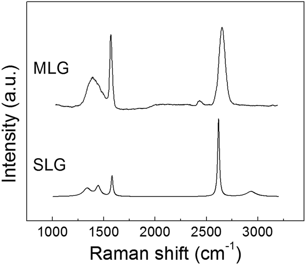 | ||
| Fig. 3 Typical Raman spectra from graphene layer that formed on liquid copper surface marked as G in Fig. 2b. | ||
We further examined the microstructure of MLG that formed on liquid copper including the uniformity of the number of graphene layers with two-dimensional Raman mapping and high-resolution SEM imaging. Optical images of MLG islands with corresponding two-dimensional I2D/IG Raman maps are shown in Fig. 4a, c and b, d, respectively. I2D/IG ranged from 0.3 to 1.3, revealing that the number of graphene layers varied across any individual MLG islands. Unlike the SLG, the characteristic of these MLG islands strongly depended on the experimental conditions. A comparison between graphene grown after 5 min and 10 min of carbon dissolution at T4 = 1300 °C, respectively, with the same 5 min of graphene formation at T3 = 1130 °C (i.e., comparing MLG islands shown in Fig. 2a with Fig. 2b) suggest that: (1) the size of the MLG islands increased with an increase in carbon dissolution time. This is reasonable since more carbon could dissolve in the liquid copper during a longer dissolution time, and therefore can generate more graphene. (2) Longer carbon dissolution at T4 before precipitation increased the variability of graphene layers within a single MLG island (cf. Fig. 2b with Fig. 4b). We noticed that a second graphene layer often nucleated additional (third or even fourth) layers.34 (3) The number density of MLG islands decreased with increasing carbon dissolution time. With longer dissolution time, more carbon could dissolve in the liquid copper. In principle, once the SLG covered the whole surface of liquid copper, excess dissolved carbon could facilitate nucleation of additional graphene layers and/or enhance the growth rate of a newly formed second graphene layer. Our experimental results suggest that the second graphene layer acted as a site for additional graphene layers to precipitate, which decreased the number density of MLG islands but increased their thicknesses. In other words, it appears that once the second graphene layer formed, the system tended to increase its area and thickness (by adding additional layers) rather than nucleating a second layer at other locations. Interestingly, the MLG islands shown in Fig. 2a and c, which were produced by identical carbon dissolution time of 5 min at T4 = 1300 °C but with different graphene formation time of 5 min and 10 min, respectively, at T3 = 1130 °C were of similar lateral size. This suggest that the growth of MLG islands ceased after approximately 5 min, likely because all supersaturated carbon had been consumed and a new thermodynamic equilibrium between graphene, dissolved carbon and graphite crucible had reached. Finally, lowering of graphene formation temperature from 1130 °C to 1090 °C while keeping carbon dissolution time the same at 5 min at T4 = 1300 °C increased the number MLG islands, albeit with significantly reduced sizes (cf. MLG islands shown in Fig. 2a with those in Fig. 2d). This suggests that lowering graphene formation temperature T3 favored MLG island nucleation but limited their lateral growth. Notice that the I2D/IG ratios obtained for MLG islands grown at the lower graphene formation temperature of 1090 °C were low but slightly larger than that for MLG islands grown at graphene formation temperature of 1130 °C (cf. Fig. 4b with Fig. 4d), suggesting that these MLG islands were thinner.
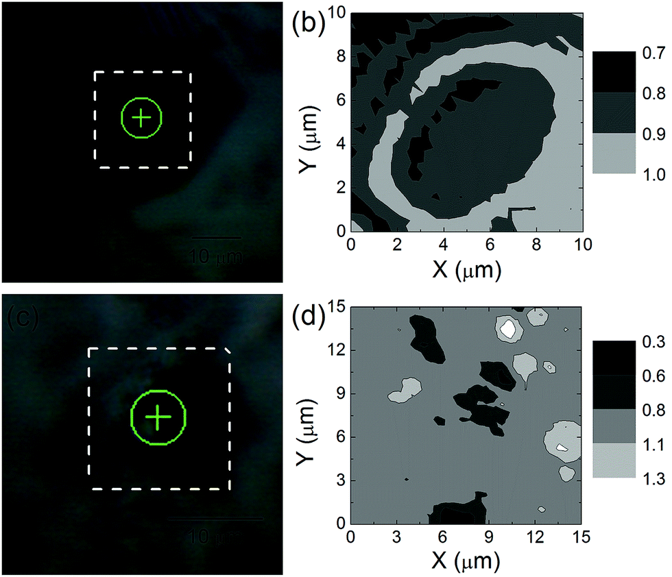 | ||
| Fig. 4 Optical images of MLG islands and corresponding two-dimensional map of the I2D/IG from the Raman spectra over a region enclosed in white dashed lined box. (a) Optical image and (b) I2D/IG map of MLG islands after 10 min of carbon dissolution at T4 = 1300 °C and 5 min of growth at T3 = 1130 °C. (c and d) The same but after 5 min of carbon dissolution at T4 = 1300 °C and 5 min of growth at T3 = 1090 °C. | ||
The cracks and wrinkles in graphene likely formed due to a mismatch in thermal expansion coefficient between copper and graphene.17 Wrinkle formation also signifies that the graphene was not strongly bonded to copper.9 The ripples possibly formed during solidification of liquid copper. Note that solidification process typically initiates planar or convective instabilities and breaks down the planar interface into cellular structure,35 which eventually develops a pattern with wavelength on the order of micrometers and generate a rippled structure.36 Carbon could then become inhomogeneously distributed on the solidified copper surface that would lead to MLG formation at the ripple edges.36 We therefore postulate that SLG first formed on liquid copper surface, then developed wrinkles and ripples upon solidification of liquid copper that eventually led to the MLG formation with large number of defects.
In contrast to the cracked, wrinkled, and rippled graphene comprised of a mixture of SLG and MLG that formed on copper, graphene that formed on silver had a smooth texture and only covered part of the silver surface. Owing to lower carbon solubility in silver than in copper at the carbon dissolution temperature T4 = 1200 °C, less carbon dissolved in liquid silver, limiting the subsequent areal dimensions of graphene. Raman spectroscopy from this graphene on silver surface, however, did not show any characteristic graphene peaks, probably due to the influence from the silver matrix. Note that since silver has a larger thermal expansion coefficient than copper,37 graphene that formed on solidified silver should display a more severe ripples and wrinkles in compared to the same on copper. Conversely, graphene has been observed to intercalate with (111) plane of silver,9 indicating some possibility of carbon–silver bonding, which might prevent the formation of ripples and wrinkles in graphene on silver surface. As a result, graphene could have followed the surface morphology of the grain structure of solidified silver. Such bonding between copper and graphene has not been observed or reported. Furthermore, graphene formed on liquid silver surface at different temperatures shared similar characteristic, i.e., a smooth graphene layer of uniform thickness, except for the distribution of impurities (cf. Fig. 2e with Fig. S1†). Since no MLG island formed due to the limited concentration of dissolved carbon, the graphene formation temperature T3 and time had no significant effect on final graphene characteristics.
Kinetics of graphene growth on liquid copper surface
Since graphene that formed on liquid copper surface had both SLG and MLG, we further investigated their formation and growth through real time and in situ observation with a high-temperature CLSM. We expect that the large SLG on liquid copper likely self-assembled from several smaller SLG rather than growing from a single SLG nucleus.26,27,38 Unfortunately, the formation and growth of SLG were not visible with CLSM since it was too thin to provide sufficient contrast from liquid copper. However, we have been able to determine indirectly SLG formation and growth from the motion of impurity oxide particles such as silica (SiO2) and alumina (Al2O3) that were floating on the liquid copper surface (Movie S1†). These impurity oxide particles were mobile on liquid copper surface during the carbon dissolution stage perhaps under the influence of natural convection and Marangoni flow. We did not observe any SLG or MLG formation at the carbon dissolution temperature T4 = 1300 °C (Fig. 5a). Upon cooling from T4 = 1300 °C to T3 = 1130 °C, movement of the particles ceased, indicating that the SLG layer had formed and fully covered the liquid copper surface within a few seconds during the cooling process and before reaching the graphene formation temperature T3. Notice that SLG did not exhibit any cracks, wrinkles, and ripples at T3 (Fig. 5b–d), further corroborating that these features developed on graphene during solidification of liquid copper. | ||
| Fig. 5 Images of formation and growth of MLG islands on liquid copper surface from in situ observations. (a) No SLG or MLG island was observed at the carbon dissolution temperature T4 = 1300 °C. Number densities and areal sizes of MLG islands after (b) 2 s, (c) 17 s, and (d) 61 s after reaching graphene formation temperature T3 = 1130 °C. | ||
In contrast, we were able to visualize directly and in situ the formation and growth of MLG islands on liquid copper surface using a high-temperature CLSM (Fig. 5 and Movie S2†). Evidently, SLG formation did not consume all of the supersaturated carbon, and the remaining carbon precipitated on the SLG at the interface between liquid copper and SLG as soon as the temperature reached the graphene formation temperature T3 to form second graphene layer (bilayer graphene, BLG) as well as small MLG precipitates (Fig. 5b). The growth of BLG was slower than that of the SLG, and surprisingly, individual MLG precipitates did not join into a uniform layer. The BLG subsequently nucleated additional MLG precipitates that eventually grew into MLG islands with a well-developed hexagonal lateral shape. Interestingly, we also observed MLG precipitate formation even before the sample reached the graphene formation temperature T3. We note that the detectable areal sizes of these MLG islands were 1–2 μm2, limited by the optical resolution of the CLSM. Occasionally, two MLG islands joined into one larger MLG island but the hexagonal shape of individual MLG islands was preserved. MLG islands slowly drifted as a unit on liquid copper surface (Fig. 5c and d), confirming that these MLG islands formed after SLG formation and were attached to the SLG layer. An increase in the carbon dissolution times typically generated fewer but thicker MLG islands. The observed morphology and size of these MLG islands with CLSM agree well with post-fabrication imaging using SEM.17,26–29 We summarize our observations of graphene morphology on liquid copper surface captured using CLSM as a schematic in Fig. 6a.
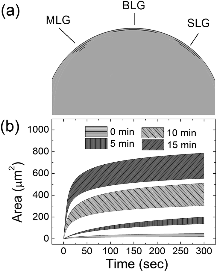 | ||
| Fig. 6 Graphene growth on liquid copper surface. (a) Schematic of the MLG island formation mechanism. (b) Growth speed observed in situ at T3 = 1130 °C with CLSM; legend gives the carbon dissolution time at T4 = 1300 °C before graphene growth. | ||
Next, we measured the growth speed of MLG islands at the graphene formation temperature T3 = 1130 °C and its dependence on carbon dissolution time at T4 = 1300 °C by measuring lateral area of individual MLG islands at various time intervals (Fig. 6b). The MLG islands grew rapidly initially, even though the growth rate was noticeably slower than those of SLG and BLG, and the growth subsequently slowed down to near zero after 5 minutes. Similar to the SLG growth, the initial growth rate of the MLG islands was faster with greater supersaturation of carbon.18
Conclusions
We report graphene formation and growth on liquid copper and liquid silver surfaces through a segregation method captured via in situ observations with a high-temperature CLSM. SLG rapidly formed on liquid metal surfaces during cooling from carbon dissolution temperature to graphene formation temperature. The SLG with a smooth texture formed and almost fully covered the liquid copper surface within seconds but only partially covered liquid silver surface. Furthermore, MLG islands with a hexagonal shape formed between liquid copper surface and SLG, and were attached underneath the SLG. Such MLG islands were not observed in graphene grown on liquid silver. The number densities, areal sizes, and thicknesses of these MLG islands can be readily tuned by varying carbon dissolution time and graphene formation temperature. For example, lowering the graphene formation temperature from 1130 °C to 1090 °C at the same carbon dissolution time of 5 min at T4 = 1300 °C resulted in a greater number of thinner and laterally smaller MLG islands. On the other hand, increasing the carbon dissolution time from 5 min to 10 min at T4 = 1300 °C while keeping the graphene formation time and temperature the same (i.e., 5 min at 1130 °C) led to fewer and thicker MLG islands with larger lateral sizes. Upon solidification of liquid metals, graphene grown on silver retained the uniform texture but the same grown on copper developed ripples and wrinkles. We postulate that our results will allow improved control of graphene growth using liquid metals.Acknowledgements
This work was supported by the US Army Research laboratory through grant W911NF-13-1-0069 (P. C. P. and M. F. I.) and US Army Research Office through grant W911NF-14-1-0651 (M. F. I.). We acknowledge the use of the Carnegie Mellon Materials Characterization Facility at Carnegie Mellon University (supported by grant MCF-677785) for SEM imaging.Notes and references
- M. Han, B. Ozyilmaz, Y. Zhang, P. Jarillo-Herero and P. Kim, Phys. Status Solidi B, 2007, 244, 4134–4137 CrossRef CAS.
- S. V. Morozov, K. S. Novoselov, M. I. Katsnelson, F. Schedin, D. C. Elias, J. A. Jaszczak and A. K. Geim, Phys. Rev. Lett., 2008, 100, 016602 CrossRef CAS PubMed.
- R. R. Nair, P. Blake, A. N. Grigorenko, K. S. Novoselov, T. J. Booth, T. Stauber, N. M. R. Peres and A. K. Geim, Science, 2008, 320, 1308 CrossRef CAS PubMed.
- K. S. Novoselov, E. McCann, S. V. Morozov, V. I. Falko, M. I. Katsnelson, U. Zeitler, D. Jiang, F. Schedin and A. K. Geim, Nat. Phys., 2006, 2, 177–180 CrossRef.
- Y. Zhang, Y. Tan, H. L. Stormer and P. Kim, Nature, 2005, 438, 201–204 CrossRef CAS PubMed.
- D. R. Forrest, I. Jasiuk, L. Brown, P. Joyce, A. Mansour and L. Salamanca-Riba, Novel Metal–Matrix Composites with Integrally-Bound Nanoscale Carbon, Nanotech. Conf. Expo., Santa Clara, CA, June, 2012 Search PubMed.
- T. Knych, P. Kwaśniewski, G. Kiesiewicz, A. Mamala, A. Kawecki and B. Smyrak, Metall. Mater. Trans. B, 2014, 45, 1196–1203 CrossRef CAS.
- T. Koltsova, L. Nasibulina, I. Anoshkin, V. Mishin, E. Kauppinen, O. Tolochko and A. Nasibulin, J. Mater. Sci. Eng. B, 2012, 2, 240–246 CAS.
- L. Salamanca-Riba, R. Isaacs, A. N. Mansour, A. Hall, D. R. Forrest, M. C. LeMieux and J. Shugart, A New Type of Carbon Nanostructure Formed Within a Metal Matrix, Nanotech. Conf. Expo., Santa Clara, CA, June, 2012 Search PubMed.
- J. V. Shugart and R. C. Scherer, US Pat., 8349759, 2013.
- J. V. Shugart and R. C. Scherer, US Pat., 8647534, 2014.
- K. S. Novoselov, A. K. Geim, S. V. Morozov, D. Jiang, Y. Zhang, S. V. Dubonos, I. V. Grigorieva and A. A. Firsov, Science, 2004, 306, 666–669 CrossRef CAS PubMed.
- S. Stankovich, D. A. Dikin, G. H. Dommett, K. M. Kohlhaas, E. J. Zimney, E. A. Stach, R. D. Piner, S. T. Nguyen and R. S. Ruoff, Nature, 2006, 442, 282–286 CrossRef CAS PubMed.
- S. Stankovich, D. A. Dikin, R. D. Piner, K. A. Kohlhaas, A. Kleinhammes, Y. Jia, Y. Wu, S. T. Nguyen and R. S. Ruoff, Carbon, 2007, 45, 1558–1565 CrossRef CAS.
- A. Reina, X. Jia, J. Ho, D. Nezich, H. Son, V. Bulovic, M. S. Dresselhaus and J. Kong, Nano Lett., 2009, 9, 3087 CrossRef CAS.
- Q. Yu, J. Lian, S. Siriponglert, H. Li, Y. P. Chen and S.-S. Pei, Appl. Phys. Lett., 2008, 93, 113103 CrossRef.
- S. Amini, J. Garay, G. Liu, A. A. Balandin and R. Abbaschian, J. Appl. Phys., 2010, 108, 094321 CrossRef.
- H. S. Mok, A. Ebnonnasir, Y. Murata, S. Nie, K. F. McCarty, C. V. Ciobanu and S. Kodambaka, Appl. Phys. Lett., 2014, 104, 101606 CrossRef.
- G. C. Dong, D. W. V. Baarle, M. J. Rost and J. W. M. Frenken, New J. Phys., 2012, 14, 1–15 CrossRef.
- Z. Peng, F. Somodi, S. Helveg, C. Kisielowski, P. Specht and A. T. Bell, J. Catal., 2012, 286, 22–29 CrossRef CAS.
- Z. Liu, Y.-C. Lin, C.-C. Lu, C.-H. Yeh, P.-W. Chiu, S. Iijima and K. Suenaga, Nat. Commun., 2014, 5, 4055 CAS.
- Z.-J. Wang, G. Weinberg, Q. Zhang, T. Lunkenbein, A. Klein-Hoffmann, M. Kurnatowska, M. Plodinec, Q. Li, L. Chi, R. Schloegl and M.-G. Willinger, ACS Nano, 2015, 9, 1506–1519 CrossRef CAS PubMed.
- W. Huafeng, Y. Chisato and H. Yoshikazu, Jpn. J. Appl. Phys., 2015, 54, 050301 CrossRef.
- E. Loginova, N. C. Bartelt, P. J. Feibelman and K. F. McCarty, New J. Phys., 2009, 11, 1–20 CrossRef.
- G. Odahara, S. Otani, C. Oshima, M. Suzuki, T. Yasue and T. Koshikawa, Surf. Sci., 2011, 605, 1095–1098 CrossRef CAS.
- D. Geng, L. Meng, B. Chen, E. Gao, W. Yan, H. Yan, B. Luo, J. Xu, H. Wang and Z. Mao, Adv. Mater., 2014, 26, 6423–6429 CrossRef CAS PubMed.
- D. Geng, B. Wu, Y. Guo, L. Huang, Y. Xue, J. Chen, G. Yu, L. Jiang, W. Hu and Y. Liu, Proc. Natl. Acad. Sci. U. S. A., 2012, 109, 7992–7996 CrossRef CAS PubMed.
- S. Amini and R. Abbaschian, Carbon, 2013, 51, 110–123 CrossRef CAS.
- S. Amini, H. Kalaantari, J. Garay, A. A. Balandin and R. Abbaschian, J. Mater. Sci., 2011, 46, 6255–6263 CrossRef CAS.
- I. Karakaya and W. T. Thompson, J. Phase Equilib., 1988, 9, 226–227 CrossRef.
- G. A. López and E. J. Mittemeijer, Scr. Mater., 2004, 51, 1–5 CrossRef.
- L. L. Oden and N. A. Gokcen, Metall. Trans. B, 1992, 23, 453–458 CrossRef.
- A. C. Ferrari, J. C. Meyer, V. Scardaci, C. Casiraghi, M. Lazzeri, F. Mauri, S. Piscanec, D. Jiang, K. S. Novoselov, S. Roth and A. K. Geim, Phys. Rev. Lett., 2006, 97, 187401 CrossRef CAS PubMed.
- A. Mohsin, L. Liu, P. Liu, W. Deng, I. N. Ivanov, G. Li, O. E. Dyck, G. Duscher, J. R. Dunlap and K. Xiao, ACS Nano, 2013, 7, 8924–8931 CrossRef CAS PubMed.
- A. R. Harutyunyan, Proc. Natl. Acad. Sci. U. S. A., 2012, 109, E2099 CrossRef CAS PubMed.
- W. W. Mullins and R. F. Sekerka, J. Appl. Phys., 1964, 35, 444–451 CrossRef.
- F. C. Nix and D. MacNair, Phys. Rev., 1942, 61, 74–78 CrossRef CAS.
- K. F. McCarty, P. J. Feibelman, E. Loginova and N. C. Bartelt, Carbon, 2009, 47, 1806–1813 CrossRef CAS.
Footnote |
| † Electronic supplementary information (ESI) available: High-resolution SEM images of graphene on liquid silver, and movies of SLG and MLG formation and growth on liquid copper surface. See DOI: 10.1039/c6ra22505e |
| This journal is © The Royal Society of Chemistry 2016 |