K. Venkata Sairama,
B. M. Gurupadayya*a,
B. Iyer Vishwanathan*a,
R. S. Chandana and
Dattatri K. Nageshab
aDepartment of Pharmaceutical Chemistry, JSS College of Pharmacy, JSS University, Mysuru 570 015, India. E-mail: bmgurupadayya@jssuni.edu.in; b.vishwanathan@gmail.com; Fax: +91 8212548359; Tel: +91 7411006136
bFaculty of Life Sciences, JSS University, Mysuru 570 015, India
First published on 4th October 2016
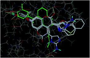
In the present study, a series of coumarin derivatives were designed, synthesized and evaluated for their antioxidant and cytotoxic properties. The title compounds, 2-(3-substituted-4-methyl-2-oxo-2H-chromen-7-yloxy)-2-methylpropanoic acid derivatives 5a–5f, were synthesized by base-catalyzed dehydrohalogenative cyclization following Hantzsch synthesis. All the newly synthesized analogues were characterized and established on the basis of mass, 1H NMR, 13C NMR and IR studies. The compounds were evaluated for their in vitro antioxidant activity and found to exhibit substantial activity. The in vitro cytotoxicity was evaluated against MCF-7, MDA-231 (human breast cancer) and HT29 (human colon adenocarcinoma) cell lines by MTT assay and the results were encouraging. Compound 5b, with lower IC50 values of 2.4 and 4.8 μM for MCF-7 and MDA-231, respectively, was considered to be potent among the series.
Recent studies indicate the beneficial role of cyclooxygenase-2 (COX-2) inhibition in cancer chemotherapy.7 PGH2 is biosynthesized from arachidonic acid by the action of COX. COX is the rate-limiting step, which in turn limits the synthesis of other PGs. Prostaglandin H2 (PGH2) is consequently converted to other PGs and thromboxane A2 by prostaglandin synthases and thromboxane synthases, respectively.
Clinical studies have indicated that prostaglandin E2 (PGE2), which is a product of PGH2, exhibits a considerable role in carcinogenesis. PGE2 elicits multiple oncogenic signals and the increased level of PGE2 in cancer tissues indicates its considerable role in regulating a number of other secondary messengers related to the cancer phenotype.8 PGE2 stimulates proliferation, leading to the expansion of tumor mass. Therefore, targeting PGE2 by COX-2 inhibition is advantageous and holds promise for the development of newer anticancer agents.9 Reports on COX-2 selective inhibitors in reducing the number and also the size of intestinal adenomas in patients diagnosed with familial adenomatous polyposis10 and reduction in the occurrence of sporadic colorectal adenomas11 have now shed light over COX-2 inhibition and support the use of this in cancer chemotherapy.
At the same time, increased production of reactive oxygen metabolites produces a state of oxidative stress. This oxidative stress can be considered as the main trigger of cellular damage and is recognised to exhibit a considerable role in carcinogenesis. Epidemiological investigations have provided enough evidence to support the role of reactive oxygen metabolites in the etiology of cancer. Under the action of the superoxide dismutase enzyme (SOD) there is an abnormal production of superoxide anions (O−˙) which results in increased production of hydrogen peroxide.12 The hydrogen peroxide is catabolized to the highly toxic hydroxyl radical (OH˙) through Fenton and Haber–Weiss reactions. This state of increased production of reactive oxygen species in the physiological system due to environmental factors and lifestyle habits, like smoking, etc., results in oxidative stress. A chronic situation of this oxidative stress causes cross-linking in lipids, proteins and nucleic acids, leading to cellular damage and resulting in alteration of the hereditary DNA, initiating cancer development, metastasis and uncontrolled proliferation. In view of the above facts, anticancer investigations are always associated with preliminary antioxidant evaluation of the tested compounds.13
The chemistry of coumarin and its fused heterocyclic derivatives has received considerable attention owing to their synthetic and pharmacological importance. Literature studies highlight that coumarin derivatives have potential to be revealed as anticancer agents. The importance of the substitution on the parent coumarin moiety towards its anticancer properties is well reported.14 The pattern of substitutions influences the coumarin’s pharmacological properties and therein its therapeutic applications. Structure–activity relationship studies on the substitution indicate the introduction of an alkyloxy function at the C7 position leads to derivatives with strong potential to reduce the plasma alkaline transferase level in hepatitis and to inhibit caspase-3 activation, indicative of its potential as a cytostatic and cytotoxic agent.15 The anticancer potential of coumarin and its active metabolite, 7-hydroxycoumarin, has been established by the growth-inhibitory cytostatic activity in human cancer cell lines, such as H727 (lung), A549 (lung), MCF-7 (breast), HL-60 (leukemia) and ACHN (renal). It is also to be noted that coumarin derivatives have been reported to demonstrate activity against prostate cancer, malignant melanoma, and metastatic renal cell carcinoma in clinical trials.16 Coumarin derivatives inhibiting sulfotransferase activity, contributing significantly to their anticancer activity against breast cancer, have also been reported.17
In the present investigation, we have designed a small library of coumarin derivatives and the designed molecules were docked onto the EGFR and COX-2 enzymes. Molecular docking studies impart knowledge about the probable structural modification of the parent moiety via potential substitutions that would result in appropriate derivatives with better interactions. The designed molecules were also screened for ADMET properties. Based on the docking scores and interactions, the best-ranked molecules were selected for synthesis. The synthesized molecules were evaluated for their in vitro antioxidant activity by 2,2′-diphenyl-1-picryl hydrazyl (DPPH), 2,2′-azino-bis(3-ethylbenzothiazoline-6-sulphonic acid) (ABTS) and superoxide anion (SA) radical scavenging assay methods. The compounds were evaluated for their cytotoxic properties using the 3-(4,5-dimethylthiazol-2-yl)-2,5-diphenyltetrazolium bromide (MTT) assay on human cancer cell lines, MCF-7, MDA-231 (human breast cancer) and HT29 (human colon adenocarcinoma). The results highlight that the coumarin moiety provides a scaffold on which pharmacophores can be arranged to afford potent and selective cytotoxic agents.
The acid derivative 2 was condensed with ethyl acetoacetate in the presence of sulphuric acid to attain the acyl derivative, 2-(3-acetyl-4-methyl-2-oxo-2H-chromen-7-yloxy)-2-methylpropanoic acid (3). The 1H NMR spectrum of the acyl derivative 3 accounts for sixteen protons, wherein the signals at δ 7.5–6.4 ppm were assigned to the aromatic protons of coumarin and a singlet at δ 2.3 ppm was attributed to the methyl protons of the acyl group (COCH3) at C3 of the coumarin moiety. In the 13C NMR spectrum of acetyl derivative 3, the chemical shifts at δ 27.2 and 191.6 ppm can be attributed to the alpha methyl carbon and the carbonyl carbon of the acetyl substituent on the C3 position of the coumarin moiety, respectively. The mass spectral characterization exhibits a molecular ion peak at 305 m/z [M+1] and an ion peak at 260 m/z, which can be attributed to the decarboxy fragment.
The intermediate, 2-(3-(2-bromoacetyl)-4-methyl-2-oxo-2H-chromen-7-yloxy)-2-methylpropanoic acid (4) was prepared by bromination of the alpha methyl group of acyl derivative 3. The bromination was executed using bromine in dioxane. In the proton NMR spectrum of the bromoacyl derivative 4, a singlet at δ 3.6 ppm was assigned to the methylene proton of the bromoacyl (COCH2Br) group. The chemical shifts at δ 32.6 and 196.4 ppm in the 13C NMR spectrum of bromoacyl derivative 4 were assigned to the methylene carbon and the carbonyl carbon of the bromoacyl substituent on the C3 position of the coumarin moiety, respectively.
The bromoacyl intermediate (4) was condensed with urea derivatives and 2-amino azoles as per the Hantzsch azole synthesis to undergo dehydrohalogenative cyclization in the presence of a catalytic amount of triethylamine and ferric chloride, affording the target molecules, 2-(3-substituted-4-methyl-2-oxo-2H-chromen-7-yloxy)-2-methylpropanoic acid derivatives 5a–5f. The IR spectrum of 2-(3-(2-aminooxazol-5-yl)-4-methyl-2-oxo-2H-chromen-7-yloxy)-2-methylpropanoic acid (5a) exhibited characteristic absorption peaks at 3445 and 1231 cm−1, accounting for the amino group and C–O–C function in the oxazole moiety, respectively. In the proton NMR spectrum of compound 5a, a singlet at δ 5.4 ppm can be attributed to the amino (NH2) proton at position C2 of the oxazole moiety. The absence of bromomethyl protons at δ 3.6 ppm and an isolated singlet at δ 7.9 ppm accounting for the CH proton at the C5 position of the oxazole moiety confirm the formation of the 2-amino oxazole moiety. The 13C NMR spectrum of compound 5a revealed three characteristic signals at δ 163.6, 123.7 and 156.2 ppm values, which can be attributed to the C2, C4 and C5 carbons of the oxazole moiety, respectively. The mass spectral characterization of compound 5a exhibits a molecular ion peak at 345 m/z [M+1], which confirms product formation.
In the proton NMR spectrum of 2-(3-(H-imidazo[1,2-a]pyridin-2-yl)-4-methyl-2-oxo-2H-chromen-7-yloxy)-2-methylpropanoic acid (5d), a characteristic doublet of doublets at δ 7.2 ppm can be attributed to the protons at the C5, C6, C7 and C8 positions of the imidazo[1,2-a]pyridine moiety. Furthermore, in the 1H NMR spectrum, an isolated singlet was observed at δ 6.9 ppm for the proton at the C3 position of the imidazo[1,2-a]pyridine moiety. The mass spectral characterization of compound 5d exhibits a molecular ion peak at 378 m/z [M+], a major ion peak at 333 m/z that can be assigned to the decarboxy derivative and another ion peak at 117 m/z, which can be attributed to the imidazo[1,2-a]pyridine fragment.
| Sl. no. | Cpd | % ABSa | TPSAc | RTBc | HBAc | HBDc | log Pc |
MW | Violationsc | log Sb |
Drug likenessb | Drug scoreb |
|---|---|---|---|---|---|---|---|---|---|---|---|---|
| a Data calculated as per eqn (1).b Molecular property as obtained from the OSIRIS property explorer software.c Molecular property as obtained from the Molinspiration online property calculation toolkit. | ||||||||||||
| 1 | 5a | 64.6 | 128.8 | 4 | 8 | 3 | 1.17 | 344.32 | 0 | −3.96 | 2.65 | 0.78 |
| 2 | 5b | 69.1 | 115.7 | 4 | 7 | 3 | 2.54 | 360.39 | 0 | −4.42 | −5.02 | 0.38 |
| 3 | 5c | 63.6 | 131.5 | 4 | 8 | 4 | 1.11 | 343.34 | 0 | −4.88 | −8.22 | 0.34 |
| 4 | 5d | 76.5 | 94.1 | 4 | 7 | 1 | 2.85 | 378.38 | 0 | −4.03 | −0.13 | 0.59 |
| 5 | 5e | 76.5 | 94.1 | 4 | 7 | 1 | 3.25 | 384.41 | 0 | −4.03 | 1.11 | 0.71 |
| 6 | 5f | 76.5 | 94.1 | 4 | 7 | 1 | 4.36 | 434.47 | 0 | −3.66 | 2.15 | 0.79 |
![]() | ||
| Fig. 1 Energy-minimized and conformationally analyzed structures aligned against common atoms interacting with 2J5F, with reference molecule 34-JAB. | ||
Site-directed mutagenesis studies have indicated a few crucial amino acid residues of EGFR, namely Leu745, Thr790, Cys797, and Met793. These amino acids are essential for binding to EGFR and have a significant part in ligand binding.18 A few of our designed coumarin derivatives exhibit interactions with the aforementioned amino acid residues, which may provide a possible lead to active molecules (Fig. 2). Among the series, compounds 5e and 5a generated good docking scores of 7.24 and 7.14 kcal M−1, respectively. In compound 5a, the amino group at position C2 of the oxazole moiety was involved in a hydrogen bond interaction with Pro794 at a distance of 2.462 Å. The carbonyl oxygen at position C2 of the coumarin moiety exhibited a hydrogen bond interaction with Met793 at a distance of 1.789 Å. The hydrogen and the hydroxyl oxygen of the carboxylic acid group of the propanoic acid fragment at position C7 of the coumarin moiety exhibited hydrogen bond interactions with the amino acid residues Glu762 and Lys748 at distances of 2.039 and 1.891 Å, respectively. Similarly, the carbonyl group of the propanoic acid fragment showed hydrogen bond interactions with Thr854 and Asp855 amino acid residues at distances of 2.089 and 2.519 Å, respectively. Compounds 5c, 5f, and 5d exhibited good docking scores of 7.08, 7.02, and 6.97 kcal M−1, respectively, higher docking scores than that of the reference ligand 34-JAB. The docking studies indicated that the carboxylic acid function of the propanoic acid fragment at position C7 of the coumarin moiety of all the derivatives was involved in hydrogen bond interactions with the amino acid residues Thr854, Asp855, Lys745 and Glu762. The carbonyl oxygen at C2 of the coumarin moiety was involved in a hydrogen bond interaction with the amino acid residue Met793. However, compound 5b exhibited a docking score of 5.58 kcal M−1, lower than that of the reference ligand 34-JAB.
All the coumarin derivatives exhibited a crash score of ≤4.4 kcal mol−1. A higher crash score indicates inappropriate penetration of the ligand into the binding site of the protein, resulting in a decreased force of interaction with the amino acids. The molecular docking study indicates that all the docked coumarin derivatives have good interactions with the enzyme and have potential scope to inhibit it.
The designed compounds, along with the reference ligand, were docked onto the target protein COX-2, 3LN1. The docking studies revealed that the designed coumarin derivatives exhibited a considerable affinity towards 3LN1. The compounds 5a–5f exhibited promising docking scores, ranging from 7.79 to 10.43 kcal M−1. Compound 5c exhibited the highest docking score among the designed compounds, with a value of 10.43 kcal M−1, and compound 5d had the lowest docking score (7.79 kcal M−1).
The docking studies on the COX-2 protein were carried out with our designed compounds along with 34-JAB (reference standard ligand for EGFR, 2J5F) to understand the comparative interaction of this with those of our designed compounds towards anticancer applications. Compounds 5c and 5f exhibited docking scores of 10.43 and 9.71 kcal M−1, respectively, higher than that of 34-JAB, indicating a better interaction to inhibit the COX-2 protein. Compound 5c was well aligned in the hydrophobic core of the 3LN1 pocket in the B-chain of the protein, which is essential for the inhibition. In compound 5c, the carbonyl oxygen of the carboxylic acid function showed hydrogen bonding with the Tyr371 residue at a distance of 2.722 Å. The 1H of the imidazole exhibited hydrogen bonding interactions with the Leu338 and Ser339 residues at distances of 2.196 and 2.402 Å, respectively. The carbonyl oxygen of the coumarin moiety at the C2 position was involved with a hydrogen bonding interaction with Phe504 at a distance of 2.558 Å (Fig. 3).
![]() | ||
| Fig. 3 Energy minimized and conformationally analyzed structures aligned against common atoms interacting with 3LN1, with reference molecule celecoxib. | ||
The hydrogen bond interaction between the carbonyl oxygen at position C2 of the coumarin moiety with the Phe504 residue and also the hydrogen bonding interaction between the nitrogen of the azole moiety and the Arg106 residue were the major and common interactions exhibited by the designed coumarin derivatives. Table 2 presents the molecular docking results of designed compounds 5a–5f along with their crash scores and polar scores.
| Sl. no. | Compound | Docking score towards 2J5Fa | Docking score towards 3LN1a | ||||
|---|---|---|---|---|---|---|---|
| Total score | Crash score | Polar score | Total score | Crash score | Polar score | ||
| a Data expressed in kcal M−1 as obtained from the docking utility of SYBYL 2.1 software. | |||||||
| 1 | 5a | 7.14 | −1.33 | 3.06 | 9.19 | −2.89 | 2.04 |
| 2 | 5b | 5.58 | −1.34 | 4.97 | 8.10 | −3.67 | 2.32 |
| 3 | 5c | 7.08 | −0.85 | 4.17 | 10.43 | −2.75 | 3.28 |
| 4 | 5d | 6.97 | −1.24 | 3.10 | 7.79 | −4.42 | 2.62 |
| 5 | 5e | 7.24 | −1.37 | 3.63 | 7.81 | −3.18 | 1.09 |
| 6 | 5f | 7.02 | −1.47 | 2.99 | 9.71 | −4.19 | 1.31 |
| 7 | 34-JAB | 6.21 | −0.67 | 2.05 | 9.32 | −4.50 | 0.74 |
| 8 | Celecoxib | — | 10.97 | −0.70 | 2.43 | ||
The establishment of a marked relationship between tissue prostaglandin levels in human breast tumor formation and the development of metastases and survival has been well reported.19,20 Significant inhibition of EGFR and COX-2 by our designed coumarins indicates their potential role in acting by a dual mechanism to suppress cancer cell proliferation.
![]() | ||
| Fig. 4 The validation of the docking protocol for the target protein. (A) Binding pose of 34-JAB onto the co-crystallized ligand 34-JAB (red) in EGFR. (B) Binding pose of celecoxib onto the co-crystallized ligand celecoxib (green) in 3LN1. | ||
The results obtained from the DPPH radical assay method indicated that the compounds exhibited significant scavenging activity ranging from 17.8 to 87.5%, in comparison to ascorbic acid (91.5% at 50 μM). The IC50 values of the compounds were substantially low, ranging from 19.8 to 34.6 μM. Compound 5b exhibited higher radical scavenging potential (87.5% at 50 μM) in comparison to the other derivatives with an IC50 value of 19.8 μM. The compound exhibited a substantial increase in the antioxidant efficacy with an increase in the test concentration. The statistical analysis of the antioxidant activity of compounds 5a–5f by the repeated measures Dunnett’s ANOVA method highlighted that the compounds exhibited a significance value of P < 0.001, indicating a confidence interval of 99.9% with respect to the reference standard ascorbic acid. The results of the antioxidant activity evaluated by the DPPH method highlighted that the derivatives with amino group substitution at position C2 of compounds 5a–5c exhibited significant activity (Fig. 5A).
![]() | ||
| Fig. 5 Mean increase in percentage antioxidant activity of the test compounds 5a–5f with increase in test concentrations (12.5, 25.0 and 50.0 μM): (A) DPPH method, (B) ABTS method, (C) SA method. | ||
The results obtained from the ABTS radical assay method indicated that the compounds exhibited considerable radical scavenging properties, with values ranging from 3.1 to 81.4% in comparison to the 87.3% antioxidant activity obtained for ascorbic acid. The results highlighted that compound 5b exhibited higher radical scavenging potential (81.3% at 50 μM) with an IC50 value of 30.9 μM (Fig. 5B). The statistical analysis of the antioxidant activity of compounds 5a–5f by the repeated measures Dunnett’s ANOVA method highlighted that the compounds were not significant with respect to the standard ascorbic acid.
The radical scavenging assay results from the SA method were not as similar as the DPPH and ABTS results. The results obtained from the SA radical assay method indicated that the compounds exhibited significant scavenging activity ranging from 9.7 to 83.5% in comparison to the 88.7% antioxidant activity obtained for ascorbic acid (Fig. 5C). Although the tested compounds exhibited significant antioxidant efficacy, compound 5c exhibited a better radical scavenging property (83.5% at 50 μM) than that of 5b (75.2% at 50 μM), in contrast to the DPPH and ABTS results.
Nevertheless, the antioxidant activity data from all three tested methods highlights that the derivatives with amino group substitution at position C2 of the azole series (5a–5c) exhibited significant activity. The antioxidant activities of the test compounds 5a–5f at the three different test concentrations by the DPPH, ABTS, and SA radical scavenging assay methods, expressed as the mean ± SEM, along with the IC50 values obtained by regression analysis, are presented in Table 3.
| Comp. | DPPH method | ABTS method | SA method | |||||||||
|---|---|---|---|---|---|---|---|---|---|---|---|---|
| Mean percentage free radical scavenging activity ± SEMa | Mean IC50 value ± SEMb | Mean percentage free radical scavenging activity ± SEMa | Mean IC50 value ± SEMc | Mean percentage free radical scavenging activity ± SEMa,c | Mean IC50 value ± SEMb | |||||||
| 12.5 μM | 25.0 μM | 50.0 μM | 12.5 μM | 25.0 μM | 50.0 μM | 12.5 μM | 25.0 μM | 50.0 μM | ||||
| a Results are expressed as the mean values from three independent experiments ± SEM.b Data obtained by DPPH method was analyzed by Dunnet’s test compared with reference drug ascorbic acid. n = 3; (†) equals P < 0.1.c Data obtained by SA method was analyzed by Dunnet’s test compared with reference drug ascorbic acid. n = 3; (**) equals P < 0.01, (*) equals P < 0.05. | ||||||||||||
| 5a | 20.87 ± 0.64 | 55.11 ± 0.19 | 79.66 ± 0.18 | 27.90 ± 0.22 | 8.44 ± 1.01 | 32.94 ± 1.12 | 69.62 ± 0.69 | 36.73 ± 0.31 | 23.33 ± 0.43 | 40.46 ± 0.43 | 73.03 ± 0.41 | 32.49 ± 0.10* |
| 5b | 37.19 ± 0.08 | 61.91 ± 0.15 | 87.54 ± 0.04 | 19.75 ± 0.08† | 13.65 ± 1.08 | 43.10 ± 0.92 | 81.38 ± 0.30 | 30.87 ± 0.24 | 26.21 ± 0.50 | 55.19 ± 0.09 | 75.15 ± 0.50 | 27.40 ± 0.17** |
| 5c | 19.76 ± 0.22 | 66.22 ± 0.04 | 81.26 ± 0.22 | 25.31 ± 0.10 | 6.36 ± 1.31 | 49.54 ± 0.56 | 72.01 ± 0.49 | 31.94 ± 0.21 | 16.69 ± 0.19 | 49.48 ± 0.34 | 83.47 ± 0.09 | 29.23 ± 0.07* |
| 5d | 21.05 ± 0.33 | 47.30 ± 0.44 | 74.05 ± 0.04 | 31.02 ± 0.12 | 3.06 ± 0.89 | 23.03 ± 0.66 | 62.09 ± 0.46 | 42.27 ± 0.14 | 13.92 ± 0.74 | 31.65 ± 0.99 | 66.34 ± 0.09 | 38.26 ± 0.13* |
| 5e | 17.82 ± 0.34 | 43.61 ± 0.26 | 67.88 ± 0.51 | 34.55 ± 0.28 | 4.09 ± 1.70 | 17.63 ± 0.29 | 53.09 ± 1.07 | 49.34 ± 0.48 | 9.73 ± 0.75 | 26.86 ± 0.57 | 58.35 ± 1.15 | 43.39 ± 0.42* |
| 5f | 25.95 ± 0.18 | 55.25 ± 0.49 | 72.79 ± 0.33 | 28.03 ± 0.06 | 8.75 ± 1.08 | 34.64 ± 0.96 | 60.25 ± 1.15 | 41.06 ± 0.30 | 14.85 ± 0.59 | 41.96 ± 1.09 | 64.71 ± 0.74 | 36.65 ± 0.06* |
| Ascorbic acid | 37.31 ± 0.15 | 64.25 ± 0.11 | 91.47 ± 0.18 | 18.87 ± 0.09 | 16.90 ± 0.82 | 46.60 ± 0.70 | 87.26 ± 0.54 | 30.73 ± 0.22 | 16.86 ± 0.34 | 52.58 ± 0.25 | 88.69 ± 0.41 | 27.70 ± 0.10 |
The cytotoxicity results towards the HT-29 cancer cell line were moderate. Compounds 5a–5c exhibited substantial cytotoxic activity with low IC50 values compared with 5-fluorouracil and doxorubicin (Fig. 6A). Compounds 5a and 5b exhibited IC50 values of 29.6 and 30.8 μM respectively.
![]() | ||
| Fig. 6 Mean IC50 values exhibited by the test compounds 5a–5f for cytotoxicity by the MTT assay method: (A) HT-29, (B) MDA-231, (C) MCF-7. | ||
The results for the cytotoxicity on breast cancer cell lines MDA-231 and MCF-7 were encouraging. Compounds 5a and 5b showed considerable cytotoxic activity with lower IC50 values (Fig. 6B and C). Compound 5b can be considered to be a potent cytotoxic agent among the tested compounds, with IC50 values of 4.84 and 2.39 μM for MDA-231 and MCF-7, respectively. Compound 5a exhibited IC50 values of 7.3 and 5.1 μM for MDA-231 and MCF-7 respectively, while compound 5c, which exhibited moderate cytotoxicity for HT-29, exhibited minimal cytotoxic action on breast cancer cell lines MDA-231 and MCF-7.
Several anticancer agents possess a serious problem of safety and effectiveness with the foremost drawback being non-selectivity. These drugs are not able to distinguish between the normal healthy cells and fast growing cancerous cells. From this aspect, it is crucial to measure the cytotoxicity towards normal cell in the process of discovery of newer antitumor drugs. As a further evaluation of the selective cytotoxicity of the lead compounds, 5a and 5b were chosen for the selectivity test over normal human cell lines, using Vero (epithelial cells) and HepG2 (hepatocytes) to ascertain their safety and efficacy towards normal cells with the MTT assay. The results indicate that compounds 5a and 5b had lower cytotoxicity towards normal cell lines. The IC50 values of all the tested compounds are given in Table 4.
| Compound | IC50 (μM) | ||||
|---|---|---|---|---|---|
| HT-29a,b,c | MDA-231a,b,c | MCF-7a,b,c | Vero cell linea | HepG2a | |
| a Results are expressed as the mean values from three parallel experiments ± SEM.b Data was analyzed by Dunnet’s test. n = 3; (***) equals P < 0.001, (**) equals P < 0.01, (*) equals P < 0.05, with respect to reference standard 5-fluorouracil.c Data was analyzed by Dunnet’s test. n = 3; (†††) equals P < 0.001, (††) equals P < 0.01, (†) equals P < 0.05, with respect to reference standard doxorubicin. NC equals not conducted. | |||||
| 5a | 29.57 ± 0.72**†† | 7.26 ± 0.24 | 5.12 ± 0.09 | 38.65 ± 1.34 | 43.13 ± 2.56 |
| 5b | 30.81 ± 0.15**††† | 4.84 ± 0.17 | 2.39 ± 0.03 | 57.89 ± 3.72 | 63.37 ± 2.84 |
| 5c | 28.46 ± 1.37***††† | 56.94 ± 1.47***††† | 44.87 ± 1.91***††† | NC | NC |
| 5d | 173.05 ± 7.16***††† | 128.19 ± 8.04***††† | 152.35 ± 8.56***††† | NC | NC |
| 5e | >200 | 181.09 ± 4.45***††† | 195.50 ± 4.62***††† | NC | NC |
| 5f | >200 | 177.15 ± 6.16***††† | 164.31 ± 9.20***††† | NC | NC |
| 5-Fluorouracil | 2.16 ± 0.07 | 1.96 ± 0.087 | 1.86 ± 0.06 | — | — |
| Doxorubicin | 3.73 ± 0.14 | 3.76 ± 0.18 | 3.24 ± 0.15 | — | — |
Our results indicated that compounds 5a and 5b induced significantly high levels of cytotoxicity as compared with the reference standards 5-fluorouracil and doxorubicin. They differ by the azole moiety; compound 5a is an oxazole derivative and 5b a thiazole derivative. The results also highlighted that the compounds with significant antioxidant potential have emerged as having considerable cytotoxic properties; this probably indicates a greater advantage with respect to the clinical aspects.
The possible differences in the cytotoxic efficacy of compounds 5a and 5b may be attributed to their partition coefficient (log10
P) values. The efficiency of a compound or drug in producing a cytotoxicity effect also depends on its accumulation in the cell. Hence, a more lipophilic nature leads to significant accumulation of the compound in the cell, resulting in considerable cytotoxicity and cell death. Compounds 5a and 5b showed clog
P values of 1.17 and 2.54, respectively. The higher log
P value of compound 5b indicates that it is more lipophilic and this may be co-related to its cytotoxic potential.
All the chemicals and solvents used in this study were purchased from Sigma-Aldrich Chemical Co., Spectrochem Ltd. and Sd fine chemicals of LR grade; the solvents were distilled prior to use. All commercially available reagents procured were used without further purification. TLC was used to assess the progress/completion of reactions and the purity of the synthesized compounds using aluminum backed sheets of silica gel 60 GF254 (Merck), with ethyl acetate and hexane (4
:
1) as a solvent system and iodine vapor as the visualizing agent. Melting points were determined in open glass capillaries and are uncorrected. The IR spectra were recorded using a Shimadzu FTIR-8400 instrument by the KBr disc pellet technique/liquid sampling using NaCl cells and only noteworthy absorption levels (cm−1) are listed. 1H NMR and 13C NMR spectra were recorded using a Bruker AC-400 MHz FT NMR spectrophotometer at 400 MHz for 1H (100 MHz for 13C) with deuterated dimethyl sulfoxide (DMSO-d6) as solvent and tetra methyl silane (TMS) as the internal standard (chemical shifts in δ, ppm). The splitting patterns were designated as follows: s: singlet; d: doublet; q: quartet; m: multiplet. The LCMS was recorded using a Shimadzu LCMS-2010A instrument by ESI. The elementary analysis was recorded using a Thermo Finnigan FLASH EA 1112 CHNS analyzer and all the compounds gave satisfactory elemental analysis. For in vitro cytotoxicity studies the cell lines were procured from the National Centre for Cell Sciences, Pune, India.
:
methanol, 60
:
40), followed by determination of purity of the synthesized compounds by ultra-fast liquid chromatography (UFLC, Shimadzu LC-20 A Series), using a PDA detector and XTerra C18 column (150 × 4.6 mm, 5 μm particle size). The mobile phase was composed of di-sodium hydrogen phosphate buffer (pH 3.6) and acetonitrile in the ratio 60
:
40, v/v, with a flow rate of 1.0 mL min−1. 20 μL of the sample was employed as the injection volume and the detection wavelength was 254 nm. All the synthesized compounds had a purity of more than or equal to 95%.
:
1); IR: 3446 (OH), 2965 (C
C), 1754, 1718 (C
O), 1234 (C–O–C); 1H NMR (400 MHz, DMSO-d6): δ 10.6 (s, 1H, OH; carboxylic acid), 7.5–6.4 (m, 3H, Ar-H), 2.3 (s, 3H, COCH3), 2.1 (s, 6H, (CH3)2C), 1.6 (s, 3H, CH3); 13C NMR (100 MHz, DMSO-d6): δ 191.6 (1C, COCH3), 173.9 (1C, COOH), 167.5 (1C, CO, C2-coumarin), 157.4 (1C, C9-coumarin), 152.1 (1C, C10-coumarin), 147.9 (1C, C7-coumarin), 142.7 (1C, C3-coumarin), 128.7 (1C, C4-coumarin), 126.9 (1C, C8-coumarin), 116.2 (1C, C6-coumarin), 113.9 (1C, C5-coumarin), 46.2 (1C, (CH3)2C), 29.4 (2C, (CH3)2C), 27.2 (1C, COCH3), 18.2 (1C, CH3); MS: m/z (%) 305 (65) [M+1], 260 (100) [C16H16O4]; anal. calc. for C16H16O6: C 63.15, H 5.30. Found: C 63.43, H 5.52.
:
1); IR: 3483 (OH), 2931 (C
C), 1761, 1724 (C
O), 1215 (C–O–C); 1H NMR (400 MHz, DMSO-d6): δ 10.4 (s, 1H, OH; carboxylic acid), 7.6–6.9 (m, 3H, Ar-H), 3.6 (s, 2H; CH2Br), 2.0 (s, 6H, (CH3)2C), 1.6 (s, 3H, CH3); 13C NMR (100 MHz, DMSO-d6): δ 196.4 (1C, COCH2Br), 176.1 (1C, COOH), 169.2 (1C, CO; C2-coumarin), 162.4 (1C, C9-coumarin), 158.7 (1C, C10-coumarin), 156.2 (1C, C7-coumarin), 145.2 (1C, C3-coumarin), 129.2 (1C, C4-coumarin), 126.2 (1C, C8-coumarin), 117.9 (1C, C6-coumarin), 115.2 (1C, C5-coumarin), 46.6 (1C, (CH3)2C), 32.6 (1C, COCH2Br), 29.6 (2C, (CH3)2C), 18.9 (1C, CH3); MS: m/z (%) 382 (100) [M+]; anal. calc. for C16H15BrO6: C 50.15, H 3.95. Found: C 50.35, H 4.21.
:
1); IR: 3445 (N–H), 3413 (OH), 3194 (C–N), 1741, 1727 (C
O), 1662 (C
N), 1231 (C–O–C); 1H NMR (400 MHz, DMSO-d6): δ 10.2 (s, 1H, OH; carboxylic acid), 7.9 (s, 1H, CH; C5-oxazole), 7.6–7.2 (m, 3H, Ar-H), 5.4 (s, 2H, NH2), 1.9 (s, 6H; (CH3)2C), 1.7 (s, 3H, CH3); 13C NMR (100 MHz, DMSO-d6): δ 177.2 (1C, COOH), 168.5 (1C, CO, C2-coumarin), 163.6 (1C, C2-oxazole), 161.5 (1C, C9-coumarin), 159.9 (1C, C10-coumarin), 156.2 (1C, C5-oxazole), 148.5 (1C, C7-coumarin), 142.7 (1C, C3-coumarin), 141.3 (1C, C4-coumarin), 126.3 (1C, C8-coumarin), 123.7 (1C, C4-oxazole), 117.6 (1C, C6-coumarin), 113.1 (1C, C5-coumarin), 46.8 (1C, (CH3)2C), 29.7 (2C, (CH3)2C), 18.3 (1C, CH3); MS: m/z (%) 345 (100) [M+1]; anal. calc. for C17H16N2O6: C 59.30, H 4.68, N 8.14. Found: C 59.42, H 4.71, N 8.33.
:
1); IR: 3426 (N–H), 3417 (OH), 3174 (C–N), 1728, 1711 (C
O), 1643 (C
N), 1265 (C–O–C); 1H NMR (400 MHz, DMSO-d6): δ 10.4 (s, 1H, OH), 7.3–6.9 (m, 3H, Ar-H), 6.3 (s, 1H, CH; C5-thiazole), 5.6 (s, 2H, NH2), 1.8 (s, 6H; (CH3)2C), 1.4 (s, 3H, CH3); 13C NMR (100 MHz, DMSO-d6): δ 178.2 (1C, COOH), 167.6 (1C, CO, C2-coumarin), 164.6 (1C, C2-thiazole), 158.1 (1C, C9-coumarin), 153.5 (1C, C10-coumarin), 148.4 (1C, C5-thiazole), 146.1 (1C, C7-coumarin), 141.8 (1C, C3-coumarin), 138.4 (1C, C4-coumarin), 129.2 (1C, C8-coumarin), 127.3 (1C, C4-thiazole), 118.3 (1C, C6-coumarin), 115.4 (1C, C5-coumarin), 48.2 (1C, (CH3)2C), 29.4 (2C, (CH3)2C), 18.7 (1C, CH3); MS: m/z (%) 360 (100) [M+]; anal. calc. for C17H16N2O5S: C 56.66, H 4.47, N 7.77, S 8.90. Found C 56.41, H 4.34, N 7.45, S 8.76.
:
1); IR: 3431, 3426 (N–H), 3385 (OH), 3197, 3183 (C–N), 1731, 1724 (C
O), 1637 (C
N), 1246 (C–O–C); 1H NMR (400 MHz, DMSO-d6): δ 11.9 (s, 1H, NH; imidazole), 10.7 (s, 1H, OH; carboxyl acid), 7.4–7.1 (m, 3H, Ar-H), 6.7 (s, 1H, CH, C5-imidazole), 6.4 (s, 2H, NH2), 1.7 ppm (s, 6H, (CH3)2C), 1.4 (s, 3H, CH3); 13C NMR (100 MHz, DMSO-d6): δ 176.9 (1C, COOH), 166.3 (1C, CO, C2-coumarin), 164.3 (1C, C2-imidazole), 159.1 (1C, C9-coumarin), 157.6 (1C, C10-coumarin), 151.1 (1C, C5-imidazole), 148.9 (1C, C7-coumarin), 143.6 (1C, C3-coumarin), 139.6 (1C, C4-coumarin), 134.9 (1C, C8-coumarin), 126.7 (1C, C4-imidazole), 122.3 (1C, C6-coumarin), 117.6 (1C, C5-coumarin), 47.9 (1C, (CH3)2C), 29.1 (2C, (CH3)2C), 18.6 (1C, CH3); MS: m/z (%) 344 (100) [M+1]; anal. calc. for C17H17N3O5: C 59.47, H 4.99, N 12.24. Found C 59.31, H 4.84, N 12.45.
:
1); IR: 3451, 3433 (N–H), 3376 (OH), 3178, 3164 (C–N), 1743, 1724 (C
O), 1671 (C
N), 1253 (C–O–C); 1H NMR (400 MHz, DMSO-d6): δ 10.6 (s, 1H, OH; carboxylic acid), 8.1–7.8 (m, 3H, Ar-H), 7.2 (dd, 4H, J = 9.2 Hz, 2J = 4.1 Hz, C5–C8; imidazo[1,2-α]pyridine), 6.9 (s, 1H, CH; C3-imidazo[1,2-α]pyridine), 1.9 (s, 6H, (CH3)2C), 1.6 (s, 3H, CH3); 13C NMR (100 MHz, DMSO-d6): δ 178.4 (1C, COOH), 171.4 (1C, CO, C2-coumarin), 170.7 (1C, C9-imidazo[1,2-α]pyridine), 168.2 (1C, C2-imidazo[1,2-α]pyridine), 164.3 (1C, C9-coumarin), 162.6 (1C, C3-imidazo[1,2-α]pyridine), 159.2 (1C, C5-imidazo[1,2-α]pyridine), 154.2 (1C, C10-coumarin), 151.3 (1C, C7-coumarin), 147.4 (1C, C8-imidazo[1,2-α]pyridine), 143.1 (1C, C3-coumarin), 138.7 (1C, C4-coumarin), 134.5 (1C, C8-coumarin), 129.7 (1C, C7-imidazo[1,2-α]pyridine), 125.6 (1C, C6-imidazo[1,2-α]pyridine), 121.8 (1C, C6-coumarin), 118.5 (1C, C5-coumarin), 48.2 (1C, (CH3)2C), 28.8 (2C, (CH3)2C), 18.6 (1C, CH3); MS: m/z (%) 378 (100) [M+]; anal. calc. for C21H18N2O5: C 66.66, H 4.79, N 7.40. Found C 66.47, H 4.88, N 7.54.
:
1); IR: 3447, 3436 (N–H), 3413 (OH), 3165, 3143 (C–N), 1762, 1741 (C
O), 1605 (C
N), 1236 (C–O–C); 1H NMR (400 MHz, DMSO-d6): δ 10.6 (s, 1H, OH; carboxylic acid), 8.2–7.9 (d, J = 8.7, 2H, C2,3-imidazo[2,1-b]thiazole), 7.8 (s, 1H, CH; C5-imidazo[2,1-b]thiazole), 7.4–7.1 (m, 3H, Ar-H), 1.9 (s, 6H; (CH3)2C), 1.6 (s, 3H, CH3); 13C NMR (100 MHz, DMSO-d6): δ 179.2 (1C, COOH), 176.7 (1C, C6-imidazo[2,1-b]thiazole), 169.4 (1C, CO, C2-coumarin), 166.2 (1C, C8-imidazo[2,1-b]thiazole), 164.7 (1C, C5-imidazo[2,1-b]thiazole), 161.8 (1C, C9-coumarin), 159.3 (1C, C2-imidazo[2,1-b]thiazole), 157.1 (1C, C3-imidazo[2,1-b]thiazole), 153.6 (1C, C10-coumarin), 147.2 (1C, C7-coumarin), 143.1 (1C, C3-coumarin), 138.7 (1C, C4-coumarin), 134.5 (1C, C8-coumarin), 118.5 (1C, C6-coumarin), 116.7 (1C, C5-coumarin), 48.7 (1C, (CH3)2C), 29.5 (2C, (CH3)2C), 18.9 (1C, CH3); MS: m/z (%) 384 (100) [M+]; anal. calc. for C19H16N2O5S: C 59.37, H 4.20, N 7.29, S 8.34. Found C 59.62, H 4.18, N 7.61, S 8.51.
:
1); IR: 3436, 3422 (N–H), 3416 (OH), 3153, 3124 (C–N), 1736, 1746 (C
O), 1617 (C
N), 1229 (C–O–C); 1H NMR (400 MHz, DMSO-d6): δ 10.2 (s, 1H, OH; carboxylic acid), 8.1–7.3 (m, 7H, Ar-H), 7.2 (s, 1H, CH; C3-imidazo[2,1-b]benzothiazol-2-yl), 1.8 (s, 6H, (CH3)2C), 1.4 (s, 3H, CH3); 13C NMR (100 MHz, DMSO-d6): δ 179.2 (1C, COOH), 176.7 (1C, C12-imidazo[2,1-b]benzothiazol-2-yl), 169.4 (1C, CO, C2-coumarin), 166.2 (1C, C11-imidazo[2,1-b]benzothiazol-2-yl), 164.7 (1C, C10-imidazo[2,1-b]benzothiazol-2-yl), 162.7 (1C, C9-coumarin), 160.4 (1C, C2-imidazo[2,1-b]benzothiazol-2-yl), 157.1 (1C, C3-imidazo[2,1-b]benzothiazol-2-yl), 154.8 (1C, C10-coumarin), 149.1 (1C, C7-coumarin), 144.6 (1C, C3-coumarin), 141.9 (1C, C8-imidazo[2,1-b]benzothiazol-2-yl), 139.4 (1C, C4-coumarin), 136.2 (1C, C5-imidazo[2,1-b]benzothiazol-2-yl), 132.7 (1C, C8-coumarin), 127.6 (1C, C7-imidazo[2,1-b]benzothiazol-2-yl), 123.4 (1C, C6-coumarin), 119.6 (1C, C6-imidazo[2,1-b]benzothiazol-2-yl), 118.3 (1C, C5-coumarin), 49.1 (1C, (CH3)2C), 29.8 (2C, (CH3)2C), 18.7 (1C, CH3); MS: m/z (%) 434 (100) [M+]; anal. calc. for C23H18N2O5S: C 63.58, H 4.18, N 6.45, S 7.38. Found C 63.71, H 4.22, N 6.64, S 7.52.| %ABS = 109 − (0.345 × TPSA) | (1) |
In order to visualize the importance of the possible anticancer efficacy of the molecules, the compounds were docked onto EGFR (PDB ID: 2J5F) and COX-2 (PDB ID: 3LN1). Crystallographic data of 2J5F and 3LN1 were used for docking studies since the data on 2J5F has been well established and has been used to understand the anticancer efficacy of the bound inhibitor.22 The protein structures of 2J5F and 3LN1 were determined at 3.0 and 2.4 Å resolutions, respectively. The bound conformations of co-crystallized ligands 34-JAB and celecoxib were used as controls in order to define the active site in EGFR and COX-2, respectively.
![]() | (2) |
All three experiments above were done in triplicate. The percentage radical scavenging activity values of the test compounds in different concentrations were compared with the negative control and ascorbic acid using the repeated measures ANOVA with Dunnet’s test. The concentrations which produced half-maximal (IC50) activity were also determined graphically from the percentage antioxidant values at various concentrations of the test compounds.
The monolayer cell cultures were trypsinized and the cell count was adjusted to 1.0 × 10⁵ cells per mL using DMEM (Dulbecco’s modified Eagle’s medium) containing 10% FBS (fetal bovine serum). To each of the wells of a 96-well microtiter plate, 100 μL of each (MCF-7, MDA-231, HT29) diluted cell suspension (approximately 10
000 cells per well) was added. After 24 h, when a partial monolayer was formed, the supernatant was flicked off. The monolayer was washed once with medium and 100 μL of different test concentrations was added per well to the partial monolayer in the microtiter plates. The plates were then incubated at 37 °C for 72 h in a 5% CO2 atmosphere. Microscopic examination was carried out and observations were recorded every 24 h for 72 h. After 72 h, the sample solutions in the wells were discarded and 20 μL of MTT (2 mg mL−1) in MEM-PR (MEM without phenol red) was added to each well. The plates were gently shaken and incubated for 3 h at 37 °C in 5% CO2 atmosphere. The supernatant was removed, 50 μL of isopropanol was added and the plates were gently shaken to solubilize the formed formazan. The absorbance was measured using a microplate reader at a wavelength of 540 nm. The percentage growth inhibition was calculated using eqn (3).
![]() | (3) |
The concentrations which produced half-maximal (IC50) activity were also determined graphically from the percentage antioxidant values at various concentrations of the test compounds. The mean IC50 values of 5-fluorouracil and tested compounds were compared with the control using the repeated measures ANOVA with Dunnet’s test.
| This journal is © The Royal Society of Chemistry 2016 |