Gene delivery using dendrimer/pDNA complexes immobilized in electrospun fibers using the Layer-by-Layer technique

Kirthiga Ramalingama, Rita Castroa, Pedro Piresa, Xiangyang Shiac, João Rodriguesa, Shili Xiao*ab and Helena Tomás*a
aCQM – Centro de Química da Madeira, MMRG, Universidade da Madeira, Campus da Penteada, 9000-390 Funchal, Portugal. E-mail: lenat@staff.uma.pt
bSchool of Textile Science and Engineering, Wuhan Textile University, Wuhan, 430073, People's Republic of China
cCollege of Chemistry, Chemical Engineering and Biotechnology, Donghua University, Shanghai 201620, People's Republic of China

Received 7th September 2016 , Accepted 1st October 2016

First published on 5th October 2016


Abstract

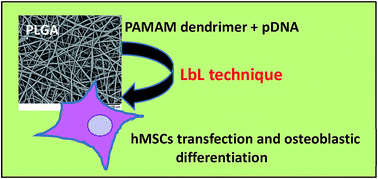
A gene delivery platform for potential use in tissue engineering applications was developed by surface functionalization of biodegradable electrospun poly(lactic-co-glycolic acid) (PLGA) fibers with nanolayers of chitosan (cationic polymer) and alginate (anionic polymer) using the Layer-by-Layer (LbL) technique. The developed system not only supported the attachment and growth of human Mesenchymal Stem Cells (hMSCs), but also was capable of delivering pDNA/dendrimer complexes and inducing cell differentiation towards the osteogenic lineage when a pDNA codifying for human Bone Morphogenetic Protein-2 (BMP-2) was used. Beyond providing a means for pDNA/dendrimer complex immobilization, the polyelectrolyte coating conferred sustained release properties to the scaffold that resulted in pDNA protection from degradation. The polyelectrolyte coating, by itself, also contributed to enhance cell differentiation.


1. Introduction

Tissue engineering involves the use of cells, materials (scaffolds) and suitable biochemical and physicochemical factors for the building of a new tissue or regeneration of a damaged one.1,2 The electrospinning technique is a well-known method that can be used for the production of 2D and 3D fiber matrices for this purpose.3–5 Electrospun fiber scaffolds can be made of many different materials (organic or hybrid organic/inorganic materials), and their characteristics, like porosity and mechanical properties, can be modulated to achieve the best conditions to allow cell attachment and growth.6,7 Furthermore, these fiber scaffolds can be “activated”, that is, endowed with the capability of delivering biochemical signals which will help to direct cell behavior (e.g. cell differentiation), such as proteins or the genes that codify them.8,9 In fact, gene delivery has recognized advantages over protein delivery, as DNA is usually less expensive and more stable than the majority of proteins.2,10 Also, proteins will be locally produced by transfected cells in physiological quantities (as proteins easily lose their activity, their direct application often implies the use of a toxic amount) and in an active form.2,10 In this scope, several studies have been performed using plasmid DNA (pDNA, encoding for the protein of interest) immobilized in electrospun substrates (by physical or chemical means) in its free form or condensed with non-viral vectors (usually, cationic polymers).11–13

Our previous studies revealed that poly(amidoamine) (PAMAM) dendrimers can effectively serve as vehicles for the delivery of the Bone Morphogenetic Protein-2 (BMP-2) gene into human Mesenchymal Stem Cells (hMSCs) and induce their osteogenic differentiation.14 PAMAM dendrimers having primary amines at the surface and tertiary amines in the interior can bind to pDNA, neutralize its charge and transport it through the membrane of hMSCs.15,16 Indeed, hMSCs can be obtained from the adult (without raising ethical concerns) and, upon certain stimuli, can differentiate into several cell types, like adipocytes, chondroblasts, and osteoblasts.17,18 These cells have also the advantage of proliferating very well in vitro and of being immunosuppressive (as such, cells from a different individual can be used in a patient).17,18 However, how to successfully immobilize the dendrimer/pDNA complexes on the surface of electrospun fibers is an issue that should be solved if one aims to apply this system in a clinical scenario.

The combination of electrospinning with the Layer-by-Layer (LbL) technique may provide a novel approach for the activation of scaffolds for tissue engineering applications. The LbL method is based on the formation of multilayers of polyelectrolytes over a substrate by the alternate adsorption of cationic and anionic polymers.19 The easiness of the process associated with the possibility of multiple surface functionalization (not only the polymers can be chemically modified but it is also possible to include molecules or particles within the layers) makes this technique very interesting in materials science, particularly in the biomedical field.20–22 Furthermore, the LbL technique can be used in many different types of surfaces (chemically and topographically different) and applied in an aqueous medium, thus supplying an optimized interface for biological interaction.20–22 In this context, the LbL technique has been mostly used in the preparation of nano and microcapsules for drug/nucleic acid delivery, as well as a coating over biomaterial surfaces in order to improve their cytocompatibility and/or obtain biologically active surfaces.20–22 Also, the studies performed until now that applied the LbL technique to immobilize pDNA in tissue engineering scaffolds also revealed that this method is very promising for this purpose.23–25

In this work, alginate (ALG, anionic polymer) and chitosan (CHI, cationic polymer) were used to form a coating over poly(lactic-co-glycolic acid) (PLGA) electrospun fibers via the LbL technique. On one hand, PLGA is reported as being biocompatible, biodegradable, and as adequate to form porous scaffolds with the right level of porosity and pore interconnectivity.26 On the other hand, both alginate and chitosan are also known for their biocompatibility and biodegradability.27,28 The original idea under test relied on the possibility to use this system for dendrimer/pDNA complexes immobilization since they can show a positive charge and interact electrostatically with the outer layer of alginate in the fibers surface. pDNA codifying the BMP-2 protein was used to induce the differentiation of hMSCs towards the osteoblastic lineage.

2. Materials and methods

2.1. Reagents and materials

Poly(D,L-lactide-co-glycolide) (PLGA) (LA/GA 50[thin space (1/6-em)]:[thin space (1/6-em)]50, Mw = 71[thin space (1/6-em)]000, 1.25 g cm−3, IV = 0.59 dL g−1) was bought from Jinan Daigang Biomaterial Co. Ltd. (China). Sodium alginate (Mw = 50–120 kDa), chitosan (from crab shells, 85% deacetylated), tetrahydrofuran (THF), dimethylformamide (DMF), 1-ethyl-3-(3-dimethylaminopropyl)carbodiimide (EDC), and N-hydroxysuccinimide (NHS) were purchased from Sigma-Aldrich. Generation 5 poly(amidoamine) (PAMAM G5) dendrimers with amino termini and an ethylenediamine core were acquired from Dendritech Inc. (USA). The dialysis membranes were from Spectrum Laboratories and the filters used for sterilization (0.22 μm) were from VWR. The Quant-iT™ PicoGreen dsDNAKit was from Invitrogen. The experiments also used Phosphate Buffer Saline solution (PBS; Gibco, without calcium or magnesium), Dulbecco's Modified Eagle's Medium (DMEM; Sigma-Aldrich), α-Minimum Essential Medium (α-MEM; Gibco), trypsin-EDTA solution (0.25% w/v, Gibco), ascorbic acid (Sigma-Aldrich), beta-glycerophosphate (Sigma-Aldrich), dexamethasone (Sigma-Aldrich), resazurin (Sigma-Aldrich), Reporter Lysis Buffer (RLB, Promega), luciferase assay system (Promega), bicinchoninic acid assay (BCA, Sigma-Aldrich), sodium chloride (Merck), sodium carbonate (Merck), sodium bicarbonate (Merck), magnesium chloride (Sigma-Aldrich), p-nitrophenyl phosphate (Sigma-Aldrich), acetic acid (Riedel-de Haen), rhodamine B (Gibco), and fluorescein isothiocyanate (FITC, Sigma-Aldrich).

2.2. Preparation of PLGA fiber mats by electrospinning

A PLGA polymer solution (27.5% w/v) was prepared by dissolving PLGA in THF/DMF (3[thin space (1/6-em)]:[thin space (1/6-em)]1) solvent mixture. The solution was left overnight for homogenization protected from light. The homogenized PLGA polymer solution was fed into the syringe of the electrospinning equipment (FM1108, Beijing Future Material Sci-tech Co. Ltd, China) and a high voltage (20 kV) was applied to the polymer solution. The flow rate was fixed at 0.004 mm s−1. During this process, a charged jet was ejected from the needle and then undergone extensive stretching and thinning along with rapid solvent evaporation. The distance between the needle tip to the collector plate was 15 cm. Throughout the process, the humidity was maintained between 38 to 40%. While the jet traveled towards the grounded collector, the fibers were formed and collected on the aluminum foil. The fiber mats were dried in the desiccator for complete evaporation of the solvent (2 days) and detached from the foil before use.

2.3. Characterization of the electrospun PLGA fiber mats

The morphology of the PLGA fibers was analyzed by scanning electron microscopy (SEM, JEOL, JSM-5600LV). Before SEM observations, the samples were sputtered with a thin gold film with a thickness of 10 nm. The porosity of the electrospun fiber mats was calculated according to the following equations:
 
ρ (g cm−3) = m (g)/d (cm) × s (cm2) (1)
 
Porosity (%) = (1 − ρ/ρ0) × 100% (2)
where ‘ρ’ is the apparent density of the electrospun scaffold, ‘m’ is the mass of the fiber, ‘d’ is the thickness of the scaffold, ‘s’ is the area of the electrospun scaffolds, and ‘ρ0’ is the density of the bulk polymer PLGA, which was assumed as being 1.25 g cm−3. The electrospun fiber mats were cut into 20 × 20 mm (8 samples) for these experiments. Each sample was weighed individually and its thickness was measured using Vernier calipers (Trade Tools Ltd., China). To enhance the accuracy of the fiber thickness, the 8 samples of the fiber mats were overlaid and then the whole thickness was measured.

2.4. Functionalization of the electrospun PLGA fiber mats using the LbL technique

Preparation of pristine sodium alginate and chitosan solutions. For the experiments, a sodium alginate solution (1 mg mL−1) was prepared in 0.15 M NaCl. The pH of the alginate solution was adjusted to 7.2 with 0.1 M NaOH. A chitosan solution (1 mg mL−1) was also prepared. The chitosan was dissolved in 0.1 M acetic acid and 0.15 M NaCl. This solution was gently stirred overnight to become homogeneous. The pH of the chitosan solution was 3.4. This solution was not adjusted to physiological pH to avoid CHI precipitation.
Preparation of rhodamine B labeled chitosan solution. Chitosan (0.2 g) was dissolved in 20 mL of 0.1 M acetic acid to prepare a 1% w/v solution and, in the next day, an equal volume of methanol was added. A rhodamine B solution was also prepared by dissolving 8 mg of the product in 4 mL of methanol. Before mixing with chitosan, rhodamine B was activated with EDC and NHS mixture for 3 hours (the concentration of EDC and NHS was two folds the concentration of rhodamine B). Then, the activated rhodamine B was added dropwise to the chitosan solution under vigorous stirring, at room temperature, in the dark, and allowed to react for 3 days. After that, the resulting solution was adjusted with NaOH (0.5 M) to pH = 10 (in order to precipitate the rhodamine B-conjugated chitosan compound), transferred to 15 mL tubes, and centrifuged for 10 minutes at 15[thin space (1/6-em)]000 rpm. The supernatant was removed and the pellet was washed using ultrapure water followed by centrifugation. This step was repeated until no fluorescence was detected in the supernatant (7 centrifugations were done). Once the washing was completed, the precipitate was dissolved in 10 mL of 0.1 M acetic acid again and dialysed against 0.1 M acetic acid solution for 3 days, followed by lyophilization to get dry rhodamine B-conjugated chitosan compound. A stock solution was prepared by dissolution of the compound in a solution of 0.1 M acetic acid and 0.15 M sodium chloride. For the experiments, a 1 mg mL−1 solution was used.
Preparation of the fluorescein isothiocyanate (FITC) labeled sodium alginate solution. An aqueous solution of sodium alginate (1% w/v; 1 g in 100 mL of water) was mixed with EDC/NHS (3[thin space (1/6-em)]:[thin space (1/6-em)]1) for the activation of the carbonyl groups on alginate, in acetic acid buffer (pH = 4.9) for 30 min, followed by addition of hexamethylenediamine (60 mg) and reaction for another 4 h. The mixture was precipitated in 2-propanol to remove the unreacted diamine. The alginate-amine derivative was then reacted with FITC (2 mg) in pH 9.0 sodium bicarbonate solution for 4 h and then dialyzed against distilled water for 2 days. The resulting alginate-FITC solution was lyophilized. For the stock solution, the alginate-FITC compound was dissolved in 0.15 M sodium chloride. The final solution concentration was brought to 1 mg mL−1 before use.
Functionalization of the PLGA fibers using the LbL technique. The PLGA fiber mat produced using the electrospinning technique and deposited onto an aluminum foil was removed after drying and was cut into pieces of 1.5 × 1.5 cm2. Glass coverslips were then covered with these fiber mats and placed in the bottom of the wells of 24-well cell culture plates. The functionalization of the PLGA fibers with polyelectrolytes was similar to what is described in the literature.19 In a typical procedure, the PLGA fiber mats were first immersed in the chitosan solution (1 mg mL−1) for 10 min, followed by rinsing with ultrapure water three times (each rinsing step took 2 minutes). Then, the substrates were immersed in the negatively charged alginate solution (1 mg mL−1) for 10 min, followed by similar rinsing steps in water. The immersion/rinsing cycles were repeated until the desired number of bilayers was achieved. In this work, it is considered that a bilayer (BL) is obtained after one deposition cycle (so, it consists in one layer of chitosan and one layer of alginate, by this order). Two bilayer-coated PLGA fibers were used in the experiments.

The polyelectrolyte deposition process over the PLGA fiber mats was followed by fluorescence spectroscopy using rhodamine B-labeled chitosan (excitation wavelength 540 nm and emission wavelength 625 nm) and FITC-labeled alginate (excitation wavelength 495 nm and emission wavelength 519 nm). To avoid possible crossing interferences in the absorption and emission processes, the study was first made with rhodamine B-labeled chitosan and pristine alginate. After, a similar study was done with pristine chitosan and FITC-labeled alginate. The fluorescence measurements were made using a microplate reader (Victor3 1420, Perkin Elmer).

2.5. Plasmid DNA (pDNA) amplification and purification

Two kinds of pDNAs were used in the present work: (a) pDNA encoding luciferase (Luc) and enhanced green fluorescent protein (EGFP) (pEGFPLuc, 6.4 kb, kindly provided by Prof. Tatiana Segura, UCLA, USA); (b) pDNA encoding the bone morphogenetic protein-2 (BMP-2) (pcDNA3.1/Hist/hBMP2, 8.5 kb, kindly donated by Prof. Yasuhiko Tabata, University of Kyoto, Japan). Both plasmids were amplified in Escherichia coli host strain, DH5α, grown overnight in Luria–Broth base medium containing adequate antibiotics (ampicillin at a final concentration of 100 μg mL−1 for pcDNA3.1/Hist/hBMP2 and kanamycin at 50 μg mL−1 for pEGFPLuc). Afterwards, the plasmids were isolated and purified using a Qiagen Plasmid Maxi Kit (Qiagen). The isolated plasmids were dissolved in a small amount of ultra-pure water and the concentration of pDNA in solution was determined from the UV absorbance at 260 nm. For evaluation of plasmid purity, the absorbance ratio at 260 nm and 280 nm was analyzed (it was always found to be in the range of 1.7 and 1.9).

2.6. Dendrimer/pDNA complex assembly and characterization

Dendrimer/pDNA complexes were prepared at several N[thin space (1/6-em)]:[thin space (1/6-em)]P ratios (where N = number of primary amines in the conjugate; P = number of phosphate groups in the pDNA backbone) by mixing adequate volumes of PAMAM G5 dendrimers (1 mg mL−1) and pDNA (0.1 μg μL−1) solutions. PBS solution (pH = 7.4) was always used to prepare these solutions. Complex solutions were vortexed and allowed to incubate for 20 minutes at room temperature prior to experiments. The N[thin space (1/6-em)]:[thin space (1/6-em)]P ratios selected to study pDNA compaction were 0, 0.5, 1, 2, 4, 6 and 8.

The ability of the dendrimers to compact pDNA was studied using the PicoGreen assay. PicoGreen is a fluorescent dye that emits light when bonded to double strand DNA. When the DNA is completely free in solution, the intensity of the fluorescence attains a maximum level. When the DNA is compacted due to complexation with the dendrimer, PicoGreen cannot bind DNA and the fluorescence intensity decreases. PicoGreen was diluted 200× in Tris–EDTA buffer (10 mM Tris, 1 mM EDTA, pH = 7.5) according to the manufacturer's instructions. Then, 100 μL of this solution was added to 100 μL of each dendrimer/pDNA complex solution. The resultant mixture was incubated for 5 minutes at room temperature in the dark. The resultant mixture was then transferred to a 96-well plate and PicoGreen fluorescence was measured using a microplate reader (Perkin Elmer, Victor3 1420) at excitation wavelength of 485 nm and emission wavelength of 535 nm. A blank was prepared with 100 μL of PBS and 100 μL of the diluted PicoGreen solution. Three independent experiments were performed and the percentage of relative fluorescence (% F) was determined using the following equation:

 
image file: c6ra22444j-t1.tif(3)

The fluorescence from free DNA was considered to be 100% (N[thin space (1/6-em)]:[thin space (1/6-em)]P ratio of 0).

The N[thin space (1/6-em)]:[thin space (1/6-em)]P ratio used when pDNA was immobilized in the electrospun fiber mats was 5. For this N[thin space (1/6-em)]:[thin space (1/6-em)]P ratio, the size of the complex was measured at 633 nm on a dynamic light scattering instrument (Zetasizer Nano ZS, Malvern Instruments) using a detection angle of 173°. The zeta potential measurement was also performed using a detection angle of 17° and was calculated using the Smoluchowsky model for aqueous suspensions. The data presented are means of three independent measurements.

2.7. Immobilization of the pDNA/dendrimer complexes on the PLGA fiber mats

Fiber mats with and without polyelectrolyte coverage (2BL, 2 bilayers of CHI/ALG polymers) were used in the experiments. The desired amount of dendrimer/pDNA complex (N[thin space (1/6-em)]:[thin space (1/6-em)]P = 5, pDNA = 1 μg per well or 2 μg per well) was dispersed in 400 μL PBS solution and added to each well of a 24-well plate containing the fiber mats (as previously described, glass coverslips were covered with the fiber mats and placed in the bottom of the wells), followed by 30 min incubation at room temperature. The amount of dendrimer/pDNA complex loaded on the PLGA fiber mats was indirectly determined by measuring the quantity remaining in solution. For that, 100 μL of the gathered supernatant was added of an equal volume of sodium alginate solution (6 mg mL−1) and shaked gently for 1 h (37 °C, 217 rpm min−1) to disrupt the electrostatic interactions between the dendrimer and the pDNA. Then the amount of pDNA in solution was quantified using the PicoGreen assay. For the quantification, a calibration curve was constructed using a series of pDNA standard solutions with increasing concentrations, in the presence of sodium alginate (6 mg mL−1).

2.8. pDNA/dendrimer complex release from the PLGA fiber mats

After loading the fiber mats with the dendrimer/pDNA complexes and quickly rinsing with ultrapure water to remove the non-bonded complexes, the release of the complexes was studied over a period of 24 h in PBS solution. The experiments were conducted at 37 °C, under shaking. An initial load of 2 μg of pDNA was used in each experiment. Samples were taken at different time intervals and analyzed using the PicoGreen assay after disruption of the electrostatic forces existent between the dendrimers and the pDNA, and using the methodology described in the previous section.

2.9. Biological assays

Cells and cell culture. All cell cultures were incubated at 37 °C, in a humidified atmosphere of 95% air and 5% carbon dioxide (Nuaire, AutoFlow IR Direct Heat CO2 incubator). NIH 3T3 cells (a cell line derived from primary mouse embryonic fibroblasts, acquired to DSMZ, Germany, ref. ACC173) were cultured in DMEM and hMSCs were cultured in α-MEM supplemented with 10% fetal bovine serum (FBS; Gibco) and 1% of an antibiotic–antimycotic solution (Gibco, with 10[thin space (1/6-em)]000 units penicillin per mL, 10 mg streptomycin per mL and 25 μg amphotericin B per mL). The primary cultures of hMSCs were established from small pieces of human trabecular bone rich in bone marrow which were obtained from patients during surgery interventions after trauma. Only bone that would have been discarded was used, with the approval of the Dr Nélio Mendonça Hospital (Funchal Central Hospital) ethics committee and informed consent from the donors. All experiments were performed in compliance with the European Union Directives 2004/23/CE, 2006/17/CE, and 2006/86/CE, also following the Code of Good Practice in Research of the University of Madeira/Centro de Química da Madeira. The trabecular bone was kept in sterilized tubes, refrigerated, in α-MEM supplemented with 10% FBS and 1% antibiotic–antimycotic solution, for 3 to 4 h. Then, primary cultures were prepared by gently shaking the tubes with the bone marrow pieces to obtain a cell suspension which was spread by cell culture dishes. The culture medium was removed and changed by new one after 1 day to remove the non-adherent hematopoietic cells. The medium was always changed twice a week until confluence was achieved. The cultures were then passaged and frozen in liquid nitrogen using the standard procedure. Cell passages were always done using trypsin (0.05% w/v) and following the standard protocol.
Cell attachment/proliferation on the PLGA fiber mats. Cell attachment and proliferation on the PLGA electrospun mats was evaluated using NIH 3T3 cells directly cultured on the electrospun PLGA fiber mats. As previously mentioned, glass coverslips were covered with the fiber mats and placed in the bottom of the wells of 24-well culture plates. For sterilization, the mats were initially left under UV radiation overnight, followed by soaking with 75% ethanol solution for 1.5 h and ultimately washed three times with PBS solution. The electrospun mats were then soaked in culture medium overnight prior to cell seeding. For the cell attachment and proliferation experiments, NIH 3T3 cells were detached from the culture dish using trypsin–EDTA solution and seeded on the top of the mats at a density of 2 × 10⁴ cells per well. Both fiber mats with and without polyelectrolyte coverage (2BL, 2 bilayers of CHI/ALG polymers) were used in the experiments. Control experiments were conducted with cells cultured directly on the plastic surface of the cell culture dishes and on the glass surface of the coverslips. Cell adhesion was evaluated after 1, 3, and 6 h of cell culture. Cell proliferation was studied from 1 until 7 days in culture. Cell adhesion/proliferation was studied based on the resazurin reduction cell metabolic assay. The resazurin reagent was prepared by mixing 0.02 g of resazurin with 200 mL of PBS solution (resazurin stock solution). This solution was sterilized with a 0.22 μm membrane inside the laminar flow hood chamber. The prepared resazurin solution was distributed into small tubes and stored at −20 °C. At the appropriate time point, the medium in each cell culture well was replaced by fresh medium (1 mL) containing 10% v/v of resazurin stock solution and the cells were further incubated for 3 h. Subsequently, 100 μL of the resultant medium was transferred to 96-well plates (white, FluoroNunc 96 well plates) and the resorufin fluorescence (λex = 530 nm, λem = 590 nm) was measured in the microplate reader (Perkin Elmer, Victor3 1420). All experiments were conducted in triplicate. Blank experiments were done using wells containing PLGA electrospun mats but without seeded cells.
In vitro gene transfection studies using the pEGFPLuc. The preparation, sterilization and pDNA/dendrimer complex loading of the PLGA fiber mats were performed according to the procedures already mentioned. In these studies, NIH 3T3 cells (2 × 10⁴ cells per well) were seeded in the wells of 24-well plates containing pDNA/dendrimer loaded fiber mats, with and without polyelectrolyte coverage. Control experiments were performed using fiber mats samples without pDNA/dendrimer loading, with and without polyelectrolyte coverage (2 bilayers). Another control experimented consisted in cells directly cultured on the glass surface of the cover slips. In all cases, cells were allowed to grow for 48 h and then cell viability was measured (using the resazurin reduction method previously described) and further harvested for luciferase activity and total protein content analysis. For cell harvesting, cells were first washed once with PBS solution, and then 100 μL of RLB was added to each well. Homogenized cell lysates were obtained after three cycles of freezing (−20 °C, 20 min)/thawing (room temperature, with shaking). Cell lysates were transferred to Eppendorf tubes for luciferase and protein analyses. Luciferase activity in cell lysates was measured using the Promega's luciferase assay reagent (the supplier's instructions were followed). For each sample, the microplate reader was set for 3 s delay with signal integration for 10 s. The amount of protein in cell lysates was determined using the bicinchoninic acid assay (BCA assay) using bovine serum albumin as a standard.
In vitro gene transfection studies using the pcDNA3.1/Hist/hBMP2. These experiments were very similar to those described in the previous subsection but used hMSCs (passage 3) and a pDNA encoding the bone morphogenetic protein-2 (BMP-2) (pcDNA3.1/Hist/hBMP2). Also, cell culture was performed along 21 days. For these experiments, α-MEM containing ascorbic acid (250 μM) and beta-glycerophosphate (5 mM) was used. Osteogenesis positive control experiments were also conducted using dexamethasone in the cell culture medium (10−9 M). For all cases, the medium was replaced every 3 days and cells were incubated at 37 °C in a humidified atmosphere with 5% CO2.

Cell viability was analyzed after 14 days of cell culture using the resazurin reduction assay. Alkaline phosphatase (ALP) activity was measured after 14 and 21 days of culture, as well as the total protein content (BCA assay). The hMSCs were first rinsed 3 times with PBS. Then, 200 μL of RLB was added to each well and homogenized cell lysates were obtained after three cycles of freezing (−20 °C, 20 min)/thawing (room temperature, 90 rpm shaking). Cell lysates were stored at −20 °C for further analysis. Alkaline phosphatase (ALP) activity was determined using a colorimetric assay involving the hydrolysis of p-nitrophenyl phosphate to p-nitrophenol (yellow). Briefly, 20 μL of the cell lysates were mixed with 200 μL of ALP substrate and incubated for 1 h at 37 °C in the dark. Then, 10 μL of 0.02 M NaOH was added to each well to stop the hydrolysis reaction. The absorbance was read at 405 nm and the p-nitrophenol content was calculated from a standard calibration curve. The results were presented using arbitrary units but expressed the number of moles of p-nitrophenol produced by unit of time and by μg of protein.

3. Results and discussion

3.1. Preparation, functionalization with polyelectrolytes and characterization of the electrospun PLGA fibers

Homogeneous electrospun fiber mats made of poly(L-lactic-co-glycolic acid) (PLGA) were successfully prepared maintaining the experimental conditions throughout the process of fiber formation. Fig. 1A and B show scanning electron microscopy (SEM) images of the obtained electrospun fiber mats before functionalization using the LbL technique, revealing fiber diameters in the micrometer range. The porosity of the fiber mats was also experimentally determined and, as expected, its value was very high (97.5 ± 0.1%). As such, the prepared electrospun materials should be able to provide a high area for cell attachment and growth and should be appropriate for tissue engineering applications. Furthermore, by detailed analysis of the SEM images, one can conclude that the pores are interconnected, which is also a very important requirement for this purpose.
image file: c6ra22444j-f1.tif
Fig. 1 SEM images of electrospun PLGA fiber mats. (A and B) Fibers without polyelectrolyte coating. (C and D) Fibers with polyelectrolyte coating. (B) and (D) are magnified images of (A) and (C), respectively.

As a mean of immobilizing the pDNA/dendrimer complexes (that are positively charged), the polymers chitosan (cationic) and alginate (anionic) were deposited as a coating over the surface of the electrospun fiber mats using the LbL technique. Two polyelectrolyte bilayers (BL) were assembled over the fiber mats (each bilayer consisted in the deposition of a first layer of chitosan and a second layer of alginate) and, as shown in Fig. 1C and D, no significant change in morphology was observed.

The success of the LbL process was confirmed by measuring the increase in intensity of the fluorescence emitted by fiber mats coated with growing bilayers of rhodamine B-labeled chitosan plus pristine alginate (Fig. 2A), and pristine chitosan plus FITC-labeled alginate (Fig. 2B). It is clear from the graphics that the fluorescence intensity increased linearly with the number of deposited polyelectrolyte bilayers, which demonstrates the effective coating of the PLGA fiber mats using the LbL technique.


image file: c6ra22444j-f2.tif
Fig. 2 (A) Fluorescence intensity of PLGA fiber mats coated with rhodamine B-labeled chitosan and pristine alginate as a function of the number of bilayers (BL); (B) fluorescence intensity of PLGA fiber mats coated with pristine chitosan and FITC-labeled alginate as a function of the number of bilayers. The data presented are means of three independent measurements (±SD).

3.2. pDNA/dendrimer complex formation, immobilization in the electrospun PLGA fiber mats and release kinetics

It is well known that generation 5 PAMAM dendrimers (PAMAM G5) with primary amines at the surface are able to bind the anionic pDNA through electrostatic interactions since they are positively charged at the physiological pH.15 When PAMAM G5 dendrimers complex with pDNA, the molecule of pDNA is simultaneously compacted (a size decrease is observed) and neutralized in terms of electric charge. Since these dendrimers also possess tertiary amines in the interior, it is believed that their buffering capability promotes the escape from the acidic endo-lysosomal compartments after cell entry through endocytosis, protecting the pDNA from degradation (this is usually described as the “proton-sponge effect”).15

In the present work, PAMAM G5 dendrimers were complexed with pDNA at different N[thin space (1/6-em)]:[thin space (1/6-em)]P ratios. Their ability to condense pDNA was evaluated by assessing the amount of “free pDNA” using the PicoGreen assay (this is a fluorescent dye that emits fluorescence when intercalated in the helix of “free” double strand DNA). From the results shown in Fig. 3A, it is clear that a maximum in pDNA compaction was achieved for a N[thin space (1/6-em)]:[thin space (1/6-em)]P ratio of 4 or higher. Hence, a N[thin space (1/6-em)]:[thin space (1/6-em)]P ratio of 5 was chosen for the subsequent experiments. At this N[thin space (1/6-em)]:[thin space (1/6-em)]P ratio, the hydrodynamic diameter and surface zeta potential of the complexes were experimentally determined in PBS solution, being 422 ± 20 nm and +6.5 ± 0.9 mV, respectively. These values are in agreement with others described in the literature and measured under similar experimental conditions (in PBS, and using the same type of plasmid (pEGFPLuc, 6.4 kb)).29 The cationic charge of the complexes was relatively low, but even so enough for its immobilization on the top of the polyelectrolyte coated electrospun fiber mats as verified in the following experiments (note that alginate was the last polymer deposited over the fiber mats during the LbL process).


image file: c6ra22444j-f3.tif
Fig. 3 Study of pDNA/dendrimer compaction at different N[thin space (1/6-em)]:[thin space (1/6-em)]P ratios (A), and amount of pDNA retained in the fiber mats (pDNA loading) as a function of the quantity of pDNA added to the wells (B and C); in (C), the amount of pDNA retained is expressed as a percentage of the quantity of pDNA added to the wells. The data presented are means of three independent measurements (±SD).

The complex loading efficiency of the electrospun fiber mats was studied for uncoated and coated samples and for two different initial pDNA concentrations in the wells (1 μg per well and 2 μg per well). The loading process consisted in the incubation of the fiber mats with a solution containing the pDNA/dendrimer complexes for 30 min (this time period was optimized for maximum loading), followed by washing to remove the complexes in excess. The amount of pDNA present in the withdrawn solution was determined using the PicoGreen assay (after disrupting the electrostatic forces within the complex with a strong negatively charged polyelectrolyte at a high concentration – alginate was also used for this purpose) and the quantity of loaded pDNA was calculated by difference from the initial amount of pDNA (in a complexed form) added to the wells.

Fig. 3B and C summarize these results. Both the polyelectrolyte coated and non-coated PLGA surfaces adsorbed pDNA/dendrimer complexes from the loading solutions. From Fig. 3B, it is evident that a higher quantity of pDNA/dendrimer complex was retained in the electrospun fiber mats when 2 μg of pDNA was added to each well for both non-coated (the mass of pDNA retained changed from 0.56 to 0.85 μg per well when the pDNA amount added changed from 1 to 2 μg per well, respectively) and coated samples (the mass of pDNA retained changed from 0.57 to 1.10 μg per well when the pDNA amount added changed from 1 to 2 μg per well, respectively). This indicates that there were still spaces available for adsorption at the surface of the fiber mats when 1 μg per well pDNA was added.

When 2 μg per well pDNA was used in the loading process, the amount of pDNA retained was higher for the coated electrospun fiber mats (1.10 μg per well) than for the non-coated ones (0.85 μg per well), whereas a similar retained amount of pDNA was obtained for the 1 μg per well loading condition (0.56 and 0.57 for the non-coated and the coated surfaces, respectively). Since less place remains available for pDNA/dendrimer complex adsorption when a higher amount of pDNA is used during the loading process, the higher negative character of the PLGA surface conferred by the outer layer of alginate then acts as an additional driving force for increasing complex adsorption. This effect is not noticeable when low amounts of pDNA are used in the loading process as the area still available for adsorption is bigger and the slight negative charge of the PLGA substrate is enough to promote adsorption.

Interestingly, the percentage of pDNA retained in the fibers in relation to the quantity initially added to the wells, was always around 50% (Fig. 3C) which may indicate that equilibrium is established between the adsorbed complexes and the ones that are kept in solution. When the concentration of the pDNA solution increases from 1 μg per well to 2 μg per well, more complexes will adsorb to the mat surface but, still, the same proportion of complexes will remain in solution (that is, changing the complex amount does not change the equilibrium constant). Similar results were obtained by other authors that used polyethylenimine (PEI)/DNA complexes immobilized on tissue engineering scaffolds made of poly(lactide-co-glycolide).30 Based on these results, a pDNA concentration of 2 μg per well was always used to load the electrospun fiber mats in the following experiments.

The in vitro release profile of pDNA from the PLGA fiber mats with and without surface modification was studied along 24 h in PBS solution at 37 °C (Fig. 4). The amount of pDNA released was monitored using the same methodology as for pDNA loading, that is, also determined using the PicoGreen assay and after disrupting the electrostatic forces within the pDNA–dendrimer complex with alginate at a high concentration. After 6 h, the quantity of pDNA released was approximately the double when the fibers were not coated with polyelectrolytes. The more sustained release observed using the coated samples might be ascribed to the strong electrostatic interaction established between the pDNA/dendrimer complexes and the negatively charged surface of the coating. After 24 hours, for the non-coated samples, the amount of pDNA detected in the PBS solution was significantly lower than at 6 h. For the coated samples, this value was not significantly different from the one at 6 h. The results at this time point were surprising since higher values of pDNA were expected for both cases. Probably, a longer incubation time makes the complexes susceptible to electrostatic disruption and pDNA degradation occurs (degraded pDNA will not be detected using the PicoGreen assay). In the coated samples, the complexes were released at a slower rate (they stayed on the PLGA coated surfaces for a longer time), and hence pDNA remained protected from degradation. Taken together, these in vitro results point out that the coated electrospun fiber mats will be more adequate as vehicles for the delivery of pDNA/dendrimer complexes due to their sustained release behavior and, thus, capability to protect the pDNA.


image file: c6ra22444j-f4.tif
Fig. 4 Cumulative release of pDNA from non-coated and coated electrospun fiber mats in PBS solution, at 37 °C. Measurements were made at times: 15 min, 30 min, 1 h, 3 h, 6 h and 24 h. The data presented are means of three independent measurements (±SD).

3.3. Cell attachment and proliferation on non-coated and coated PLGA fiber mats

NIH 3T3 cells (a fibroblast cell line) were used to study the attachment and proliferation of cells on the PLGA fiber mats for both coated and non-coated samples. The metabolic activity of cells was assessed using the resazurin reduction assay which is based on the capacity of metabolic active cells (living cells) to transform the non-fluorescent compound resazurin into the fluorescent compound resorufin. The fluorescence intensity is, then, directly proportional to the cell metabolic activity and, thus, to the number of viable cells, allowing an indirect measure of cell attachment and proliferation over the fiber substrates.

Fig. 5A shows the results of cell attachment as a function of time (until 6 h of cell culture) and of the type of surface (cells were seeded on the top of the plastic surface of cell culture wells and coverslips, as well as on the top of non-coated and coated electrospun fiber mats). Apparently, cells attached better to the control samples (plastic surface, cover slips) then on the top of the fiber mats. However, one should notice that the resorufin produced during the assay used for cell attachment quantification may be absorbed by PLGA fibers and its detection in solution can be partially reduced.31 Even assuming that this is not important, it was clear that non-coated and polyelectrolyte coated electrospun fiber mats were capable of supporting cell attachment in a great extent and without significant differences between them. The fluorescence intensity grew along the first 6 hours of cell culture revealing a progressive cell attachment process. The scanning electron microscopic pictures in Fig. 6A and B also corroborate this finding. Furthermore, cells appear with a normal morphology and show cytoplasmatic extensions that interact with fibers.


image file: c6ra22444j-f5.tif
Fig. 5 NIH 3T3 cells attachment (A) and proliferation (B) on the plastic surface of the culture dishes, on the glass surface of coverslips, and on non-coated and 2 bilayer-coated electrospun fiber mats. The values represent means from 4 replicates ± SD.

image file: c6ra22444j-f6.tif
Fig. 6 Scanning electron microscopy images of NIH 3T3 cells on non-coated (A and C) and 2 bilayer-coated electrospun fiber mats (B and D). Images were taken after 6 h (A and B) and 3 days (C and D) in culture.

Fig. 5B shows that cells were able to growth in all the four substrates studied (plastic, coverslip, PLGA fiber, 2BL coated PLGA fiber) until seven days in culture. At days 3 and 7, there was a minor difference between the number of cells present on the top of coated and non-coated samples. Likely, this is due to an electrostatic repulsion between the negative charge conferred by the outer layer of alginate on the surface and the also negative charge of the cell membrane that is covered with glycolipids and glycoproteins. The observation of the cultures by scanning electron microscopy techniques also revealed a good coverage of the surfaces by NIH 3T3 cells after 3 days of culture (Fig. 6C and D) in accordance with the quantitative data obtained through the resazurin reduction assay.

3.4. In vitro gene transfection studies

Two types of gene transfection studies were performed with 2BL polyelectrolyte coated and non-coated electrospun fiber mats by immobilizing pDNA/dendrimer cationic complexes over their surface. The first one used a pDNA encoding for the luciferase enzyme and NIH 3T3 cells (Fig. 7). The second one used a pDNA encoding for bone morphogenetic protein-2 (BMP-2) and human mesenchymal stem cells (hMSCs) (Fig. 8), that is, conditions that are more close to the envisaged application.
image file: c6ra22444j-f7.tif
Fig. 7 NIH 3T3 cells viability (A) and luciferase activity normalized for the protein content (B) after 48 h of culture and for the pDNA/dendrimer complex non-loaded and loaded electrospun fiber mats. The values represent means from 4 replicates ± SD.

image file: c6ra22444j-f8.tif
Fig. 8 hMSCs viability at day 14 (A) and normalized alkaline phosphatase (ALP) activity at days 14 and 21 (B) for pDNA/dendrimer complex non-loaded and loaded electrospun fiber mats, with and without polyelectrolyte coating. The alkaline phosphatase activity was normalized for the protein content. “Positive control” means that dexamethasone was present in the cell culture media whereas “Negative control” imply its absence. The values represent means from 4 replicates ± SD.
Transfection experiments using the luciferase reporter gene. After 48 h of cell culture, the transfection efficiency achieved by the pDNA coated electrospun fiber mats was assessed. During this time period, NIH 3T3 cells viability was not significantly different in the 4 different types of surface studied, that is, in pDNA/dendrimer complex coated and non-coated, and polyelectrolyte coated and non-coated electrospun fiber mats (Fig. 7A).

As can be observed in Fig. 7B, the luciferase enzyme activity was significantly higher when cells were cultured over the electrospun fiber mats coated with pDNA/dendrimer complexes when compared with the results obtained when the pDNA/dendrimer complex was not present (surfaces used as controls). Furthermore, the level of luciferase activity was enhanced for the surfaces coated with polyelectrolytes as a mean of immobilizing the pDNA/dendrimer complexes. These findings are in agreement with the previous results that revealed a slightly higher pDNA loading in this type of surfaces and, also, a sustained release of the loaded pDNA which may be beneficial for its stability and cellular internalization. Indeed, the transfection efficiency achieved by polyelectrolyte coated electrospun fiber mats was about 1.5 times higher than that observed when the polyelectrolyte layers were not present.

Transfection experiments using the BMP-2 gene. Even if the levels of luciferase activity previously obtained were not impressive, it was hypothesized that they could be sufficient for a clinical relevant application (e.g. tissue engineering) such as the differentiation of hMSCs towards the osteoblastic lineage. The same methodology for cell transfection was followed with prolonged time having in consideration the lower growth rate of this type of cells and the characteristic behavior of the osteogenesis process occurring in vitro. Hence, the cells were cultured along 21 days on the surface of electrospun fiber mats loaded with pDNA/dendrimer complexes with and without polyelectrolyte coating. As positive controls, cells were also cultured on the top of the plastic surface of culture dishes and of electrospun fiber samples (with and without polyelectrolyte coating) in the presence of dexamethasone which is a well known inducer of osteogenesis.32 Experiments were further done simply using hMSCs cultured in the top of the plastic surface of the cell culture dishes and of the electrospun fiber mats (with and without polyelectrolyte coating) that were considered negative controls of the osteogenesis process.

At day 14 of cell culture (Fig. 8A), hMSCS viability was analyzed using the resazurin reduction assay. The results clearly showed that the presence of dexamethasone did not affect cell viability at this time point. Also, cell viability was apparently higher when cells were cultured on the plastic surface of the culture dishes than when electrospun fiber mats were used. This is in agreement with the results obtained with NIH 3T3 cells and the justification for this finding is the same as presented before, that is, the probable absorption of resorufin by the fibers.31 Furthermore, cell viability was not significantly different among the four situations where electrospun fiber mats were used.

Alkaline phosphatase (ALP) activity was measured at days 14 and 21 after cell seeding. This parameter is well established as an early marker of the osteogenesis process in vitro and is known to present high values between 2 to 3 weeks of cell culture, before the occurrence of mineralization.33 In the present work, cells were lysed and ALP activity was determined in their extracts. The results were normalized for the total protein content to get an indication of the ALP activity per cell, that is, of the extent of cellular differentiation. Fig. 8B reveals that hMSCs differentiation towards the osteoblastic lineage was strongly enhanced in the experiments done in the presence of dexamethasone (positive control) when compared with those performed in its absence and in the absence of pDNA/dendrimer complexes (negative control, without differentiation factors). When pDNA/dendrimer complexes were immobilized in the electrospun fiber mats (with and without polyelectrolyte coating), the values of ALP activity were always increased in relation to the correspondent controls without pDNA/dendrimer complexes (negative controls).

Furthermore, when pDNA/dendrimer complexes were immobilized in the electrospun fiber mats without polyelectrolyte coating, the values of ALP activity were even higher than those of the positive control (with dexamethasone). However, when pDNA/dendrimer complexes were immobilized in the electrospun fiber mats with polyelectrolyte coating, the extent of ALP activity stayed in the middle of the correspondent positive and the negative control. These results can be explained by the fact that dexamethasone is a biochemical factor that is immediately available in the cell culture medium whereas that does not happen with BMP-2 when pDNA/dendrimer complexes are used. Indeed, BMP-2 will only be produced after complex internalization, release of the pDNA from the dendrimer, entry of pDNA in the cell nucleus, transcription of the gene, and translation of the mRNA into the active protein. It is, thus, a slower process that, in any case, is expected to have longer time biological effects (due to the sustained release of complexes by the fibers) and less side effects (in comparison to the use of massive quantities of differentiation factors or other drugs that can be used for the same purpose). Because complex release is faster in the electrospun fiber mats without polyelectrolyte coating, an enhanced ALP activity in respect to the positive control is observed for those samples. Interestingly, when analyzing the results obtained for both positive and negative controls, it seems that the simple presence of the polyelectrolytes in the electrospun fiber mats has a positive effect on differentiation. So, even if complex release from polyelectrolyte coated electrospun fiber mats was slower, the positive effect on differentiation caused by the presence of the polyelectrolyte layers likely conducted to non-significant differences of ALP activity (at both days 14 and 21) between polyelectrolyte non-coated and coated substrates having immobilized complexes. In vivo, however, one should expect prolonged biological effects when polyelectrolyte coated electrospun fiber mats are used due to their sustained release properties.

4. Conclusions

The merging of the electrospinning and the LbL techniques constitutes a promising approach for the development of biologically active surfaces for biomedical applications, namely for tissue engineering. In this work, electrospun PLGA fiber mats with fiber diameters in the micrometer range were prepared and functionalized at the surface with chitosan and alginate polyelectrolytes to serve as a platform for the immobilization of pDNA/dendrimer complexes. This approach provided a system for the controlled release of pDNA/dendrimer complexes along the time, simultaneously protecting pDNA from degradation. The functionalized fiber mats not only supported the attachment and growth of hMSCs, but also contributed for an enhancement of cell differentiation towards the osteoblastic lineage when pDNA codifying BMP-2 was used.

Acknowledgements

This work was supported by funds of Fundação para a Ciência e a Tecnologia (FCT), namely through S. Xiao Post-Doc grant (SFRH/BPD/75420/2010), and the CQM project (PEst UID/QUI/00674/2013, Portuguese Government funds). X. Shi gratefully acknowledges FCT and Santander bank for the Invited Chair in Nanotechnology.

References

  1. V. Marx, Tissue engineering: organs from the lab, Nature, 2015, 522, 373–377 CrossRef CAS PubMed.
  2. J. L. Santos, D. Pandita, J. Rodrigues, A. P. Pêgo, P. L. Granja and H. Tomás, Non-viral gene delivery to mesenchymal stem cells: methods, strategies and application in bone tissue engineering and regeneration, Curr. Gene Ther., 2011, 11, 46–57 CrossRef CAS PubMed.
  3. J.-H. Jang, O. Castano and H.-W. Kim, Electrospun materials as potential platforms for bone tissue engineering, Adv. Drug Delivery Rev., 2009, 61, 1065–1083 CrossRef CAS PubMed.
  4. T. J. Sill and H. A. von Recum, Electrospinning: applications in drug delivery and tissue engineering, Biomaterials, 2008, 29, 1989–2006 CrossRef CAS PubMed.
  5. G. C. Rutledge and S. V. Fridrikh, Formation of fibers by electrospinning, Adv. Drug Delivery Rev., 2007, 59, 1384–1391 CrossRef CAS PubMed.
  6. J. Lannutti, D. Reneker, T. Ma, D. Tomasko and D. Farson, Electrospinning for tissue engineering scaffolds, Mater. Sci. Eng., C, 2007, 27, 504–509 CrossRef CAS.
  7. S. N. Jayasinghe, Cell electrospinning: a novel tool for functionalising fibres, scaffolds and membranes with living cells and other advanced materials for regenerative biology and medicine, Analyst, 2013, 138, 2215–2223 RSC.
  8. W. Ji, Y. Sun, F. Yang, J. J. J. P. Beucken, M. Fan, Z. Chen and J. A. Jansen, Bioactive electrospun scaffolds delivering growth factors and genes for tissue engineering applications, Pharm. Res., 2011, 28, 1259–1272 CrossRef CAS PubMed.
  9. Y. Yang, T. Xia, F. Chen, W. Wei, C. Liu, S. He and X. Li, Electrospun fibers with plasmid bFGF polyplex loadings promote skin wound healing in diabetic rats, Mol. Pharmaceutics, 2012, 9, 48–58 CrossRef CAS PubMed.
  10. M. Heyde, K. A. Partridge, R. O. Oreffo, S. M. Howdle, K. M. Shakesheff and M. C. Garnett, Gene therapy used for tissue engineering applications, J. Pharm. Pharmacol., 2007, 59, 329–350 CrossRef CAS PubMed.
  11. S. O'Rorke, M. Keeney and A. Pandit, Non-viral polyplexes: scaffold mediated delivery for gene therapy, Prog. Polym. Sci., 2010, 35, 441–458 CrossRef.
  12. Q. Xie, L. Jia, H. Xu, X. Hu, W. Wang and J. Jia, Fabrication of Core-Shell PEI/pBMP2-PLGA Electrospun Scaffold for Gene Delivery to Periodontal Ligament Stem Cells, Stem Cells Int., 2016, 5385137 Search PubMed.
  13. W. Ji, Y. Sun, F. Yang, J. J. J. P. van den Beucken, M. Fan, Z. Chen and J. A. Jansen, Bioactive Electrospun Scaffolds Delivering Growth Factors and Genes for Tissue Engineering Applications, Pharm. Res., 2011, 28, 1259–1272 CrossRef CAS PubMed.
  14. J. L. Santos, E. Oramas, A. P. Pêgo, P. L. Granja and H. Tomás, Osteogenic differentiation of mesenchymal stem cells using PAMAM dendrimers as gene delivery vectors, J. Controlled Release, 2009, 134, 141–148 CrossRef CAS PubMed.
  15. C. Dufès, I. F. Uchegbu and A. G. Schätzlein, Dendrimers in gene delivery, Adv. Drug Delivery Rev., 2005, 57, 2177–2202 CrossRef PubMed.
  16. D. K. Smith, Dendrimers and the double helix – from DNA binding towards gene therapy, Curr. Top. Med. Chem., 2008, 8, 1187–1203 CrossRef CAS PubMed.
  17. C. Nombela-Arrieta, J. Ritz and L. E. Silberstein, The elusive nature and function of mesenchymal stem cells, Nat. Rev. Mol. Cell Biol., 2011, 12, 126–131 CrossRef CAS PubMed.
  18. S. Ma, N. Xie, W. Li, B. Yuan, Y. Shi and Y. Wang, Immunobiology of mesenchymal stem cells, Cell Death Differ., 2014, 21, 216–225 CrossRef CAS PubMed.
  19. G. Decher, Fuzzy Nanoassemblies: Toward Layered Polymeric Multicomposites, Science, 1997, 277, 1232–1237 CrossRef CAS.
  20. A. L. Becker, A. P. Johnston and F. Caruso, Layer-by-layer-assembled capsules and films for therapeutic delivery, Small, 2010, 6, 1836–1852 CAS.
  21. P. T. Hammond, Building biomedical materials layer-by-layer, Mater. Today, 2012, 15, 196–206 CrossRef CAS.
  22. J. L. Santos, A. Nouri, T. Fernandes, J. Rodrigues and H. Tomás, Gene delivery using biodegradable polyelectrolyte microcapsules prepared through the layer-by-layer technique, Biotechnol. Prog., 2012, 28, 1088–1094 CrossRef CAS PubMed.
  23. C. M. Jewell, J. Zhang, N. J. Fredin and D. M. Lynn, Multilayered polyelectrolyte films promote the direct and localized delivery of DNA to cells, J. Controlled Release, 2005, 106, 214–223 CrossRef CAS PubMed.
  24. C. Li, S. Y. Tzeng, L. E. Tellier and J. J. Green, (3-Aminopropyl)-4-methylpiperazine end-capped poly(1,4-butanediol diacrylate-co-4-amino-1-butanol)-based multilayer films for gene delivery, ACS Appl. Mater. Interfaces, 2013, 5, 5947–5953 CAS.
  25. J. Blacklock, Y. Z. You, Q. H. Zhou, G. Mao and D. Oupický, Gene delivery in vitro and in vivo from bioreducible multilayered polyelectrolyte films of plasmid DNA, Biomaterials, 2009, 30, 939–950 CrossRef CAS PubMed.
  26. Z. Pan and J. Ding, Poly(lactide-co-glycolide) porous scaffolds for tissue engineering and regenerative medicine, Interface Focus, 2012, 2, 366–377 CrossRef PubMed.
  27. R. Jayakumar, D. Menon, K. Manzoor, S. V. Nair and H. Tamura, Biomedical applications of chitin and chitosan based nanomaterials – a short review, Carbohydr. Polym., 2010, 82, 227–232 CrossRef CAS.
  28. K. Y. Lee and D. J. Mooney, Alginate: properties and biomedical applications, Prog. Polym. Sci., 2012, 37, 106–126 CrossRef CAS PubMed.
  29. S. Xiao, R. Castro, J. Rodrigues, X. Shi and H. Tomás, PAMAM Dendrimer/pDNA Functionalized-Magnetic Iron Oxide Nanoparticles for Gene Delivery, J. Biomed. Nanotechnol., 2015, 11, 1370–1384 CrossRef CAS PubMed.
  30. J. H. Jang, Z. Bengali, T. L. Houchin and L. D. Shea, Surface adsorption of DNA to tissue engineering scaffolds for efficient gene delivery, J. Biomed. Mater. Res., Part A, 2006, 77, 50–58 CrossRef PubMed.
  31. R. Qi, M. Shen, X. Cao, R. Guo, X. Tian, J. Yu and X. Shi, Exploring the dark side of MTT viability assay of cells cultured onto electrospun PLGA-based composite nanofibrous scaffolding materials, Analyst, 2011, 136, 2897–2903 RSC.
  32. F. Langenbach and J. Handschel, Effects of dexamethasone, ascorbic acid and β-glycerophosphate on the osteogenic differentiation of stem cells in vitro, Stem Cell Res. Ther., 2013, 4, 117 CrossRef PubMed.
  33. L. F. Fernandes, M. A. Costa, M. H. Fernandes and H. Tomás, Osteoblastic behavior of human bone marrow cells cultured over an adsorbed collagen layer, over the surface of collagen gels and inside collagen gels, Connect. Tissue Res., 2009, 50, 336–346 CrossRef CAS PubMed.

This journal is © The Royal Society of Chemistry 2016
Click here to see how this site uses Cookies. View our privacy policy here.