Shubhrajyotsna Bhardwaja,
Nirma Mauryaa,
Ashok Kumar Singh*a,
Ritu Varshneyb and
Partha Royb
aDepartment of Chemistry, Indian Institute of Technology – Roorkee, Roorkee 247667, India. E-mail: akscyfcy@iitr.ernet.in
bDepartment of Biotechnology, Indian Institute of Technology – Roorkee, Roorkee 247667, India
First published on 30th September 2016
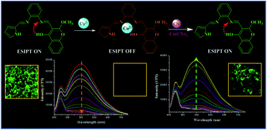
An excited state intramolecular proton transfer (ESIPT) process-based novel chromogenic and fluorogenic probe (2) was synthesized with the aim of sequential in situ detection of Cu2+ and CN− ions under aqueous and biological conditions. The probe revealed chelating affinity for transition metal cations, amidst which only Cu2+ efficiently quenches the emission intensity. Further in situ addition of CN− results in a metal displacement reaction and a turn on fluorescence response. This quencher displacement sensing strategy results in “ON–OFF–ON” type of fluorescence changes with high selectivity and great affinity in nanomolar detection of [CN−] and outlines the working principle of the IMPLICATION logic gate. The in vitro cytotoxic activity studied via the MTT assay revealed that in situ-formed Cu complexes worked as potential anticancer chemotherapeutic agents towards MCF-7 (human breast adenocarcinoma) cell lines (2 + Cu2+ IC50 = 5.69 ± 0.26 μg mL−1, 3 + Cu2+ IC50 = 7.36 ± 0.29 μg mL−1). The selective detection of Cu2+ and CN− ions in biological systems was also explained by intracellular bioimaging studies in MCF-7 cell lines.
Copper is an essential trace element in humans and is involved in many biological and environmental processes.8 Deficiency and increased levels of Cu2+ cause serious problems to human health. Its homeostasis is key to severe diseases such as Menke's, Wilson's, Parkinson's and Alzheimer's diseases. High concentration levels cause the accumulation of Cu2+ in the liver and correspondingly are responsible for idiopathic copper toxicosis syndrome.9 In addition, Cu2+ takes part actively in the pathogenesis of cardiovascular disease, gastrointestinal disorders and diabetes.10 According to the US Environmental Protection Agency (EPA), [Cu2+] less than 1.3 ppm is acceptable in drinking water. In contrast with toxic metal ions, cyanide can cause death in a few minutes, even at low amounts, by suppressing the central nervous system. It directly affects all aerobic organisms by breaking the electron transport chain in the mitochondrial membrane and prevents respiration.11 Chronic exposure to CN− leads to many diseases, including those of the cardiac, vascular, endocrine, visual and metabolic systems.12 Repeated exposure to low concentrations of cyanides over a long period causes nausea, muscle cramps, weakness, loss of appetite, paralysis of the arms and legs, and memory deficit.13 That is why it is one of the most lethal poisons known. Diverse detecting principles have been reported for cyanide recognition, using mechanisms such as nucleophilic addition of the anionic species onto activated carbonyl groups, hydrogen bonding interactions, Michael acceptor type activated CC double bond, formation of cyanohydrin derivatives and demetallation of metal chelates.14–16 Still, most of these sensory systems face interference from other anionic species, resulting in lack of selectivity and sensitivity for cyanide. This problem can effectively be avoided by utilizing copper–cyanide affinity. Use of high copper–cyanide affinity has become a valuable strategy to establish highly effective chemosensors for cyanide in aqueous media.
As the metal chelating entities were simply conjugated to the fluorophore units, the emission was not absolutely quenched after Cu2+ coordination, which is a hitch for quantitative investigations. So superior fluorophore units that bind Cu2+ directly to form strong, non-fluorescent Cu2+ complexes are desirable.17 Based on this prescription, we project and synthesize the excited state intramolecular proton transfer (ESIPT) process-based novel multifunctional chemosensors 3-((E)-((2-((E)-((1H-pyrrol-2-yl)methylene)amino)phenyl)imino)methyl)-2-methoxy-2H-chromen-4-ol (probe 2) and 3-((E)-((2-((E)-((1H-pyrrol-2-yl)methylene)amino)phenyl)imino)methyl)-2-ethoxy-2H-chromen-4-ol (probe 3) for finding both Cu2+ and CN− ion concentrations [Scheme S1†]. The structures of the ligands were confirmed by elemental analysis, IR spectroscopy, UV-vis spectroscopy and 1H NMR (Fig. S1 and S3†) and 13C NMR spectroscopy (Fig. S2 and S4†). All characterisation techniques confirmed the presence of a methoxy group in probe 2 and an ethoxy group in probe 3. This pointed towards in situ nucleophilic substitution via the solvent used. Mechanistic detail of the formation of probe 2 is shown in Fig. 1.
ESIPT has been developed as an emerging signalling mechanism; it is a rapid process with timescales ranging from fractions of picoseconds to tens of picoseconds, as the swiftness of proton transfer is far greater than that of electron transfer in the excited state.18,19 One of the greatest advantages of ESIPT is large wavelength shifts that avoid overlap between absorption and emission spectra. ESIPT-based probe 2 behaves as a dual emitter as a result of the participation of the ground state and excited state of keto–enol tautomers [Fig. 2]. Absorption originates from the ground state of the stable enol form (E), potentially stabilised by intramolecular hydrogen bonding, and emission takes place from the preferred excited state of the keto form (K*), resulting in enhanced wavelength shift. Photoexcitation at 410 nm results in rapid conversion from the excited state of the enol (E*) to the excited state of the keto (K*) tautomer by ESIPT within a picosecond. Decay from K* to K (ground state of keto tautomer) gives rise to an emission band at 550 nm. At ground state level the less stable keto form (K) reverts to the more stable enol form (E) via reverse proton transfer. Molecules in the E* state that do not undergo ESIPT generate an emission band at 475 nm.
Fig. 3 shows the absorbance and emission intensity changes of 2 (2 μM) in aqueous environment with the inclusion of Cu2+ ions. Remarkably, the fluorescence emission intensity around 550 nm was absolutely quenched when one equivalent of Cu2+ ions was added, presumably as a result of MLCT-based heavy metal ion effect, signalling gross formation of the 2–Cu complex.20 Job's plot measurements and molar ratio analysis (Fig. S6†) of absorbance at 410 nm also supported the 1:1 stoichiometry between 2 and Cu2+ in situ. Fluorescence studies of the novel synthesized probe at different [Cu2+] revealed the lower detection limit to be 5.12 × 10⁻⁷ mol L−1 as shown in Fig. S7.† The binding process of Cu2+ with probe were also investigated by optimized minimum energy structure (Density Functional Theory, DFT) calculation methods in the gas phase by applying the Gaussian 09W computational program with a B3LYP job using the basis set 6-31G (d, p) for probe and LANL2DZ for possible Cu2+ complexes respectively. The feasible Cu2+ complexes generated contained structural changes in the prospective site of probe. The bond lengths corresponding to –CN (pyrrole), –CN (benzene), –OH changed from 1.300 Å, 1.400 Å, 1.304 Å, 1.371 Å to 1.326 Å, 1.427 Å, 1.314 Å, 1.327 Å respectively. All the above bond length increases and –OH decreases are due to loss of ESIPT process upon binding with Cu2+. The energy band gap between HOMO–LUMO of the probable 2–Cu2+ complex was decreased (1.470 eV to 1.034 eV) owing to charge transfer between probe and Cu2+ ion, which also supports the red shift in the absorption spectra upon addition of Cu2+ (Fig. 4).
Thus, the response of sensor, developed in situ by adding 1.0 equiv. of Cu2+ to ligand solution, to a variety of anions (sodium salts) including CN−, F−, Cl−, Br−, I−, NO3−, PO43−, SO32−, SO42−, S2−, HPO42− was subsequently investigated. Specific fluorescence output of the probe in the presence of Cu2+ is not affected by the various counter anions with different shape and size, except CN− ion as shown in Fig. 5(ii). CN− coordinates with Cu2+ and forms an extremely stable [Cu(CN)x]n− complex having a lower solubility product constant (K = 1.27 × 10⁻²⁷). Addition of CN− ion concomitantly enhances the fluorescence intensity (λem = 550) as a function of time and almost restores the original state of the probe, as shown in Fig. 6. Titration experiment at various [CN]− revealed these ESIPT-based dual fluorescence sensors are capable of nanomolar detection of CN− in aqueous and biological systems (Fig. S8†).
Fig. 5 Selectivity studies of probe 2 (2 μM). Probe 2 in aqueous solution (10 mM PBS buffer, 1.0% DMSO) with tested metal ions (i) and in situ formed 2 + Cu2+ with tested anions (ii). |
As the quencher displacement strategy causes consequent changes in emission intensity at 550 nm, the resultant system works as a molecular switch at this fluorescence intensity, and can be used to perform Boolean logic operations. Molecular logic function was performed with the synthesized probe 2 along with Cu2+ (In-1) as well as CN− (In-2) as inputs in the emission mode. In the existing system, the strong fluorescence at 550 nm is assigned as the ON state (output = 1) while the weak fluorescence is defined as the OFF state (output = 0). The threshold value of fluorescence intensity is specified as 15000 CPS. Addition of Cu2+ to the probe 2 solution leads to fluorescence quenching to below the threshold level, whereas in situ addition of excess [CN−] almost restores the fluorescence intensity. The strong fluorescence intensity above the threshold label is recognized in the absence (0, 0) and presence (1, 1) of both inputs and also CN− alone. These results simply establish the IMPLICATION logic gate, “→”, i.e., “p implies q” or “if p then q”, so the OFF state or 0 output is obtained only when the input is copper acetate. The working principle of the IMPLICATION logic gate is outlined in Fig. 7.
Assessment of cytotoxicity of probes 2, 3, 2 + Cu2+ and 3 + Cu2+ complexes on human cell lines MCF-7 (breast cancer cell line), Hep G2 (human liver cancer cell line), HeLa (immortal cell line) and HEK-293 (Human Embryonic Kidney 293) was done via MTT assay. The cell lines were exposed for 24 h to medium containing the tested probes and complexes at 3 μg mL−1. Screening for cytotoxicity showed that compounds have remarkable activity towards the MCF-7 cell line. In vitro anticancerous activity of the tested probes and respective complexes towards the MCF-7 cell line as well as HEK-293 cell lines (control experiment) was studied at various concentrations (0–60 μg mL−1). In vitro anticancerous activity was calculated in terms of cell viability (%) via the following formula:
Cell viability (%) = [(mean OD of treated cells × 100)/mean OD of vehicle-treated cells] |
A concomitant decrease in cell viability (%) was observed on subsequent increments of concentrations of the tested probes and their respective complexes. At higher concentrations, penetration of the tested probes and complexes inside the cells is increased, leading to more potent anticancerous activity.
Evaluation of the cytotoxicity of the tested compounds towards the MCF-7 cell line demonstrated that both the Cu2+ complexes exhibit potent cytotoxicity as compared with their respective parent probes. The 2 + Cu2+ complex showed best potency against the MCF-7 cell line with lowest IC50 value (IC50 = 5.69 ± 0.26 μg mL−1) while the 3 + Cu2+ complex also displayed very pronounced potency (IC50 = 7.36 ± 0.29 μg mL−1). Probe 2 (IC50 = 35.48 ± 0.34 μg mL−1) and probe 3 (IC50 = 48.99 ± 0.30 μg mL−1) also exhibited moderate cytotoxicity against the MCF-7 cell line. However, the results clearly indicate a noncytotoxic effect on the HEK-293 cell line (normal cell line). The morphological changes discovered in the MCF-7 and HEK-293 cell lines exposed to the tested probes and complexes (30 μg mL−1) for 24 h are presented in Fig. 8 and S9,† respectively. The MCF-7 cell line exhibited reduction of morphology and cell adhesion ability after this exposure.
To establish the sensing ability of 2 towards Cu2+ and CN− ions in living cells, bioimaging studies with the MCF-7 cell line were performed. When the cells preloaded with probe 2 (1 μM) were treated with equimolar cupric acetate, an immediate decrease in fluorescence intensity was observed inside the cells. When these cells were treated with various concentrations of NaCN (1–100 μM), fluorescence was marginally visible after 20 min of incubation prior to the NaCN treatment. The time dependent and concentration dependent increase of fluorescence intensity observed inside the cells apparently explains the chemodosimetric sensing of Cu2+ and CN− ions, as shown in Fig. 9.
Footnote |
† Electronic supplementary information (ESI) available. See DOI: 10.1039/c6ra22352d |
This journal is © The Royal Society of Chemistry 2016 |