Effect of the substitution position (2, 3 or 8) on the spectroscopic and photophysical properties of BODIPY dyes with a phenyl, styryl or phenylethynyl group

Angel Orte*a, Elke Debroyeb, Maria J. Ruedas-Ramaa, Emilio Garcia-Fernandeza, David Robinsonc, Luis Crovettoa, Eva M. Talaveraa, Jose M. Alvarez-Peza, Volker Leenb, Bram Verbelenb, Lucas Cunha Dias de Rezendebd, Wim Dehaenb, Johan Hofkensb, Mark Van der Auweraerb and Noël Boens*b
aDepartment of Physical Chemistry, Faculty of Pharmacy, University of Granada, Cartuja Campus, 18071 Granada, Spain. E-mail: angelort@ugr.es; Tel: +34-958243825
bDepartment of Chemistry, KU Leuven (Katholieke Universiteit Leuven), Celestijnenlaan 200f, 3001 Leuven, Belgium. E-mail: Noel.Boens@kuleuven.be
cSchool of Chemistry, University of Nottingham, University Park, Nottingham, NG7 2RD, UK
dFaculdade de Ciências Farmacêuticas de Ribeirão Preto, Universidade de São Paulo, Av. Do Café s/n, Ribeirão Preto, SP 14040-903, Brazil

Received 6th September 2016 , Accepted 21st October 2016

First published on 21st October 2016


Abstract

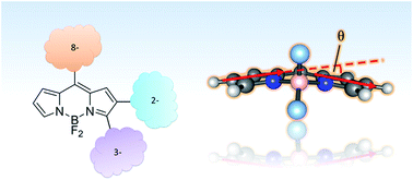
A very active branch of organic chemistry is putting great effort into tailoring fluorescent dyes for a myriad of applications, from technological to bioanalytical and biomedical applications. Among the major families of fluorophores, those derived from 4,4-difluoro-4-bora-3a,4a-diaza-s-indacene (BODIPY dyes) are undergoing a recent boost thanks to the simplicity and robustness of the chemistry involved. The BODIPY core can be modified with numerous side groups, the 8-position being a modification place with important effects on the spectroscopic and photophysical properties of the resulting dyes. Likewise, previous work has shown that the addition of groups attached at the 3- and 2-positions can result in dyes with very different properties. Herein, we generalize the effect of the substituent side groups by studying nine BODIPY dyes substituted with a phenyl, styryl or phenylethynyl moiety at the 2-, 3- or 8-position of the BODIPY scaffold. Within the class of phenyl- or phenylethynyl-substituted dyes, substitution at the 2-position always leads to dyes with the broadest bandwidths and the largest Stokes shifts. We investigate the solvent effect on the spectroscopic properties of the dyes, using four empirical solvent scales (dipolarity, polarizability, acidity and basicity: Catalán, J. Phys. Chem., 2009, 113, 5951). These analyses identify solvent dipolarity and polarizability as critical parameters accounting for the observed solvent-dependent shifts of the absorption and emission maxima. Finally, time-dependent density functional theory calculations provide insights into the structural and energetic issues concerning the spectroscopic properties of these fluorophores.


Introduction

The family of 4,4-difluoro-4-bora-3a,4a-diaza-s-indacene derivatives, better known as BODIPY (boron dipyrromethene or boron dipyrrin,1–3 Chart 1) dyes, entail a rapidly growing class of compounds, characterized by extremely favorable fluorescent features, such as tunable excitation/emission wavelengths in the visible spectral range, narrow spectral bandwidths and good (photo)chemical stability. Likewise, relatively high molar absorption coefficient, ε(λ), and fluorescence quantum yield, Φ, values lead to a high brightness of these dyes.4 A very active branch of scientific development is putting great effort in fine-tuning the BODIPY properties by introducing suitable chemical groups onto the central core. As a result, the BODIPY family is expanding constantly and has found applications in wide-ranging fields, from technological (organic light-emitting diodes,5–7 dye-sensitized solar cells8,9) to analytical and in vivo imaging applications.10,11
image file: c6ra22340k-c1.tif
Chart 1 Representation of the “BODIPY core” and its IUPAC numbering system. Common BODIPYs have two fluorine atoms bound to the boron atom (4-position).

Recent advances in organic synthesis have expanded the possibilities of controlled modification of the BODIPY moiety, including substitution by tailored groups at every position of the core.12 The direct conjugation of fused π-systems, extended π-bonds, the addition of alkyl or aryl groups, or the insertion of electron withdrawing or donating substituents have different effects on the spectroscopic and photophysical properties of the resulting dyes [fluorescence quantum yield (Φ) and lifetime (τ), absorption and emission energies (λabs and λem), Stokes shift (Δ[small nu, Greek, macron]), full width at half maximum of the absorption and emission band (fwhmabs and fwhmem), etc.]. For instance, fusing rigid π-conjugated carbocycles to the BODIPY core results in near-infrared emitting dyes.13–16

Modification at the 8- (or meso-) position (Chart 1) results in a variety of substituent-promoted effects, as quantum chemical calculations have demonstrated the presence of a node in the HOMO at this position and a significant increase of the electron density upon excitation.17 Alkylation at the meso-position usually has a mild effect, resulting in boron dipyrromethenes with emission maxima around 500 nm and large fluorescence quantum yield values.17,18 Nevertheless, we recently found that a tert-butyl group at the 8-position causes an important decrease in the fluorescence quantum yield Φ and lifetime τ of the resulting dye, as well as a significant red shift of the fluorescence and, consequently, a striking 1560 cm−1 Stokes shift Δ[small nu, Greek, macron], much larger than those of other meso-alkylated analogues.19 Boron dipyrromethenes, modified at the meso-position with strong electron withdrawing groups, possess large red shifts,20,21 because the LUMO is highly stabilized3 compared to unsubstituted BODIPY.3,22–24 In contrast, electron donating groups at the meso-position cause a blue-shift in the absorption and emission spectra, while keeping high fluorescence quantum yields and lifetimes.25–28 Certain modifications at the meso-position may result in almost nonfluorescent compounds, as for meso-alkenyl-29 or meso-formylBODIPYs.30 Likewise, a meso-phenylBODIPY dye is also weakly fluorescent.31,32 However, meso-phenylBODIPYs can display bright fluorescence, depending on the dihedral angle and steric hindrance of the rotation of the phenyl group with respect to the BODIPY core plane. For instance, while phenyl or p-tert-butylphenyl substituents at the meso-position cause low Φ and τ values, o-tolyl- or mesityl groups result in boron dipyrromethene dyes with high Φ (>0.90).32 The enhanced nonradiative deactivation in 8-phenyl- or p-tert-butylphenyl-substituted BODIPYs is attributed to the population of a distorted conformation of the BODIPY framework in the excited state, leading to loss of planarity.32 Another example of the effect of the dihedral angle of the meso-phenyl substituent on the fluorescence properties of the dyes can be found in the comparison between meso-phenyl-3,5-dimethylBODIPY,33 meso-p-methylphenyl-3,5-dimethylBODIPY and meso-p-methylphenyl-1,3,5,7-tetramethylBODIPY.34 While the 3,5-dimethyl substitution gives rise to moderate Φ values (0.17–0.42 for meso-phenyl- and 0.11–0.29 for meso-p-methylphenyl-substituted boron dipyrromethenes, depending on the solvent), the addition of two methyl groups at the positions 1- and 7- causes an increase in the fluorescence, with Φ up to the range 0.46–0.72.34

Although substituents at the 8-position have normally the largest effect on the photophysical properties of BODIPY derivatives, modifications at the 3- and 2-positions also give rise to dyes with diverse properties. Extending the conjugation with double bonds at the 3-position yields fluorophores with a high brightness. However, when the conjugation of the BODIPY framework is extended at the 2-position, dyes with a large Stokes shift Δ[small nu, Greek, macron], but lower Φ value, are obtained. In a preliminary study, our group described this effect for styryl and triazolyl substituents introduced at either the 3- or the 2-position.35

In this work, we generalize the effect of the substituent at the 2-, 3- or 8-position of the BODIPY core. Hence, we investigate nine 4,4-difluoro-4-bora-3a,4a-diaza-s-indacene dyes substituted with a phenylethynyl or styryl moiety at the 2, 3 or 8-position (2-Ethyn, 3-Ethyn, 8-Ethyn, 2-Styryl, 3-Styryl and 8-Styryl), a p-tert-butylphenyl group at the 2- or 3-position (2-Ph and 3-Ph) and phenyl at the 8-position (8-Ph) (Chart 2). The full characterization of the 8-phenylethynyl derivative (8-Ethyn) has been reported previously27 and has been included for discussion purposes. The spectroscopic and photophysical characteristics of these compounds have been investigated in a series of solvents by UV-vis spectrophotometry and steady-state and time-resolved fluorescence spectroscopy. These experiments allow us to determine the relevant spectroscopic and photophysical properties: spectral shape and maxima [λabs(max) and λem(max)], Stokes shifts (Δ[small nu, Greek, macron]), bandwidths (fwhmabs and fwhmem), fluorescence quantum yields (Φ), fluorescence lifetimes (τ) and rate constants of fluorescence (kf) and nonradiative (knr) deactivation processes. We investigate the solvent effect on the spectroscopic properties of the dyes, according to a generalized procedure based on a set of four empirical solvent scales: dipolarity, polarizability, acidity and basicity of the medium.36 Time-dependent density functional theory (TD-DFT) calculations provide insights into the structural and energetic properties concerning the spectroscopic parameters of the dyes investigated.


image file: c6ra22340k-c2.tif
Chart 2 Chemical structures of the BODIPY derivatives studied in this work.

Results and discussion

Synthesis reactions

Nine BODIPY derivatives (Chart 2) having three types of substituents, at either the 2-, 3- or 8-position, have been prepared for this study: phenyl derivatives (2-Ph, 3-Ph and 8-Ph), phenylethynyl derivatives (2-Ethyn, 3-Ethyn and 8-Ethyn) and styryl derivatives (2-Styryl, 3-Styryl and 8-Styryl). The synthesis of 2-Ph, 3-Ph, 2-Ethyn and 3-Ethyn was carried out by following our previously reported procedure,37 whereas the synthesis of 8-Ph was performed as described by Kee et al.32 Also 8-Ethyn26 and 3-Styryl35 were synthesized prepared according to our protocols. Novel synthetic procedures for BODIPY derivatives 2-Styryl and 8-Styryl are described next.

2-Styryl [4,4-difluoro-5,7,8-trimethyl-2-(E)-styryl-4-bora-3a,4a-diaza-s-indacene] was prepared as follows (Scheme 1): to a stirred solution of BODIPY 1 (117 mg, 0.33 mmol, prepared as in ref. 37) in toluene (5 mL) at room temperature, (E)-styrylboronic acid (64 mg, 0.43 mmol) and tetrakis(triphenylphosphine) palladium (4 mg, 3.4 μmol) were added, followed by the addition of 1.5 mL of Na2CO3 1 M (aq). The solution was heated to reflux and after 3 h the reaction mixture was poured in Et2O (300 mL) and dried over magnesium sulfate. Compound 2-Styryl (24 mg, 0.072 mmol, 22% yield) was obtained as a dark purple solid powder after purification by silica gel column chromatography [petroleum ether/dichloromethane, 2[thin space (1/6-em)]:[thin space (1/6-em)]1–1[thin space (1/6-em)]:[thin space (1/6-em)]1 (v/v)]. Mp 219 °C. NMR spectra (Fig. S1 and S2 in the ESI): 1H NMR (600 MHz, CDCl3) δ 7.77 (s, 1H), 7.45 (d, J = 7.4 Hz, 2H), 7.34 (t, J = 7.4 Hz, 2H), 7.23 (t, J = 7.4 Hz, 1H), 7.16 (s, 1H), 6.98 (d, J = 16.3 Hz, 1H), 6.91 (d, J = 16.3 Hz, 1H), 6.17 (s, 1H), 2.58 (s, 3H), 2.57 (s, 3H), 2.43 (s, 3H) ppm. 13C NMR (75 MHz, CDCl3) δ 160.5, 145.6, 141.5, 137.7, 137.1, 134.9, 134.6, 129.5, 128.8, 127.6, 127.4, 126.2, 123.1, 120.6, 119.4, 77.2, 17.1, 16.6, 15.1 ppm. LRMS (EI): 336, 316 (M − F).


image file: c6ra22340k-s1.tif
Scheme 1 Schematic representation of the synthesis of 2-Styryl.

8-Styryl [4,4-difluoro-8-(E)-styryl-4-bora-3a,4a-diaza-s-indacene] was prepared as follows (Scheme 2): to a solution of BODIPY 2 (310 mg, 1.5 mmol, prepared as in ref. 38) in toluene (15 mL) under continuous stirring at 110 °C, benzaldehyde 3 (200 μL, 1.97 mmol), acetic acid (1.2 mL, 20.9 mmol) and piperidine (1.0 mL, 10.1 mmol) were added. Instantaneous formation of a red-colored compound was observed. After 5 min, the solution was cooled to room temperature and the solvent was evaporated. After silica column purification [petroleum ether/dichloromethane, 1[thin space (1/6-em)]:[thin space (1/6-em)]9 (v/v)], 8-Styryl (127 mg, 0.43 mmol, 29% yield) was obtained as a dark solid. Mp 140 °C. NMR spectra (Fig. S3 and S4): 1H NMR (600 MHz, CDCl3) δ 7.89 (s, 2H), 7.62 (d, J = 6.6 Hz, 2H), 7.52 (d, J = 15.9 Hz, 1H), 7.48–7.42 (m, 4H), 7.37 (s, 2H), 6.56 (s, 2H) ppm. 13C NMR (75 MHz, CDCl3) δ 144.71, 143.75, 143.02, 135.53, 133.83, 130.54, 129.17, 128.28, 127.98, 121.13, 117.89 ppm. HRMS (EI) calculated for C17H13BF2N2 294.1140, found 294.1143. LRMS (EI): 294, 273 [M − F].


image file: c6ra22340k-s2.tif
Scheme 2 Schematic representation of the synthesis of 8-Styryl.

All BODIPY dyes synthesized for this study are solid powders with a strongly colored metallic shine and form intensely colored solutions. When irradiated in solution, a generally bright fluorescence can be observed. In the following sections, we describe the spectroscopic and photophysical features of these dyes, including solvent effects.

Spectroscopic and photophysical properties

The UV-vis absorption and fluorescence emission spectra of the nine compounds dissolved in 18 solvents (see Table S1 in the ESI) have been measured. The dyes exhibit typical BODIPY-like absorption spectra with a narrow band due to the S1 ← S0 transition and a shoulder at lower wavelengths. A blue-shifted, weak S2 ← S0 transition band is also detectable in some of the spectra.

Among the phenyl-substituted BODIPY dyes (2-Ph, 3-Ph and 8-Ph), some differences in absorption behavior are observed. The presence of the p-tert-butylphenyl moiety shifts the absorption maximum λabs(max) from 516–530 nm when this group is at the 2-position (2-Ph) to 513–527 nm when it is at the 3-position (3-Ph). In both cases, λabs(max) is more red-shifted in the more polarizable solvents toluene and chlorobenzene. In contrast, the values of λabs(max) of 8-Ph (with a phenyl substituent at the meso-position) are blue-shifted in relation to those of 2-Ph and 3-Ph and are close to those of unsubstituted boron dipyrromethene,3,22–24 ranging from 489 nm in acetonitrile to 497 nm in toluene and chlorobenzene: the solvent dependence correlating with the refractive index. This absorption energy range is in good agreement with that of other BODIPYs substituted at the meso-position with a weak electron acceptor or donor.33,39 One should also note that the absorption spectrum of 8-Ph is similar to BODIPY dyes substituted by a mesityl or o-tolyl moiety in 8-position. This is due to the large dihedral angle (60°) between the BODIPY and phenyl moiety found for 8-Ph by X-ray diffraction,32 which reduces the delocalization of the electron cloud of the BODIPY moiety over the phenyl group.27

For the phenylethynylBODIPYs (2-Ethyn, 3-Ethyn and 8-Ethyn), the absorption spectra exhibit also the characteristic features of typical BODIPYs. 2-Ethyn possesses an absorption maximum between 503 and 525 nm, whereas the λabs(max) of 3-Ethyn varies between 525 and 545 nm (Fig. 1A). For both dyes, the lowest λabs(max) value is found in acetonitrile and the largest in cyclohexane. Remarkably, the effect of the substitution position (2 vs. 3) on λabs(max) is much larger for the phenylethynyl moiety than for the phenyl group and, moreover, is opposite: moving the p-tert-butylphenyl substituent from the 2- (2-Ph) to the 3-position (3-Ph) causes a 3 nm blue shift of λabs(max), whereas the analogous change from 2-Ethyn to 3-Ethyn results in a ca. 20 nm red shift of λabs(max). The previously reported 8-Ethyn27 is bathochromically shifted with respect to 2-Ethyn and 3-Ethyn, with λabs(max) ranging from 537 to 547 mm (see Table S2) parallel with an increasing refractive index. As the HOMO of the BODIPY core has a node in the 8-position, these data suggest that the 8-phenylethynyl moiety acts as an electron acceptor stabilizing the LUMO. This rationale also helps to account for the difference between 2-Ethyn and 3-Ethyn. The resonance forms of BODIPY suggest that, as in the 8-position, the coefficient of the HOMO in the 3-position is smaller than in the 2-position, the opposite being the case for those of the LUMO. Hence if the phenylethynyl moiety stabilizes the LUMO rather than destabilizing the HOMO, its effect and hence the red shift vs. unsubstituted BODIPY, will be most outspoken for 3-Ethyn and 8-Ethyn. Strikingly, the λabs(max) values of the 8-Ph are blue shifted with respect to those of 3-Ph and 2-Ph.


image file: c6ra22340k-f1.tif
Fig. 1 (A) Normalized absorption spectra of 3-Ethyn in a selection of solvents. (B) Corresponding normalized fluorescence emission spectra upon excitation at 488 nm.

Styryl-substituted BODIPYs (2-Styryl, 3-Styryl and 8-Styryl) exhibit different absorption properties. 3-Styryl displays a main, narrow S1 ← S0 transition band (Fig. 2A) similar to other 3-substituted boron dipyrromethenes.35,37 The λabs(max) values range from 549 to 561 nm, with a typical red shift from acetonitrile to chlorobenzene. The extended conjugation provided by the styryl functional group causes an extra bathochromic shift of around 20 nm with respect to 3-Ethyn, and ca. 30 nm with respect to 3-Ph. In contrast, 2-Styryl and 8-Styryl exhibit a clear dual-band absorption and emission behavior (Fig. S5). The relative abundance of each band varies with the solvent, and the absorption and the excitation spectra do not match. Multi-exponential fluorescence decay traces were also found for both dyes. Likewise, both BODIPY dyes undergo changes in the relative absorption of each band with time (Fig. S6) and completely discolor after 180 min in solvents such as THF and cyclohexanone, even when the solution is kept in the dark. This effect is especially observed for 2-Styryl, but can also be detected for 8-Styryl. This is indicative of (photo-)instability of the dyes. Because of this (photo-)instability, no further experiments were performed with 2-Styryl and 8-Styryl.


image file: c6ra22340k-f2.tif
Fig. 2 (A) Normalized absorption spectra of 3-Styryl in a selection of solvents. (B) Corresponding normalized fluorescence emission spectra upon excitation at 510 nm.

The most interesting spectroscopic features are related to the fluorescence emission of the BODIPY derivatives. Herein, we compare the maxima of the emission spectra [λem(max)], Stokes shifts (Δ[small nu, Greek, macron]), fluorescence quantum yields (Φ) and lifetimes (τ), and other fluorescence-related properties of the dyes studied.

A comparison of the phenyl-substituted BODIPY dyes (2-Ph, 3-Ph and 8-Ph) already highlights very remarkable differences (Tables 1 and 2). 3-Ph and 8-Ph display the characteristic emission features of boron dipyrromethenes: (a) a mirror image-shaped, narrow emission band (fwhmem averaged over all the solvents tested equals 1060 ± 50 cm−1 and 1390 ± 110 cm−1 for 3-Ph and 8-Ph, respectively); (b) a small Stokes shift Δ[small nu, Greek, macron] [in the 859–1111 cm−1 range for 3-Ph (average Δ[small nu, Greek, macron] = 990 ± 67 cm−1) and in the 633–856 cm−1 range for 8-Ph (average Δ[small nu, Greek, macron] = 726 ± 62 cm−1)]; (c) emission maxima λem(max) that are red-shifted in the more polarizable solvents [λem(max) moves from 544 nm in acetonitrile to 554 nm in chlorobenzene and toluene for 3-Ph, whereas for 8-Ph λem(max) varies from 507 nm in methanol to 517 nm in toluene and 518 nm in cyclohexane]. The fwhmem results are intermediate between those of 8-methylBOBIPY (fwhmem around 950 cm−1)19 and those of meso-halogenated BODIPYs (fwhmem around 1400 cm−1), as is the trend in the emission maxima λem(max) with the different solvents. However, the Stokes shifts Δ[small nu, Greek, macron] are larger than those reported for meso-alkylated and meso-halogenated BODIPY dyes, which are in the 400 cm−1 range.17,33,40 For instance, meso-p-methylphenyl-3,5-dimethylBODIPY and meso-p-methylphenyl-1,3,5,7-tetramethylBODIPYs exhibit Δ[small nu, Greek, macron] values between 513 and 627 cm−1,34 and between 447 and 560 cm−1 for meso-phenyl-3,5-dimethylBODIPY.33 Concerning the solvent dependence of these spectroscopic parameters, one can observe that while for 3-Ph Δ[small nu, Greek, macron], fwhmabs and fwhmem increase more or less parallel with the dielectric constant ε of the solvent, this relation is quite erratic for Δ[small nu, Greek, macron] and fwhmem of 8-Ph.

Table 1 Spectroscopic and photophysical data of 3-Ph and 2-Ph as a function of solvent. The solvents are numbered according to increasing refractive index n as in Table S1
Product   Solvent λabs(max) [nm] λem(max) [nm] Δ[small nu, Greek, macron] [cm−1] fwhmabs [cm−1] fwhmem [cm−1] Φa τb [ns] kfc [108 s−1] knrc [108 s−1]
a Fluorescence quantum yield ± one standard uncertainty. Φ determined vs. rhodamine 6G in water (Φr = 0.76) as a reference.b Fluorescence lifetimes obtained with λex = 488 nm, and λem (3-Ph) = 560, 565, 570 nm or λem (2-Ph) = 560, 570, 580 nm. The standard errors are obtained from the diagonal elements of the covariance matrix available from the global analysis fit of decay traces recorded at three emission wavelengths λem and are between 6 and 10 ps for 3-Ph and between 7 and 17 ps for 2-Ph.c The propagated errors are calculated using the uncertainty (standard deviation) of Φ and the standard error of τ.d EtOAc = ethyl acetate, PrCN = butanenitrile, Bu2O = dibutyl ether, THF = tetrahydrofuran, c-C6H12 = cyclohexane, c-C6H10O = cyclohexanone, PhCl = chlorobenzene.
3-Ph 1 CH3OH 517 547 1061 1680 1110 0.81 ± 0.01 5.13 1.58 ± 0.02 0.37 ± 0.02
2 CH3CN 513 544 1111 1700 1170 0.895 ± 0.001 5.08 1.76 ± 0.01 0.21 ± 0.01
3 (C2H5)2O 522 549 942 1530 1040 1.00 ± 0.02 5.11 1.96 ± 0.04 0.00 ± 0.04
4 Acetone 518 547 1023 1630 1110 0.895 ± 0.005 5.09 1.76 ± 0.01 0.21 ± 0.01
5 EtOAcd 519 547 986 1630 1090 0.86 ± 0.02 4.89 1.76 ± 0.04 0.29 ± 0.04
6 2-Propanol 520 550 1049 1620 1080 0.91 ± 0.01 4.96 1.84 ± 0.02 0.18 ± 0.02
7 PrCNd 518 548 1057 1670 1090 0.99 ± 0.03 4.90 2.02 ± 0.06 0.02 ± 0.06
8 Bu2Od 525 551 899 1520 1010 0.96 ± 0.08 4.84 1.98 ± 0.17 0.08 ± 0.17
9 THFd 522 551 1008 1570 1060 0.877 ± 0.009 4.71 1.86 ± 0.02 0.26 ± 0.02
10 1-Pentanol 523 552 1005 1600 1040 0.899 ± 0.007 4.81 1.87 ± 0.02 0.21 ± 0.02
11 1,4-Dioxane 521 550 1012 1650 1060 1.00 ± 0.05 4.65 2.15 ± 0.11 0.00 ± 0.11
12 CH2Cl2 521 551 1045 1660 1100 0.87 ± 0.01 4.99 1.74 ± 0.02 0.26 ± 0.02
13 c-C6H12d 527 552 859 1470 960 0.99 ± 0.07 4.68 2.12 ± 0.15 0.02 ± 0.15
14 1-Octanol 525 553 964 1550 1020 0.92 ± 0.04 4.76 1.93 ± 0.08 0.17 ± 0.08
15 CHCl3 524 553 1001 1630 1050 0.94 ± 0.09 4.99 1.88 ± 0.18 0.12 ± 0.18
16 c-C6H10Od 522 551 1008 1640 1070 0.99 ± 0.06 4.54 2.18 ± 0.13 0.02 ± 0.13
17 Toluene 527 554 925 1580 1000 0.97 ± 0.02 4.39 2.21 ± 0.05 0.07 ± 0.05
18 PhCld 527 554 925 1610 1030 0.94 ± 0.09 4.36 2.16 ± 0.21 0.14 ± 0.21
2-Ph 1 CH3OH 519 582 2086 2300 2320 0.359 ± 0.005 4.23 0.85 ± 0.01 1.52 ± 0.01
2 CH3CN 516 581 2168 2260 2440 0.442 ± 0.005 4.89 0.90 ± 0.01 1.14 ± 0.01
3 (C2H5)2O 525 573 1596 2190 1960 0.58 ± 0.02 5.98 0.97 ± 0.03 0.70 ± 0.03
4 Acetone 519 583 2115 2270 2310 0.47 ± 0.01 5.11 0.92 ± 0.02 1.04 ± 0.02
5 EtOAcd 521 576 1833 2250 2110 0.56 ± 0.01 5.47 1.02 ± 0.02 0.80 ± 0.02
6 2-Propanol 522 581 1945 2240 2160 0.61 ± 0.02 5.08 1.20 ± 0.04 0.77 ± 0.04
7 PrCNd 520 582 2049 2230 2280 0.36 ± 0.02 5.18 0.69 ± 0.04 1.24 ± 0.04
8 Bu2Od 527 571 1462 2160 1770 0.58 ± 0.06 5.87 1.0 ± 0.1 0.7 ± 0.1
9 THFd 525 581 1836 2190 2090 0.571 ± 0.004 5.34 1.07 ± 0.01 0.80 ± 0.01
10 1-Pentanol 525 580 1806 2180 2050 0.61 ± 0.01 5.24 1.16 ± 0.02 0.74 ± 0.02
11 1,4-Dioxane 524 575 1693 2140 1990 0.57 ± 0.02 5.76 0.99 ± 0.04 0.75 ± 0.04
12 CH2Cl2 523 581 1909 2160 2150 0.57 ± 0.02 5.53 1.03 ± 0.04 0.78 ± 0.04
13 c-C6H12d 528 566 1272 2180 1500 0.72 ± 0.05 5.80 1.24 ± 0.09 0.48 ± 0.09
14 1-Octanol 527 579 1704 2200 2030 0.50 ± 0.02 5.56 0.90 ± 0.04 0.90 ± 0.04
15 CHCl3 526 582 1829 2140 2000 0.52 ± 0.04 5.75 0.90 ± 0.07 0.83 ± 0.07
16 c-C6H10Od 523 583 1968 2240 2180 0.43 ± 0.02 5.19 0.83 ± 0.04 1.10 ± 0.04
17 Toluene 530 575 1477 2110 1800 0.63 ± 0.02 5.50 1.15 ± 0.04 0.67 ± 0.04
18 PhCld 529 582 1721 2150 1960 0.53 ± 0.06 5.40 1.0 ± 0.1 0.9 ± 0.1


Table 2 Spectroscopic and photophysical data of 8-Ph as a function of solvent. The solvents are numbered according to increasing refractive index n
Product   Solvent λabs(max) [nm] λem(max) [nm] Δ[small nu, Greek, macron] [cm−1] fwhmabs [cm−1] fwhmem [cm−1] Φa τb [ps] kfc [108 s−1] knrc [108 s−1]
a Fluorescence quantum yield ± one standard uncertainty. Φ determined vs. rhodamine 6G in water (Φr = 0.76) as a reference.b The standard error on the fluorescence lifetime is 1 ps [obtained from the diagonal elements of the covariance matrix available from the global analysis fit of decay traces recorded at three emission wavelengths λem (515, 520, 525 nm)]. λex = 488 nm.c The propagated errors are calculated using the uncertainty (standard deviation) of Φ and the standard error of τ.d See Table 1.
8-Ph 1 CH3OH 490 507 684 1180 1340 0.044 ± 0.004 189 2.3 ± 0.2 50.6 ± 0.4
2 CH3CN 489 510 842 1320 1420 0.041 ± 0.004 181 2.3 ± 0.2 53.0 ± 0.4
3 (C2H5)2O 492 509 679 1000 1240 0.050 ± 0.006 235 2.1 ± 0.3 40.4 ± 0.3
4 Acetone 491 509 720 1180 1270 0.045 ± 0.006 180 2.5 ± 0.3 53.1 ± 0.5
5 EtOAcd 491 509 720 1090 1460 0.056 ± 0.004 220 2.5 ± 0.2 42.9 ± 0.3
6 2-Propanol 492 511 756 1040 1340 0.082 ± 0.004 284 2.9 ± 0.1 32.3 ± 0.2
7 PrCNd 491 511 797 1180 1460 0.06 ± 0.02 240 2.5 ± 0.8 39.2 ± 0.9
8 Bu2Od 494 512 712 950 1440 0.074 ± 0.004 301 2.5 ± 0.1 30.8 ± 0.2
9 THFd 493 511 715 1080 1250 0.065 ± 0.007 250 2.6 ± 0.3 37.4 ± 0.3
10 1-Pentanol 494 511 673 1040 1430 0.12 ± 0.01 375 3.2 ± 0.3 23.5 ± 0.3
11 1,4-Dioxane 494 511 673 1040 1360 0.12 ± 0.01 342 3.5 ± 0.3 25.7 ± 0.3
12 CH2Cl2 493 512 753 1260 1230 0.07 ± 0.01 329 2.1 ± 0.3 28.3 ± 0.3
13 c-C6H12d 496 518 856 860 1570 0.12 ± 0.02 274 4.4 ± 0.7 32.1 ± 0.7
14 1-Octanol 495 512 671 990 1390 0.24 ± 0.03 430 5.6 ± 0.7 17.7 ± 0.7
15 CHCl3 495 512 671 1070 1340 0.10 ± 0.01 456 2.2 ± 0.2 19.7 ± 0.2
16 c-C6H10Od 495 511 633 1030 1390 0.07 ± 0.01 259 2.7 ± 0.4 35.9 ± 0.4
17 Toluene 497 517 778 980 1570 0.096 ± 0.007 434 2.2 ± 0.2 20.8 ± 0.2
18 PhCld 497 516 741 1070 1530 0.10 ± 0.01 473 2.1 ± 0.2 19.0 ± 0.2


In order to better understand the differences between different of BODIPYs, we also analyzed the relation between Δ[small nu, Greek, macron] and fwhmem or fwhmabs more quantitatively. If only low frequency molecular vibrations (vib < kT) contribute to Δ[small nu, Greek, macron] and fwhmem or fwhmabs, the relation between both properties is given by eqn (1):41–43

 
(fwhm)2 = 16(ln[thin space (1/6-em)]2)ERkT (1)
where ER is the excess energy with which a vertical transition from the minimum of the ground state reaches the excited state potential energy surface (PES) upon absorption of a photon (and analogously for emission). Hence the Stokes shift Δ[small nu, Greek, macron] is 2ER. For T = 290 K, eqn (1) can be expressed as: Δ[small nu, Greek, macron] = 8.9514 × 10⁻⁴ (fwhm)2 (in cm−1). For 3-Ph and 8-Ph the average fwhmem is respectively 1060 ± 50 cm−1 and 1390 ± 110 cm−1, which yields a respective average Stokes shift Δ[small nu, Greek, macron] of 1010 and 1730 cm−1 using eqn (1). For 3-Ph this matches the average experimental Δ[small nu, Greek, macron] value of 990 cm−1. However, for 8-Ph the average Δ[small nu, Greek, macron] value calculated according to eqn (1) is more than twice as large as the experimental value, which is only 726 cm−1. When instead of the average values of fwhmem the average values of fwhmabs are used, one finds 2320 cm−1 and 1030 cm−1 for the average calculated Δ[small nu, Greek, macron] of 3-Ph and 8-Ph, respectively. Now the average value of the experimental Δ[small nu, Greek, macron] of 3-Ph is overestimated by more than a factor of 2, whereas the agreement with that of 8-Ph is marginal. While for 3-Ph the fwhmabs is significantly broader than fwhmem (1610 vs. 1060 cm−1), the reverse trend is observed for 8-Ph (1080 vs. 1390 cm−1). This would suggest that for 3-Ph the PES is steeper in the excited state than in the ground state, while for 8-Ph the opposite is suggested. The steep PES for the ground state could be due to steric hindrance. In contrast to what was observed earlier for molecules with a strongly dipolar excited state,43 the values of the Stokes shift estimated from the fwhmabs and fwhmem are overestimated and the deviation becomes stronger for broader bands. Especially when the bands of the BODIPY derivatives get broader, fwhmabs and fwhmem are no longer uniquely determined by low frequency (torsional) vibrations but also envelope the 0–1 high frequency vibronic band. However, the absorption and emission maxima whose position determines the Stokes shift always corresponds with the 0–0 vibronic band of the high frequency vibration for the compounds studied here. The emission maximum of 2-Ph varies with the solvent, ranging from 566 nm in cyclohexane to 583 nm in cyclohexanone and acetone. The spectral emission features of 2-Ph differ significantly from those of 3-Ph and 8-Ph. 2-Ph exhibits a much broader emission band (average fwhmem = 2050 ± 240 cm−1) than 3-Ph and 8-Ph do, as well as a larger Stokes shift Δ[small nu, Greek, macron] (average Δ[small nu, Greek, macron] = 1799 ± 243 cm−1). Moreover, fwhmabs of 2-Ph (2200 ± 50 cm−1) is similar to fwhmem and hence quite broader than the average fwhmabs of 3-Ph (1610 cm−1) and 8-Ph (1080 cm−1). As observed for 8-Ph, the average Δ[small nu, Greek, macron] of 2-Ph, as calculated from fwhmem using eqn (1), is much larger than the experimental value (3750 cm−1 vs. 1799 cm−1). When the values of fwhmabs are used to calculate the average Stokes shift of 2-Ph an even larger value of 4340 cm−1 is found. However, in contrast to what was observed for 8-Ph, but in accordance to what was found for 3-Ph, fwhmem, fwhmabs and Δ[small nu, Greek, macron] of 2-Ph increase roughly parallel with the dielectric constant ε of the solvent. 3-Ph exhibits large Φ values, from 0.81 to 1.00, which are in good agreement with previously reported ones,35,37 except for 3-Ph in toluene that shows a slightly larger Φ in this work. Regarding the rate constants of radiative (kf) and nonradiative (knr) S1 deactivation, 3-Ph shows an increase of kf with solvent refractive index n, from 1.6 × 10⁸ s−1 in methanol to 2.2 × 10⁸ s−1 in toluene, whereas knr ranges from negligible values in diethyl ether and 1,4-dioxane to 0.4 × 10⁸ s−1 in methanol. 2-Ph shows remarkable differences in the Φ values as a function of the solvent used, ranging from 0.359 to 0.72. For 2-Ph, there is no clear trend in the solvent dependence of kf with an average value of (1.0 ± 0.1) × 108 s−1. For knr an increase with the dielectric constant ε of the solvent can be observed. The average knr value of 2-Ph amounts to (0.9 ± 0.2) × 108 s−1. The larger values of knr observed for 2-Ph compared to 3-Ph, correlate with its larger values of fwhmem and Δ[small nu, Greek, macron], suggesting a larger displacement of the PES of the ground state and the excited state. This larger displacement leads to a better Franck–Condon factor between the zeroth vibrational level of the excited state and an iso-energetic vibrational level of the ground state.33 In contrast to 2-Ph and 3-Ph, substitution at the meso-position in 8-Ph diminishes drastically the Φ values (similar to the values observed earlier in toluene),32 so that 8-Ph is a much less bright fluorescent dye than its 2- and 3-phenyl-substituted equivalents. The average kf value of 8-Ph is (2.8 ± 0.9) × 108 s−1, in the same range as for the 2- and 3-substituted analogues. However, knr is much larger, with an average value of (33 ± 12) × 108 s−1, due to a different equilibrium position along the rotational coordinate in the ground and excited state.32,33 This makes the fluorescence lifetime τ much shorter for 8-Ph than for 2-Ph and 3-Ph (Fig. S7). For kf of 8-Ph there is no trend in the solvent dependence. The highest knr values of 8-Ph are found for the highly polar, nonpolarizable solvents methanol, acetonitrile and acetone while the lowest knr values are observed for the nonpolar, highly polarizable solvents chloroform, toluene and chlorobenzene. There is however no clear relation with solvent polarity of polarizability. A previous publication suggested that this could be due to the effect of the environment viscosity.32 Tables 1 and 2 compile the spectroscopic and photophysical data of 2-Ph, 3-Ph and 8-Ph as a function of solvent.

Fluorescence features similar to those of 2-Ph and 3-Ph were found for the corresponding 2- and 3-substituted phenylethynyl BODIPY dyes (2-Ethyn and 3-Ethyn). 3-Ethyn showed fluorescence properties comparable to those of 3-Ph, i.e., a narrow emission band (fwhmem = 950 ± 50 cm−1) with λem(max) in the range from 547 nm (in acetonitrile and acetone) to 557 nm (in toluene and chlorobenzene) and a small Stokes shift (Δ[small nu, Greek, macron] = 552 ± 101 cm−1, averaged over all solvents). In analogy to what is found for 3-Ph, fwhmabs of 3-Ethyn (1260 ± 160 cm−1) is significantly larger than fwhmem. Using eqn (1) and the average value of fwhmem, an average Stokes shift Δ[small nu, Greek, macron] of 800 cm−1 is obtained for 3-Ethyn, which agrees marginally with the experimental average value of Δ[small nu, Greek, macron]. However, based on the average value of fwhmabs an average value of Δ[small nu, Greek, macron] of 1430 cm−1 is calculated using eqn (1), which is ca. three times larger than the experimental value. Fig. 1B shows the fluorescence emission spectra of 3-Ethyn in a selection of solvents. 3-Ethyn exhibits large Φ values, from 0.77 (in cyclohexanone) to 1.00 in several solvents, which is in good agreement with previous reports.35,37 Likewise, the fluorescence lifetime τ decreases from 5.17 ns in methanol to 4.35 ns in chlorobenzene (Fig. S8). These Φ and τ values involve an increase in kf in more polarizable solvents, ranging from (1.6–1.7) × 108 s−1 in acetonitrile and methanol to (2.2–2.3) × 108 s−1 in chlorobenzene and toluene, combined with low values for knr. As found for 3-Ph, the higher kf values of 3-Ethyn are found for highly polarizable, nonpolar solvents (e.g., toluene and chlorobenzene) whereas polar, nonpolarizable solvents (e.g., methanol and acetonitrile) yield the lowest kf values. Also in analogy with 3-Ph, the knr values of 3-Ethyn do not show a clear solvent dependence, but they always remain smaller than kf. 2-Ethyn displays a fluorescence behavior similar to that of 2-Ph: a broad emission band (fwhmem = 1960 ± 210 cm−1) with λem(max) varying between 556 nm (in cyclohexane) and 569 nm (in butanenitrile) and a 3-fold larger Stokes shift Δ[small nu, Greek, macron] (1720 ± 301 cm−1, averaged over all solvents) than the corresponding 3-Ethyn[small nu, Greek, macron] = 552 ± 101 cm−1) and 8-Ethyn[small nu, Greek, macron] = 464 ± 33 cm−1). In parallel with what was observed for 2-Ph, fwhmabs of 2-Ethyn (2210 ± 110 cm−1) is only slightly larger than fwhmem. Using eqn (1) and the average value of fwhmem, an average Δ[small nu, Greek, macron] value of 3440 cm−1 is calculated for 2-Ethyn, which is again twice the experimental value. Based on the average value of fwhmabs, an average Δ[small nu, Greek, macron] value of 4360 cm−1 is found using eqn (1), which is again more than twice the experimental value. The Φ values range from 0.20 (in cyclohexanone) to 0.72 (in toluene). The latter is slightly higher than the previously reported value of 0.61.35 Our results for Φ of 2-Ethyn in THF, methanol and acetonitrile are in good agreement with previously reported data.35 In similarity with 2-Ph, the fluorescence decay rate constant kf of 2-Ethyn, with a similar average value of (1.1 ± 0.3) × 108 s−1, does not display any clear solvent dependent trend. The solvent effect on knr of 2-Ethyn is similar to that of 2-Ph. As discussed for 2-Ph, the larger value of knr of 2-Ethyn compared to 3-Ethyn can be related to the broader absorption and emission bands and the larger Stokes shift. As observed for 2-Ph and 3-Ph, the values of kf of 2-Ethyn [(1.1 ± 0.3) × 108 s−1] amount to about 50% of those of 3-Ethyn [(1.9 ± 0.2) × 108 s−1]. With these kf and knr values, the fluorescence lifetime τ of 2-Ethyn is in the range from 3.70 to 5.09 ns (Fig. S8). Table 3 compiles the spectroscopic and photophysical properties of 2-Ethyn and 3-Ethyn. These data confirm the different performance of BODIPY dyes substituted at the 2- and 3-positions. For 8-Ethyn, the average Stokes shift calculated using eqn (1) with the average value of fwhmem, 1390 cm−1, amounts to 1720 cm−1, which is nearly four times the experimental value of 464 cm−1. However, when the average fwhmabs value, 960 cm−1, is used an average value of 820 cm−1 is calculated for Δ[small nu, Greek, macron], which is still nearly twice the experimental value. When the phenyl-substituted BODIPYs are compared to their ethynylphenyl-substituted counterparts, the average values of Δ[small nu, Greek, macron], fwhmabs and fwhmem are largest for the 2-substituted BODIPYs. For 3-substituted BODIPYs, fwhmabs is larger than fwhmem, which indicates a steeper PES in the excited state compared to the ground state for the rotation of the substituent, while the opposite (i.e., steeper PES in the ground state than in the excited state) occurs for the 8-substituted BODIPYs for which fwhmabs is smaller than fwhmem. For the 2-substituted BODIPYs, fwhmabs and fwhmem are nearly the same, but are much larger than those observed for the 3- and 8-substituted BODIPYs indicating a larger difference in the equilibrium position of the rotation angle of the substituent. The Stokes shifts Δ[small nu, Greek, macron] are the smallest for the 8-substituted analogues and the largest for the 2-substituted ones.

Table 3 Spectroscopic and photophysical data of 3-Ethyn and 2-Ethyn, as a function of solvent. The solvents are numbered according to increasing refractive index n
Product   Solvent λabs(max) [nm] λem(max) [nm] Δ[small nu, Greek, macron] [cm−1] fwhmabs [cm−1] fwhmem [cm−1] Φa τb [ns] kfc [108 s−1] knrc [108 s−1]
a Fluorescence quantum yield ± one standard uncertainty. Φ determined vs. rhodamine 6G in water (Φr = 0.76) as a reference.b Fluorescence lifetime. The standard errors are obtained from the diagonal elements of the covariance matrix available from the global analysis fit of decay traces recorded at three emission wavelengths λem and are between 11 and 14 ps for 3-Ethyn and between 11 and 14 ps for 2-Ethyn. For both 3-Ethyn and 2-Ethyn: λex = 488 nm, and λem = 560, 563, 566 nm.c The propagated errors are calculated using the uncertainty (standard deviation) of Φ and the standard error of τ.d See Table 1.
3-Ethyn 1 CH3OH 530 548 620 1330 990 0.86 ± 0.01 5.17 1.66 ± 0.02 0.27 ± 0.02
2 CH3CN 525 547 766 1640 1000 0.83 ± 0.01 5.14 1.61 ± 0.02 0.33 ± 0.02
3 (C2H5)2O 536 551 508 1090 910 0.87 ± 0.03 5.15 1.69 ± 0.06 0.25 ± 0.06
4 Acetone 529 547 622 1430 1000 0.88 ± 0.01 5.12 1.72 ± 0.02 0.23 ± 0.02
5 EtOAcd 532 548 549 1280 940 0.84 ± 0.01 4.92 1.71 ± 0.02 0.33 ± 0.02
6 2-Propanol 535 552 576 1230 920 0.845 ± 0.001 4.95 1.71 ± 0.01 0.31 ± 0.01
7 PrCNd 530 549 653 1460 1000 0.91 ± 0.03 4.96 1.83 ± 0.06 0.18 ± 0.06
8 Bu2Od 540 553 435 1030 860 0.868 ± 0.003 4.87 1.78 ± 0.01 0.27 ± 0.01
9 THFd 534 551 578 1250 960 0.84 ± 0.01 4.76 1.76 ± 0.02 0.34 ± 0.02
10 1-Pentanol 538 554 537 1220 920 0.86 ± 0.02 4.82 1.78 ± 0.04 0.29 ± 0.04
11 1,4-Dioxane 535 552 576 1380 990 1.00 ± 0.02 4.65 2.15 ± 0.04 0.00 ± 0.04
12 CH2Cl2 535 554 641 1380 940 0.90 ± 0.01 4.78 1.88 ± 0.02 0.21 ± 0.02
13 c-C6H12d 545 554 298 1100 870 1.0 ± 0.1 4.70 2.1 ± 0.2 0.0 ± 0.2
14 1-Octanol 540 555 501 1160 950 0.90 ± 0.04 4.71 1.91 ± 0.09 0.21 ± 0.09
15 CHCl3 539 554 502 1110 880 0.86 ± 0.02 4.77 1.80 ± 0.04 0.29 ± 0.04
16 c-C6H10Od 534 552 611 1330 1010 0.77 ± 0.08 4.60 1.7 ± 0.2 0.5 ± 0.2
17 Toluene 543 557 463 1110 940 1.00 ± 0.05 4.43 2.3 ± 0.1 0.0 ± 0.1
18 PhCld 542 557 497 1220 960 0.95 ± 0.09 4.35 2.2 ± 0.2 0.1 ± 0.2
2-Ethyn 1 CH3OH 509 566 1979 2270 2210 0.38 ± 0.02 3.70 1.03 ± 0.05 1.68 ± 0.05
2 CH3CN 503 567 2244 2520 2320 0.337 ± 0.004 4.05 0.83 ± 0.01 1.64 ± 0.01
3 (C2H5)2O 516 560 1523 2160 1880 0.59 ± 0.03 5.09 1.16 ± 0.06 0.81 ± 0.06
4 Acetone 506 565 2064 2200 2190 0.47 ± 0.02 4.32 1.09 ± 0.05 1.23 ± 0.05
5 EtOAcd 509 562 1853 2220 2020 0.547 ± 0.004 4.63 1.18 ± 0.01 0.98 ± 0.01
6 2-Propanol 513 566 1825 2260 2030 0.55 ± 0.03 4.36 1.26 ± 0.07 1.03 ± 0.07
7 PrCNd 507 569 2149 2270 2190 0.24 ± 0.01 4.27 0.56 ± 0.02 1.78 ± 0.02
8 Bu2Od 521 559 1305 2080 1710 0.61 ± 0.05 4.93 1.2 ± 0.1 0.8 ± 0.1
9 THFd 513 562 1700 2190 1980 0.588 ± 0.001 4.58 1.28 ± 0.01 0.90 ± 0.01
10 1-Pentanol 516 565 1681 2240 1970 0.55 ± 0.03 4.47 1.23 ± 0.07 1.01 ± 0.07
11 1,4-Dioxane 513 559 1604 2140 1880 0.65 ± 0.02 4.83 1.35 ± 0.04 0.72 ± 0.04
12 CH2Cl2 513 566 1825 2220 2070 0.55 ± 0.02 4.54 1.21 ± 0.04 0.99 ± 0.05
13 c-C6H12d 525 556 1062 2110 1450 0.71 ± 0.04 4.92 1.44 ± 0.08 0.59 ± 0.08
14 1-Octanol 517 565 1643 2270 1920 0.56 ± 0.04 4.69 1.19 ± 0.09 0.94 ± 0.09
15 CHCl3 517 565 1643 2270 1730 0.45 ± 0.04 4.74 0.95 ± 0.08 1.16 ± 0.09
16 c-C6H10Od 511 567 1933 2160 2100 0.20 ± 0.03 4.34 0.46 ± 0.07 1.84 ± 0.07
17 Toluene 521 562 1400 2040 1750 0.72 ± 0.02 4.68 1.54 ± 0.04 0.60 ± 0.04
18 PhCld 520 565 1532 2090 1880 0.61 ± 0.06 4.55 1.3 ± 0.1 0.9 ± 0.1


In contrast to 8-Ph, phenylethynyl substitution at the meso-position (8-Ethyn) did not result in such a large enhancement of nonradiative deactivation, and 8-Ethyn exhibited large Φ (0.550–0.715) and τ (6.72–7.60 ns) values (Table S2).27 In parallel with 3-Ph and 3-Ethyn, highly polarizable, nonpolar solvents (e.g., toluene and chlorobenzene) yield higher kf values of 8-Ethyn than polar, nonpolarizable solvents do (e.g., methanol and acetonitrile), with polarizability being the major factor determining the magnitude of kf. The larger values of Φ and smaller values of knr observed for 8-Ethyn in contrast to 8-Ph are caused by the extended conjugation of the π-electrons within the triple bond of the meso-substituent. Due to the absence of steric hindrance (in contrast to 8-Ph) there is no exciton phonon coupling with the rotation of the substituent, which could induce a radiationless decay. The most red-shifted λabs(max) of 8-Ethyn in the series of phenylethynyl compounds are in contrast to the most blue-shifted λabs(max) of 8-Ph in the series of phenyl-substituted dyes.

Finally, 3-Styryl – substituted at the 3-position with the (E)-styryl group – also shows fluorescence emission spectral features in line with the other 3-substituted dyes. 3-Styryl reveals a narrow S0 ← S1 emission band (fwhmem = 740 ± 30 cm−1), mirror image of the absorption band with λem(max) undergoing a bathochromic shift from 559 nm in acetonitrile to 570 nm in chlorobenzene. The styryl substituent at the 3-position causes an additional red shift in the absorption spectra compared to 3-Ph and 3-Ethyn. However, this effect is less present in the emission spectra, leading to very small Stokes shift values (Δ[small nu, Greek, macron] = 297 ± 30 cm−1, averaged over all the solvents). The average Δ[small nu, Greek, macron] values calculated from the average value of fwhmem (740 cm−1) using eqn (1) amounts to 480 cm−1 which is close to the average experimental value 297 cm−1. Using the average value of fwhmabs (800 cm−1) the value of 570 cm−1 is obtained for Δ[small nu, Greek, macron]. When compared to the other 3-substituted derivatives, the phenyl substitution in 3-Ph is causing the largest Stokes shifts (Δ[small nu, Greek, macron] = 990 ± 67 cm−1), followed by 3-Ethyn[small nu, Greek, macron] = 552 ± 101 cm−1) and finally 3-Styryl[small nu, Greek, macron] = 297 ± 30 cm−1). The different conjugation of the substituents investigated at the 3-position can be rationalized since phenyl substitution creates the largest relaxation of the excited state, whereas styryl substitution produces further stabilization of the ground state. Fig. 2B shows selected examples of 3-Styryl emission spectra. The Φ and τ values for 3-Styryl decline in more polarizable solvents: Φ decreases from 0.66 in acetonitrile to 0.42 in cyclohexanone, whereas τ changes from 4.66 ns in diethyl ether to 3.89 ns in chlorobenzene. These results can be rationalized as the value of kf remains largely invariable in the different solvents [average kf = (1.3 ± 0.1) × 108 s−1], whereas knr of 3-Styryl increases in more polarizable solvents (from 0.75 × 10⁸ s−1 in acetonitrile to 1.26 × 10⁸ s−1 in chlorobenzene). The Φ values reported herein are slightly lower than previously published values in methanol, acetonitrile, THF and toluene;35 however, our results are self consistent between many other different solvents. The spectroscopic properties of 3-Styryl are presented in Table 4. As mentioned above, 2-Styryl and 8-Styryl are unstable compounds, exhibiting time-dependent, dual-band emission features (see ESI and Fig. S5 and S6 for more details).

Table 4 Spectroscopic and photophysical data of 3-Styryl as a function of solvent. The solvents are numbered according to increasing refractive index n
Product   Solvent λabs(max) [nm] λem(max) [nm] Δ[small nu, Greek, macron] [cm−1] fwhmabs [cm−1] fwhmem [cm−1] Φa τb [ns] kfc [108 s−1] knrc [108 s−1]
a Fluorescence quantum yield ± one standard uncertainty. Φ determined vs. rhodamine 6G in water (Φr = 0.76) as a reference.b Fluorescence lifetime. The standard errors are obtained from the diagonal elements of the covariance matrix available from the global analysis fit of decay traces recorded at three emission wavelengths λem (560, 565 and 570 nm) and are between 12 and 16 ps. λex = 532 nm.c The propagated errors are calculated using the uncertainty (standard deviation) of Φ and the standard error of τ.d See Table 1.
3-Styryl 1 CH3OH 550 560 325 860 760 0.60 ± 0.02 4.62 1.30 ± 0.04 0.87 ± 0.04
2 CH3CN 549 559 326 890 780 0.66 ± 0.02 4.54 1.45 ± 0.04 0.75 ± 0.05
3 (C2H5)2O 553 561 258 760 730 0.58 ± 0.01 4.66 1.24 ± 0.02 0.90 ± 0.02
4 Acetone 550 560 325 860 770 0.60 ± 0.02 4.40 1.36 ± 0.05 0.91 ± 0.05
5 EtOAcd 551 561 324 830 750 0.61 ± 0.02 4.53 1.35 ± 0.04 0.86 ± 0.05
6 2-Propanol 554 563 289 820 750 0.57 ± 0.01 4.42 1.29 ± 0.02 0.97 ± 0.02
7 Bu2Od 556 564 255 750 710 0.48 ± 0.03 4.56 1.05 ± 0.07 1.14 ± 0.07
8 1-Butanol 555 565 319 820 740 0.59 ± 0.04 4.38 1.35 ± 0.09 0.94 ± 0.09
9 THFd 556 566 318 840 770 0.55 ± 0.01 4.22 1.30 ± 0.02 1.07 ± 0.03
10 1-Pentanol 556 566 318 810 750 0.48 ± 0.01 4.30 1.12 ± 0.02 1.21 ± 0.03
11 1,4-Dioxane 555 564 288 800 740 0.56 ± 0.02 4.26 1.31 ± 0.05 1.03 ± 0.05
12 CH2Cl2 556 565 286 820 720 0.59 ± 0.02 4.29 1.38 ± 0.05 0.96 ± 0.05
13 c-C6H12d 558 565 222 660 650 0.53 ± 0.01 4.32 1.23 ± 0.02 1.09 ± 0.03
14 CHCl3 559 569 314 780 730 0.53 ± 0.08 4.24 1.3 ± 0.2 1.1 ± 0.2
15 c-C6H10Od 555 565 319 790 750 0.42 ± 0.02 4.08 1.03 ± 0.05 1.42 ± 0.05
16 Toluene 560 569 282 760 710 0.52 ± 0.02 3.93 1.32 ± 0.05 1.22 ± 0.05
17 PhCld 561 570 281 770 720 0.51 ± 0.01 3.89 1.31 ± 0.03 1.26 ± 0.03


Solvatochroism

It is useful to determine the origin of the solvent-dependent spectral changes by applying the most recent, comprehensive treatment of the solvent effect (based on a set of four empirical, complementary, mutually independent solvent scales, i.e., dipolarity, polarizability, acidity and basicity of the medium) described by Catalán.36 In this method, the polarizability and dipolarity of a particular solvent are characterized by the parameters SP and SdP, respectively, whereas solvent acidity and basicity are described by the scales SA and SB, respectively. The {SA, SB, SP, SdP} values for a large number of solvents can be found in the literature.36 Mathematically, the solvent effect on the physicochemical observable y can be expressed by the multilinear eqn (2):
 
y = y0 + aSASA + bSBSB + cSPSP + dSdPSdP (2)
where y0 denotes the physicochemical property of interest in the gas phase; aSA, bSB, cSP and dSdP are regression coefficients that describe the sensitivity of the property y to the various solvent–solute interaction mechanisms; and {SA, SB, SP, SdP} are independent solvent parameters (indices) accounting for the various types of solvent–solute interactions.

The spectroscopic observables y analyzed in this paper are the absorption maxima [small nu, Greek, macron]abs [= 1/λabs(max)] and the fluorescence emission maxima [small nu, Greek, macron]em [= 1/λem(max)], both expressed in cm−1. The results of the fits of y = [small nu, Greek, macron]abs and y = [small nu, Greek, macron]em according to eqn (2) are compiled in Table S3 for 3-Ethyn, 2-Ethyn and 8-Ethyn, Table S4 for 3-Ph, 2-Ph and 8-Ph, and Table S5 for 3-Styryl. Making use of the Catalán solvent scales {SA, SB, SP, SdP} [eqn (2)] gives excellent fits of y = [small nu, Greek, macron]abs of 3-Ph, 2-Ph, 8-Ph, 3-Ethyn, 2-Ethyn, 8-Ethyn and 3-Styryl for the solvents listed in the respective Tables 1–4 and S2, using the correlation coefficient r as goodness-of-fit criterion [r ≥ 0.928, Tables S3–S5, ESI]. Similar high-quality fits were obtained for the multilinear analysis of y = [small nu, Greek, macron]em according to eqn (2) (r ≥ 0.844, Tables S3–S5, ESI).

The unique, extra benefit of the generalized (i.e. Catalán) treatment of the solvent effect is that it allows one to separate the relative contributions of dipolarity, polarizability, acidity and basicity of the medium. Therefore, we utilized the new methodology to resolve which solvent properties are primarily responsible for the observed shifts of [small nu, Greek, macron]abs and [small nu, Greek, macron]em. The relative importance of each of the {SA, SB, SP, SdP} solvent scales was studied by omitting in turn one or two solvent scales from the regression analysis [eqn (2)]. These analyses (Tables S3–S5, ESI) clearly identify solvent dipolarity and polarizability as critical parameters accounting for the experimental solvatochromic shifts of [small nu, Greek, macron]abs and [small nu, Greek, macron]em of 3-Ethyn, 2-Ethyn, 8-Ethyn, 3-Ph, 2-Ph, 8-Ph and 3-Styryl (see ESI for details).

Some general trends in the results of the Catalán analyses can be observed. For the absorption spectra ([small nu, Greek, macron]abs) of all the compounds, dSdP is positive and ranges from 200 to 700, while cSP has much larger, negative values ranging between ca. −1100 and −1450. This indicates that the major effect of the solvent on the absorption spectra is related to the polarizability: an increasing solvent polarizability induces a red shift of λabs(max). For the emission spectra ([small nu, Greek, macron]em) of the 3- and 8-substituted BODIPYs, the solvent dependence resembles that of the absorption spectra: cSP is negative with values between −670 and −1570, while dSdP is positive, but with smaller values ranging from 130 to 710. For these dyes, the effect of solvent polarity on λem(max) is the same as for λabs(max): a higher polarizability induces a red shift of λem(max). The situation for the emission spectra of the 2-substituted BODPYs 2-Ph and 2-Ethyn is different: negative values are recovered for both cSP and dSdP. How λem(max) of these dyes moves as a function of the solvent used depends on the relative weights of solvent polarizability (cSP) and dipolar polarity (dSdP). The seemingly erratic dependence of λem(max) of 2-Ph and 2-Ethyn as a function of solvent refractive index n (Table S1) can be rationalized by the substantial influence of solvent dipolarity (ε). The correlation of λem(max) of 2-Ph or 2-Ethyn with solvent polarizability is less obvious as compared to that of the corresponding λabs(max). The negative cSP and dSdP produce, besides a red shift of λem(max) with increasing n (polarizability), also a red shift with increasing ε (dipolarity).

Since the first report on the use of the solvent scales {SA, SB, SP, SdP},36 the physicochemical observables y in eqn (2) have been almost exclusively the spectroscopic parameters [small nu, Greek, macron]abs and [small nu, Greek, macron]em. A plausible reason may be that the accurate determination of the absorption [λabs(max) = 1/[small nu, Greek, macron]abs] and emission [λem(max) = 1/[small nu, Greek, macron]em] maxima is straightforward when a wavelength-calibrated spectrophotometer and a wavelength-calibrated, fully corrected – both for the excitation and emission channel – spectrofluorometer are utilized. In principle, however, any solvent-influenced, physicochemical observable can be analyzed using the Catalán approach to determine which solvent property is primarily responsible for its experimental, solvent-dependent behavior. As an example, Fig. S10 in the ESI shows that solvent dipolarity, SdP, is an important factor in the variation of fwhmabs and fwhmem. In a pioneering paper, we analyzed the solvent-dependent behavior of the kinetic parameters kf and knr of three boron dipyrromethene dyes.44 Since kf (= Φ/τ) and knr [(1 − Φ)/τ] are calculated from the values of fluorescence quantum yield (Φ) and lifetime (τ), obtaining accurate values of kf and knr depends critically on the accuracy with which Φ and τ are measured. These measurements are generally more challenging than those of [small nu, Greek, macron]abs and [small nu, Greek, macron]em. One can expect a higher degree of uncertainty of the kf and knr values, even when the measurements of Φ and τ have been carefully executed by skilled researchers.

The analyses of the kf values of 2-Ph, 2-Ethyn, 8-Ph and 3-Styryl using {SA, SB, SP, SdP} [eqn (2)], and as a function of ε [eqn (3)] and f(n2) [eqn (4)] indicate that there is no clear trend in the dependence of kf on solvent (di)polarity (or ε) or polarizability (or n) for these compounds. Conversely, analyses of y = kf of 3-Ph, 3-Ethyn and 8-Ethyn according to eqn (2) recover positive cSP and significantly (4–5-fold) smaller, negative dSdP estimated values (Table S6). Hence, higher polarizability leads to higher kf values whereas higher polarity yields lower kf values. This is in agreement with the observation that the higher kf values of these dyes are found for highly polarizable, nonpolar solvents (e.g., toluene and chlorobenzene) whereas polar, nonpolarizable solvents (e.g., methanol and acetonitrile) yield the lowest kf values. Extra linear fits of y = kf as a function of ε [(di)polarity, eqn (3)] and as a function of f(n2) [polarizability, eqn (4)] corroborate that polarizability (n) is more effective than polarity (ε) in influencing the value of kf (Table S6).

 
y = y0 + (3)
 
y = y0 + bf(n2) with f(n2) = (n2 − 1)/(2n2 + 1) (4)

Analyses of knr of 3-Ph and 3-Ethyn as a function of {SA, SB, SP, SdP} [eqn (2)], ε [eqn (3)] and f(n2) [eqn (4)] do not show any clear solvent dependence. The solvent effect on kf, knr, λabs(max) and λem(max) of 3-Ph is in complete agreement with that of 3-Ethyn. Fitting the knr values of 8-Ethyn according to eqn (2) gives a good fit with positive, nearly equal cSP and dSdP estimates (Table S6), indicating that polarity and polarizability have equal importance in influencing the value of knr. The multilinear fit of y = knr of 2-Ph according to eqn (2) recovers a small, negative cSP and a 3-fold larger positive dSdP value (Table S6), accounting for the large knr (>108 s−1) values in the more polar solvents (methanol, acetonitrile, acetone, 2-propanol, butanenitrile and cyclohexanone) with ε > 18 (Table 1). That solvent dielectric constant ε is more crucial than solvent refractive index n in affecting the knr values of these dyes is corroborated by the significantly superior fit of y = knr as a function of ε [eqn (3)] compared to that as a function f(n2) [eqn (4)] (Table S6). Very similar results are obtained for 2-Ethyn (Table S6). The solvent effect on kf, knr, λabs(max) and λem(max) of 2-Ph is completely similar to that of 2-Ethyn. An excellent multilinear fit of y = knr of 8-Ph according to eqn (2) was found with a positive dSdP estimate and a ca. 8-fold higher, negative cSP estimate (Table S6). Higher polarity thus leads to higher knr values whereas higher polarizability yields lower knr values. These results are in agreement with the higher knr values of 8-Ph obtained in highly polar, nonpolarizable solvents (e.g., methanol, acetonitrile, acetone) compared to those in nonpolar, highly polarizable solvents (e.g., chloroform, toluene, chlorobenzene). That solvent polarizability decreases the knr values more than that solvent polarity increases them (i.e., polarizability has a higher weight than polarity) is confirmed by the better linear fit of y = knr as a function of f(n2) [eqn (4)] than as a function of ε [eqn (3)] (Table S6). Finally, the satisfactory fit of y = knr of 3-Styryl according to eqn (2) yields a positive cSP estimate and a ca. 10-fold smaller, negative dSdP value (Table S6), accounting for the highest knr values observed for the most polarizable, nonpolar solvents. That polarizability is the key factor influencing the knr values of 3-Styryl is confirmed by the satisfactory linear fit of y = knr as a function of f(n2) [eqn (4)] compared to the unacceptable linear fit vs. ε [eqn (3)].

For a visual inspection of the general influence of ε and f(n2) on the kf, knr, λabs(max) and λem(max) values for all the studied derivatives, correlation plots have been included in Fig. S11–S14 (ESI).

Quantum chemical calculations

Computational approaches have been proven useful in determining the excited-state properties of BODIPY dyes. In a previous study, we demonstrated that the BODIPY core is nonplanar at the relaxed S1 geometry.45 We have also demonstrated that bulky substituents at the 8-position on the BODIPY ring lead to very distorted, nonplanar geometries in the S1 state, leading to a fast nonradiative decay via a conical intersection.19 Lindsey, Holten and coworkers employed the SAC-CI method to investigate phenyl substituents at the 8-position.32 They found that in the S1 state, rotation around the bond connecting the phenyl group to the BODIPY ring was barrierless, allowing an efficient coupling to a radiationless deexcitation mechanism, giving rise to the low fluorescence quantum yields observed experimentally. Mukherjee and Thilagar investigated substitutions around the BODIPY core with respect to relative stability of the electronic ground states.46 They found that alkyl substitutions at the 3/5-positions contributed to a stabilization of the ground-state energy coupled with planar relaxed S0 geometries.

Given in Table 5 are the angles between the pyrrole rings describing the planarity of the BODIPY core (Fig. 3). All of the substituted boron dipyrromethenes considered follow the same trend of becoming less planar in the S1 state compared to S0, with five of the nine BODIPY derivatives having an angle greater than 10° at the S1 relaxed geometry. 8-Styryl shows significant distortion in the ground state but particularly in the S1 excited state, with an angle of 40°. By comparison to meso-tert-butylBODIPY,19 we can predict that 8-Styryl will deexcite via radiationless decay.

Table 5 Angle between the planes of the pyrrole rings of the BODIPY core at the S0 and S1 relaxed geometries, calculated using ωB97X/6-311G(d)
BODIPY molecule S0 S1
2-Ph
3-Ph 14°
8-Ph 11°
2-Ethyn
3-Ethyn
8-Ethyn 11°
2-Styryl 11°
3-Styryl
8-Styryl 14° 40°


image file: c6ra22340k-f3.tif
Fig. 3 Schematic diagram of the planarity of the BODIPY core, defined as planar when the angle, θ, is equal to zero.

Simulated emission spectra calculated from ab initio molecular dynamics (AIMD) are given in Fig. 4. The styryl substituted BODIPYs show large shifts in their calculated emission maxima, with 8-Styryl showing a large shift and a very broad peak. It is worth noting that the calculated emission profiles (and therefore the broadening) are based on the assumption of a normalized emission intensity (i.e., we assume Φ = 1 for each given geometry). Given the nonradiative relaxation mechanism expected for 8-Styryl, we would expect the calculated profile to be somewhat different to that given in Fig. 3, although we cannot calculate the fluorescence quantum yield directly. For the phenyl-substituted dyes the experimentally observed effect of the substitution position on the emission maximum (Tables 1 and 2) agrees well with the simulated emission spectra. Phenyl substitution at the 8-position (8-Ph) exhibits the shortest emission maximum, whereas the longest emission maximum corresponds to the 3-Ph. However, for the phenylethynyl-substituted BODIPYs, the effect of the substitution position is inverted in the simulated spectra when compared to the experimentally obtained spectra. Whereas 2-Ethyn was found to exhibit the shortest emission maximum in the simulation, the experimental spectra showed that this is the compound with the reddest emission among the phenylethynyl-substituted dyes (Table 3). On the other hand, 3-Ethyn, the BODIPY with the shortest emission maximum in the experimental spectra (Table 3) shows the largest red shift in the simulation. Furthermore, while for the phenyl-substituted dyes the range of the emission maxima in the simulation is about 50 nm, corresponding to what was found experimentally, this range is for the ethynylphenyl substituted BODIPYs much larger in the simulation (110 nm) than found experimentally (5 nm in toluene).


image file: c6ra22340k-f4.tif
Fig. 4 Simulated emission spectra calculated from AIMD trajectories with ωB97X/6-311G(d) of (A) phenyl-, (B) phenylethynyl-, and (C) styrylBODIPYs, substituted at the positions 2 (solid lines), 3 (dashed lines), and 8 (dotted lines).

Besides the emission spectra the calculations also yield information on the dipole moments of the different species involved: relaxed ground state, Franck Condon S1 excited state, relaxed S1 excited state, Franck Condon ground state (Table 6). In all cases, except for 2-Styryl, the relaxed S0 dipole moment is higher than the relaxed S1 dipole moment calculated using DFT. For all compounds studied we observe upon excitation, which occurs at the equilibrium geometry of the S0 state, a decrease of the permanent dipole moment. This explains the positive values obtained for dSdP for [small nu, Greek, macron]abs of all compounds. For all compounds studied we observe upon emission, which occurs at the equilibrium geometry of the S1 state, an increase of the permanent dipole moment. This explains the positive values obtained for dSdP recovered for [small nu, Greek, macron]em of the 3- and 8-substituted BODIPYs. The increase of the dipole moment upon emission is however in contradiction with the negative values obtained for dSdP for 2-Ph and 2-Ethyn. In absorption, the spectral shift of [small nu, Greek, macron]abs is proportional with μ0(μ0μE), all dipole moments at the S0 equilibrium (relaxed) geometry. Conversely, for emission the spectral shift of [small nu, Greek, macron]em is proportional with μE(μ0μE), all dipole moments at the S1 equilibrium (relaxed) geometry.47–49 In spite of these predictions we could not find any quantitative correlation between dSdP and either μ0(μ0μE) or μE(μ0μE).

Table 6 Dipole moments, in debye, calculated using ωB97X/6-311G(d). The second column refers to the ground-state relaxed dipole moment, while the fifth column refers to the excited-state relaxed dipole moment. The third column refers to the excited-state dipole moment calculated at the relaxed ground-state geometry, while the fourth column refers to the ground-state dipole moment calculated at the relaxed excited-state geometry
BODIPY molecule S0 equilibrium (relaxed) geometry S1 equilibrium (relaxed) geometry
S0 dipole moment S1 dipole moment S0 dipole moment S1 dipole moment
2-Ph 4.92 3.67 4.94 3.84
3-Ph 4.04 2.13 4.27 2.64
8-Ph 5.72 3.53 5.94 3.44
2-Styryl 5.13 4.02 5.51 5.32
3-Styryl 4.05 2.45 4.06 2.71
8-Styryl 6.57 2.97 8.47 3.61
2-Ethyn 5.34 3.35 5.55 4.69
3-Ethyn 4.28 2.08 4.45 1.82
8-Ethyn 6.98 3.03 7.42 2.93


While solvent polarity is important, there are some interesting features that cannot be assigned to the dielectric constant ε alone. 3-Ph, for example, displays a fluorescence quantum yield in acetone of 0.895, while in butanenitrile Φ increases to 0.99, despite ε being approximately equal for the two solvents (20.7 for acetone and 20.3 for butanenitrile). 2-Ethyn exhibits a Φ value of 0.47 in acetone, and only 0.24 in butanenitrile. In Table 6 the calculated dipole moments of the BODIPY derivatives are given, along with those for acetone and butanenitrile. In all cases, the relaxed S0 dipole moment is higher than the relaxed S1 dipole moment using DFT, except for 2-Styryl. For almost all of the cases observed, high Φ values are seen when the relaxed S1 dipole moment is lower than the dipole moment of the solvent, with the exception of 3-Ph. We speculate that where the relaxed S1 dipole moment is close to (or less than) the dipole moment of the solvent, the conical intersection can be stabilized and hence Φ becomes significantly less than 1. Inspection of the experimental data above confirms that these BODIPY derivatives do indeed show a very solvent dependent behavior with respect to the fluorescence quantum yield.

Conclusions

In this work, we have synthesized and characterized, by UV-vis spectrophotometry and steady-state and time-resolved fluorescence spectroscopy, nine BODIPY derivatives substituted with a phenyl, styryl or phenylethynyl group at the 2-, 3- or 8-position, in a large number of solvents. Our data confirm that 3-substitution is beneficial for producing boron dipyrromethenes with sharp absorption bands and high fluorescence quantum yields. All three 3-substituted derivatives (3-Ph, 3-Ethyn and 3-Styryl) showed these highly favorable features. Conversely, substitution at the 2-position yields BODIPY dyes with large Stokes shifts and broad bands, as found in 2-Ph and 2-Ethyn. Substitution at the meso-position produces dyes with features similar to the 3-substituted ones, except for meso-phenylBODIPY 8-Ph. Spectroscopically, the behavior of both 3-Ph and 8-Ph was in general very similar. However, 8-Ph exhibited less red-shifted emission bands and, importantly, phenyl-substitution at the meso-position remarkably decreased both the quantum yield and fluorescence lifetime through the enhancement of the nonradiative deactivation processes.

Experimental

Instrumentation

Absorption spectra were collected using a Perkin-Elmer Lambda 650 UV/Vis spectrophotometer. Spectrally corrected steady-state fluorescence emission spectra were collected on a JASCO FP-6500 or an Edinburgh Instruments FL 980 spectrofluorometer. All absorption and fluorescence emission measurements were carried out using undegassed samples in 5 × 10 mm cuvettes (with 10 mm optical path length for absorption and a 90° angle setup for fluorescence collection through the shortest side), in a Peltier temperature-controlled cell holder set at 20 °C. Fluorescence quantum yield (Φx) determination was performed according to eqn (5):50,51
 
image file: c6ra22340k-t1.tif(5)
where x and r refer respectively to sample x (i.e., BODIPY derivatives) and reference (standard) fluorophore r with known quantum yield Φr; F denotes the integrated fluorescence spectra, with spectral and inner-filter correction (the latter was minimized by keeping the absorbance below 0.1, but in cases of small Stokes shift, the correction was needed); A(λex) stands for the absorbance at the corresponding excitation wavelength; and n is the refractive index of the solvent. Rhodamine 6G dissolved in water was employed as the fluorescence quantum yield reference (Φr = 0.76).52 For each quantum yield determination, eight independent Φx measurements were performed, using (2 conc. of sample x) × (2 conc. of reference r) × (2 excitation wavelengths λex).

Fluorescence lifetimes, τ, were obtained from a χ2-minimization by iterative reconvolution fitting of the convolution of the δ-response function with the instrumental response function to fluorescence decay traces, recorded using a FluoTime200 fluorometer (PicoQuant GmbH) working in single photon timing mode.53–55 The pulsed excitation source was either a 485 nm or a 532 nm diode laser (LDH series from PicoQuant GmbH), operated with a PDL-800 driver (PicoQuant) at a pulse repetition rate of 20 MHz, except for 8-Ph, whose decay traces were collected at a 40 MHz repetition rate. For each compound in a given solvent, three different fluorescence decay traces were collected at different emission wavelengths, selected by a grating monochromator, after a polarizer set at the ‘magic angle’. The fluorescence decay traces were collected over 1320 channels, with a time increment of 36 ps or 18 ps per channel (for 20 MHz or 40 MHz repetition rates, respectively), until they reached 2 × 10⁴ counts in the peak channel. Histograms of the instrument response functions were collected using a LUDOX scatterer.

Quantum chemical calculations

Structures of the ground (S0) and the first singlet excited (S1) state were optimized using unrestricted DFT with the ωB97X functional and the 6-311G(d) basis set. In order to study the first singlet excited state within Kohn–Sham DFT, the maximum overlap method (MOM) was employed to converge the self-consistent field (SCF) procedure to an excited-state solution. In this procedure, an initial set of orbitals for the ground state is generated, then a β electron is excited from the HOMO to the LUMO; the MOM procedure then prevents the variational collapse to the ground state within the subsequent SCF calculation. This approach has the advantage that the orbitals are specifically optimized for the state of interest and the transition energies can be calculated using a ΔSCF approach. This approach is accurate for a large number of states; however, the excitation energy to valence orbitals (i.e. non-Rydberg) leading to open-shell singlet states is usually underestimated. The reason for this deficiency is associated with the use of a single determinant describing a mixed-spin state. The computed excitation energies (and thus gradients) can be improved significantly by applying the Ziegler post-SCF spin-purification correction, eqn (6):
 
E = 2ESET (6)
where E is the energy of the spin-purified (true) singlet state, ES is the energy of the spin-mixed state and ET is the energy of the corresponding triplet state. This approach has been successfully applied previously to the BODIPY core.19,45 Solvation was taken into account using the polarizable continuum model (PCM), with a dielectric constant set to 37.5 (acetonitrile).

Ab initio molecular dynamics (AIMD) simulations were performed in both the gas phase and solvated phase, using the PCM. All AIMD simulations were run for a total of 104 steps, with a time step of 10 a.u. Fock matrix extrapolation was employed, using the last 10 Fock matrices and extrapolated using a 5th-order polynomial. Ground-state and excited-state potential energy surfaces were explored using the MOM method outlined above. All DFT calculations were performed with the Q-Chem software.

Author contributions

NB, MVdA, JH, WD and AO designed research; ED, VL, BV, LCDdR and WD synthesized the compounds; AO, ED, MJRR, EGF, LC, EMT and JMAP performed the spectroscopic measurements; NB and MVdA performed the solvatochroism study; DR performed the quantum chemical calculations; AO and NB wrote the draft manuscript; all authors reviewed and edited the final version.

Acknowledgements

We acknowledge financial support from the Fonds voor Wetenschappelijk Onderzoek (FWO, grant G.0962.13) and a postdoctoral fellowship to E. D. The authors also thank Belspo for funding through IAP VII-05.

References

  1. R. P. Haugland, The Molecular Probes® Handbook—A Guide to Fluorescent Probes and Labeling Technologies, Life Technologies Corp., Eugene OR, USA, 11th edn, 2010 Search PubMed.
  2. G. Ulrich, R. Ziessel and A. Harriman, Angew. Chem., Int. Ed., 2008, 47, 1184–1201 CrossRef CAS PubMed.
  3. A. Loudet and K. Burgess, Chem. Rev., 2007, 107, 4891–4932 CrossRef CAS PubMed.
  4. S. E. Braslavsky, Pure Appl. Chem., 2007, 79, 293–465 CrossRef CAS.
  5. L. Bonardi, H. Kanaan, F. Camerel, P. Jolinat, P. Retailleau and R. Ziessel, Adv. Funct. Mater., 2008, 18, 401–413 CrossRef CAS.
  6. R. Ziessel, L. Bonardi, P. Retailleau and F. Camerel, C. R. Chim., 2008, 11, 716–733 CrossRef CAS.
  7. N. J. Findlay, J. Bruckbauer, A. R. Inigo, B. Breig, S. Arumugam, D. J. Wallis, R. W. Martin and P. J. Skabara, Adv. Mater., 2014, 26, 7290–7294 CrossRef CAS PubMed.
  8. M. Benstead, G. H. Mehl and R. W. Boyle, Tetrahedron, 2011, 67, 3573–3601 CrossRef CAS.
  9. S. P. Singh and T. Gayathri, Eur. J. Org. Chem., 2014, 2014, 4689–4707 CrossRef CAS.
  10. N. Boens, V. Leen and W. Dehaen, Chem. Soc. Rev., 2012, 41, 1130–1172 RSC.
  11. T. Kowada, H. Maeda and K. Kikuchi, Chem. Soc. Rev., 2015, 44, 4953–4972 RSC.
  12. N. Boens, B. Verbelen and W. Dehaen, Eur. J. Org. Chem., 2015, 2015, 6577–6595 CrossRef CAS.
  13. A. B. Descalzo, H.-J. Xu, Z.-L. Xue, K. Hoffmann, Z. Shen, M. G. Weller, X.-Z. You and K. Rurack, Org. Lett., 2008, 10, 1581–1584 CrossRef CAS PubMed.
  14. L. Zeng, C. Jiao, X. Huang, K.-W. Huang, W.-S. Chin and J. Wu, Org. Lett., 2011, 13, 6026–6029 CrossRef CAS PubMed.
  15. D.-J. Kang, D.-H. Eom, J.-T. Mo, H.-S. Kim, P. Sokkalingam, C.-H. Lee and P.-H. Lee, Bull. Korean Chem. Soc., 2010, 31, 507–510 CrossRef CAS.
  16. S. Yamazawa, M. Nakashima, Y. Suda, R. Nishiyabu and Y. Kubo, J. Org. Chem., 2016, 81, 1310–1315 CrossRef CAS PubMed.
  17. J. Bañuelos, I. J. Arroyo-Córdoba, I. Valois-Escamilla, A. Alvarez-Hernández, E. Peña-Cabrera, R. Hu, B. Z. Tang, I. Esnal, V. Martínez and I. López Arbeloa, RSC Adv., 2011, 1, 677–684 RSC.
  18. J. Bañuelos-Prieto, A. R. Agarrabeitia, I. Garcia-Moreno, I. Lopez-Arbeloa, A. Costela, L. Infantes, M. E. Perez-Ojeda, M. Palacios-Cuesta and M. J. Ortiz, Chem.–Eur. J., 2010, 16, 14094–14105 CrossRef PubMed.
  19. L. Jiao, C. Yu, J. Wang, E. A. Briggs, N. A. Besley, D. Robinson, M. J. Ruedas-Rama, A. Orte, L. Crovetto, E. M. Talavera, J. M. Alvarez-Pez, M. Van der Auweraer and N. Boens, RSC Adv., 2015, 5, 89375–89388 RSC.
  20. G. Sathyamoorthi, J. H. Boyer, T. H. Allik and S. Chandra, Heteroat. Chem., 1994, 5, 403–407 CrossRef CAS.
  21. S. Choi, J. Bouffard and Y. Kim, Chem. Sci., 2014, 5, 751–755 RSC.
  22. A. Schmitt, B. Hinkeldey, M. Wild and G. Jung, J. Fluoresc., 2009, 19, 755–758 CrossRef CAS PubMed.
  23. I. J. Arroyo, R. Hu, G. Merino, B. Z. Tang and E. Peña-Cabrera, J. Org. Chem., 2009, 74, 5719–5722 CrossRef CAS PubMed.
  24. B. R. Groves, S. M. Crawford, T. Lundrigan, C. F. Matta, S. Sowlati-Hashjin and A. Thompson, Chem. Commun., 2013, 49, 816–818 RSC.
  25. C. A. Osorio-Martínez, A. Urías-Benavides, C. F. A. Gómez-Durán, J. Bañuelos, I. Esnal, I. López Arbeloa and E. Peña-Cabrera, J. Org. Chem., 2012, 77, 5434–5438 CrossRef PubMed.
  26. V. Leen, P. Yuan, L. Wang, N. Boens and W. Dehaen, Org. Lett., 2012, 14, 6150–6153 CrossRef CAS PubMed.
  27. N. Boens, L. Wang, V. Leen, P. Yuan, B. Verbelen, W. Dehaen, M. Van der Auweraer, W. D. De Borggraeve, L. Van Meervelt, J. Jacobs, D. Beljonne, C. Tonnelé, R. Lazzaroni, M. J. Ruedas-Rama, A. Orte, L. Crovetto, E. M. Talavera and J. M. Alvarez-Pez, J. Phys. Chem. A, 2014, 118, 1576–1594 CrossRef CAS PubMed.
  28. B. Dhokale, T. Jadhav, S. M. Mobin and R. Misra, Dalton Trans., 2015, 44, 15803–15812 RSC.
  29. B. Dhokale, T. Jadhav, S. M. Mobin and R. Misra, Chem. Commun., 2014, 50, 9119–9121 RSC.
  30. K. Krumova and G. Cosa, J. Am. Chem. Soc., 2010, 132, 17560–17569 CrossRef CAS PubMed.
  31. C. Yu, L. Jiao, H. Yin, J. Zhou, W. Pang, Y. Wu, Z. Wang, G. Yang and E. Hao, Eur. J. Org. Chem., 2011, 2011, 5460–5468 CrossRef CAS.
  32. H. L. Kee, C. Kirmaier, L. Yu, P. Thamyongkit, W. J. Youngblood, M. E. Calder, L. Ramos, B. C. Noll, D. F. Bocian, W. R. Scheidt, R. R. Birge, J. S. Lindsey and D. Holten, J. Phys. Chem. B, 2005, 109, 20433–20443 CrossRef CAS PubMed.
  33. W. Qin, M. Baruah, M. Van der Auweraer, F. C. De Schryver and N. Boens, J. Phys. Chem. A, 2005, 109, 7371–7384 CrossRef CAS PubMed.
  34. Y. Chen, L. Wan, D. Zhang, Y. Bian and J. Jiang, Photochem. Photobiol. Sci., 2011, 10, 1030–1038 CAS.
  35. V. Leen, T. Leemans, N. Boens and W. Dehaen, Eur. J. Org. Chem., 2011, 2011, 4386–4396 CrossRef CAS.
  36. J. Catalán, J. Phys. Chem. B, 2009, 113, 5951–5960 CrossRef PubMed.
  37. V. Leen, E. Braeken, K. Luckermans, C. Jackers, M. Van der Auweraer, N. Boens and W. Dehaen, Chem. Commun., 2009, 4515–4517 RSC.
  38. D.-C. Wang, H.-P. Wang, S. Gao, T.-Y. Zhang and X.-J. Peng, Acta Crystallogr., Sect. E: Struct. Rep. Online, 2007, 63, 2238–2239 Search PubMed.
  39. W. Qin, M. Baruah, A. Stefan, M. Van der Auweraer and N. Boens, ChemPhysChem, 2005, 6, 2343–2351 CrossRef CAS PubMed.
  40. E. Palao, A. R. Agarrabeitia, J. Bañuelos-Prieto, T. A. Lopez, I. Lopez-Arbeloa, D. Armesto and M. J. Ortiz, Org. Lett., 2013, 15, 4454–4457 CrossRef CAS PubMed.
  41. R. A. Marcus, J. Chem. Phys., 1965, 43, 1261–1274 CrossRef CAS.
  42. B. S. Brunschwig, S. Ehrenson and N. Sutin, J. Phys. Chem., 1987, 91, 4714–4723 CrossRef CAS.
  43. G. Verbeek, S. Depaemelaere, M. Van der Auweraer, F. C. De Schryver, A. Vaes, D. Terrell and S. De Meutter, Chem. Phys., 1993, 176, 195–213 CrossRef CAS.
  44. A. Filarowski, M. Kluba, K. Cieślik-Boczula, A. Koll, A. Kochel, L. Pandey, W. M. De Borggraeve, M. Van der Auweraer, J. Catalán and N. Boens, Photochem. Photobiol. Sci., 2010, 9, 996–1008 CAS.
  45. E. A. Briggs, N. A. Besley and D. Robinson, J. Phys. Chem. A, 2013, 117, 2644–2650 CrossRef CAS PubMed.
  46. S. Mukherjee and P. Thilagar, RSC Adv., 2015, 5, 2706–2714 RSC.
  47. V. E. Lippert, Z. Naturforsch., A: Astrophys., Phys. Phys. Chem., 1955, 10, 541–545 Search PubMed.
  48. N. Mataga, Y. Kaifu and M. Koizumi, Bull. Chem. Soc. Jpn., 1955, 28, 690–691 CrossRef CAS.
  49. N. Mataga, Y. Kaifu and M. Koizumi, Bull. Chem. Soc. Jpn., 1956, 29, 465–470 CrossRef CAS.
  50. B. Valeur and M. N. Berberan-Santos, Molecular Fluorescence. Principles and Applications, Wiley-VCH Verlag GmbH, Weinheim, Germany, 2nd edn, 2012 Search PubMed.
  51. J. R. Lakowicz, Principles of Fluorescence Spectroscopy, Springer, 3rd edn, 2006 Search PubMed.
  52. J. Olmsted, J. Phys. Chem., 1979, 83, 2581–2584 CrossRef CAS.
  53. H. Lemmetyinen, N. V. Tkachenko, B. Valeur, J.-i. Hotta, M. Ameloot, N. P. Ernsting, T. Gustavsson and N. Boens, Pure Appl. Chem., 2014, 86, 1969–1998 CrossRef CAS.
  54. M. vandeVen, M. Ameloot, B. Valeur and N. Boens, J. Fluoresc., 2005, 15, 377–413 CrossRef CAS PubMed.
  55. W. Becker, Advanced Time-Correlated Single Photon Counting Techniques, Springer, Berlin, Germany, 2005 Search PubMed.

Footnote

Electronic supplementary information (ESI) available: Fig. S1–S14, Tables S1–S6, details on the spectral features of 2-Styryl and 8-Styryl, and details on the solvatochroism study. See DOI: 10.1039/c6ra22340k

This journal is © The Royal Society of Chemistry 2016
Click here to see how this site uses Cookies. View our privacy policy here.