DOI:
10.1039/C6RA22198J
(Paper)
RSC Adv., 2016,
6, 97882-97889
Preparation of one-dimensional Fe3O4@P(MAA-DVB)–Pd(0) magnetic nanochains and application for rapid degradation of organic dyes†
Received
5th September 2016
, Accepted 8th October 2016
First published on 10th October 2016
Abstract
One dimensional (1D) magnetic Fe3O4@P(MAA-DVB)–Pd(0) nanochains are successfully prepared through distillation precipitation of methacrylic acid (MAA) and divinylbenzene (DVB) over Fe3O4 nanochains procured from magnetic-field-induction of hollow magnetic nanoparticles. 1D magnetic Fe3O4@P(MAA-DVB)–Pd(0) nanochains were subsequently obtained by complexation of Pd2+ and carboxyl groups on the surface of nanochains and late reduction by NaBH4. The Pd content is approximately 3.69 wt% determined by TGA, and the saturation magnetization of Fe3O4@P(MAA-DVB)–Pd(0) nanochains with average length of 20 μm is 38 emu g−1. The as-prepared nanochains can be used as a magnetic stir to catalytically reduce dyes like rhodamine B (RhB). The color of the solution fades within 30 s. Furthermore, the nanochains exhibits excellent reusability. Thus, this kind of 1D magnetic nanochains shows potential application value in ultra-fast and highly efficient degradation of dyes.
Introduction
1D magnetic nanochains with a peapod-like morphology are an emerging magnetic material with unique properties.1–4 Usually, the magnetic nanoparticles self-assemble one by one through dipolar interaction due to the colinearity of magnetic moments along the chain.5,6 Up to now, the primary methods for the assembly of magnetic nanoparticles as a building block include dipole-induced method,7,8 template-induced method,9–11 magnetic-field-induced method.12–14 The shell materials, which are used for fixing the Fe3O4 nanoparticles, contain SiO2,15,16 TiO2,17 K4Nb6O17,18 carbon19 and dopamine,20 etc. Hydrolysis method,14,16,21 distillation precipitation polymerization22,23 and deposition method,20,24 are the main methods for the coating of the shells. The magnetic nanochains of coated materials possess a unique anisotropy due to the spatial orientation and orderly arrangement compared with single magnetic nanoparticles.25,26 Thus, the kind of materials has been applied in catalysis,27–30 biomedicine31–34 and environment35–37 fields, which can be ascribed to their charming and unique properties.
1D magnetic nanochains with the same properties as magnetic stirrer can rotate individually at their local position under external magnetic field. This property satisfies the demands of microliter bioassay and lab-on-chip applications because of the small size and rapid and efficient blending ability of 1D magnetic nanochains. Chen et al.15 successfully prepared the nano-sized magnetic stir bar through external magnetic field induction. The 1D magnetic nanochains coated with silicon exhibits a good magnetic property and can mix the micro droplet system solution rapidly in a common magnetic stir plate. Yang et al.38 obtained the different shapes of palladium loaded magnetic stir by dipole-induced method, which can be applied to heterogeneous catalysis in microscopic systems. They have found that use of the magnetic nanochains enhances the reaction rate and accelerates the reaction processes. A low-cost, high-efficient, facile operation of micro-sized “stirrer” has been so far given much focus by researchers from different fields.
Previously, the researchers have prepared a series of 1D magnetic nanochains with polymer cladding through external magnetic-field-induction and distillation precipitation polymerization.23,39,40 It has been shown that this route is more controllable, innocuous and cast. It is easier to regulate the thickness of polymer shell and magnetic content by distillation precipitation. Based on the results from the previous studies, this paper has provided a facile and efficient way to the preparation of palladium loaded 1D peapod-like hollow magnetic nanochains (Fe3O4@P(MAA-DVB)–Pd(0)), which can be used as micro-stirrers for dye catalytic reductions. The formation of Fe3O4@P(MAA-DVB)–Pd(0) is schematically illustrated in Scheme 1. First, the hollow Fe3O4 nanoparticles were prepared by hydrothermal method. Second, under the external magnetic field, the hollow magnetic nanoparticles self-assembled along the magnetic induction lines, and then were coated by poly(MAA-DVB) through distillation precipitation. Finally, the palladium nanoparticles were loaded on the surface of Fe3O4@P(MAA-DVB) nanochains to prepare Fe3O4@P(MAA-DVB)–Pd(0) by complexation and subsequent reduction of palladium ions.41 Furthermore, the catalytic activity of the as-prepared 1D magnetic nanochains were evaluated through the catalytic reduction of different dyes (Scheme 2).
 |
| Scheme 1 The preparation progress of 1D hollow magnetic nanochains Fe3O4@P(MAA-DVB)–Pd(0). | |
 |
| Scheme 2 Hydrogenation of rhodamine B (RhB) in microcontainer with Fe3O4@P(MAA-DVB)–Pd(0) as magnetic stirring bars and catalyst. | |
Experimental sections
Materials
Ferric chloride (FeCl3·6H2O, AR), urea (AR), divinylbenzene (DVB, AR) and sodium borohydride (NaBH4, AR) were purchased from Sinopharm Chemical Reagent Co., Ltd. Sodium citrate (AR) and methacrylic acid (MAA, AR) were purchased from Tianjin Fuchen Chemical Reagents Factory. Polyacrylamide (PAM, AR) was obtained from Tianjin Kemiou Chemical Reagent Co., Ltd. Acetonitrile (AR) was obtained from Tianjin Hongyan Chemical Reagent Factory. 2,2′-Azobisisobutyronitrile (AIBN, AR) was obtained from Chengdu Kelong Chemical Reagents Factory. Palladium acetate (Pd(C2H3O2)2, AR) was obtained from J&K Scientific Ltd.
The preparation of hollow magnetic nanoparticles
Fe3O4 hollow nanoparticles were prepared following the previously reported method with a slight modification.42 In a typical synthesis, 5 mmol of FeCl3·6H2O, 10 mmol of sodium citrate were dissolved in 80 mL of water with stirring. Then 1 g urea was added in the above solution, followed by the addition of 0.6 g PAM. After a vigorous stir, the solution was transferred into a 100 mL Teflon-lined stainless-steel autoclave and then heated at 200 °C for 12 h. The product was collected with a magnet and washed by distilled water and ethanol for several times. Finally, the sample was dried in a freezer dryer.
The preparation of 1D magnetic nanochains
The 1D magnetic nanochains were formed by modified distillation precipitation polymerization under external magnetic field.40 In a typical synthesis, 0.03 g of hollow Fe3O4 nanoparticles were dispersed in 40 mL acetonitrile in a 150 mL three-necked flask and then a mixture solution of MAA (0.5 g), DVB (0.5 g) and AIBN (0.02 g) was added into the three-necked flask. After even mixing, the solution was heated up to 80 °C. After reacting for 2 h, the product was collected with a magnetic and washed with ethanol for several times. Finally, the product was dried in a in a freezer dryer.
The preparation of 1D Fe3O4@P(MAA-DVB)–Pd(0) magnetic nanochains
1D Fe3O4@P(MAA-DVB)–Pd(0) magnetic nanochains were formed by complexation effect between carboxyl and palladium ion.43 In a typical synthesis, the above product of 1D magnetic nanochains was dispersed in 60 mL ethanol in a 250 mL three-necked flask with mechanical stir. Then, Palladium acetate (Pd(C2H3O2)2, 4.45 × 10⁻³ mol L−1, 10 mL) was added dropwise. After the above solution being stirred at room temperature for 10 h, 30 mL NaBH4 solution (2.7 × 10⁻² mol L−1) was added in it and the reaction was maintained for 2 h. Finally, the product was collected using magnet and washed with water and ethanol for several times and then dried in a freezer dryer.
Catalytic reduction
The catalytic ability of the palladium loaded 1D magnetic nanochains was evaluated through catalytic reduction of dye like RhB. Typically, RhB solution (20 mg L−1, 200 μL) was added into microcontainer, and then fresh NaBH4 solution (3.78 g L−1, 50 μL) was added. Subsequently, 50 μL aqueous dispersion of Fe3O4@P(MAA-DVB)–Pd(0) (0.9 mg L−1, 50 μL) was added, the magnetic stirrer was turned on and the track of time was kept. After reduction, 200 μL solution was mixed with 3 mL distilled water and then was recorded by UV-vis absorption spectra in different reaction time. For catalytic performance of different catalytic concentration and the catalytic reduction of other dyes, the procedures were similar to the above reduction progress. In addition, the magnification experiment of RhB solution (20 mg L−1, 3 mL) was catalyzed in catalysis (0.9 mg L−1, 0.5 mL) with NaBH4 solution (3.78 g L−1, 0.5 mL), the procedures are similar to the above reduction progress.
Characterization
1D Fe3O4@P(MAA-DVB)–Pd(0) magnetic nanochains was characterized by a transmission electron microscope (TEM, JEOL-3010). A scanning electron microscope (SEM, JSM-6700F) was used to record the scanning electron microscope (SEM) and (EDS), respectively. Powder X-ray diffraction (XRD, XRD-7000) was used to characterize the phase structure of the samples. Fourier transform infrared spectroscopy (FT-IR) was measured on a TENSOR27 spectrometer. The shell thickness structure was investigated by thermal gravimetric analysis (TGA, Q50) from room temperature to 800 °C with a heating rate of 10 °C min−1 under the atmosphere of nitrogen. The magnetic properties of samples were examined by vibrating sample magnetometer (VSM) on a LakeShore 7307. Optical microscope was used to investigate the magnetic response on DMM-330C microscope. UV-vis diffuse reflectance absorption spectra was measured on a Lab-Tech spectrophotometer.
Results and discussion
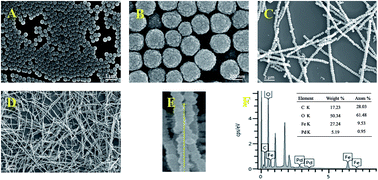
Superparamagnetic hollow nanoparticles were prepared by hydrothermal method with sodium citrate as reductant, urea as alkaline environment, and PAM as stabilizer at 200 °C. Nanoparticles subsequently self-assembled into 1D nanochains by the external magnetic field and were coated with polymer P(MAA-DVB) under the initiation of AIBN by distillation precipitation polymerization. Finally, the palladium loaded 1D peapod-like magnetic nanochains (Fe3O4@P(MAA-DVB)–Pd(0)) were obtained through complexation and late reduction by NaBH4. The preparation process is illustrated in Scheme 1. The morphology and size of the nanoparticles and nanochains were characterized by SEM and TEM images. It can be easily seen from the SEM images (Fig. 1A and B) that, nanoparticles with the average diameter of approximate 260 nm have a uniform gibbous spherical shape and a narrow size distribution. Likewise, the TEM image (Fig. 2A) shows that nanoparticles with a rough surface have good mono-dispersion in water and are composed of irregular shaped primary particles. And Fig. 2A further demonstrates that the nanoparticle has offwhite center while the edge is relatively dark, which firmly confirms the hollow structure of the obtained nanoparticles. Besides, Fig. 1C clearly displays that one-dimensional nanochains, just like ‘Tanghulu’, have a uniform diameter of about 380 nm and an average length of 20 μm. Furthermore, the TEM image shows that the 1D nanochains have a core–shell structure with an average shell thickness of about 56 nm after coated with polymer and the visible interparticle spacing between adjacent particles filled with polymer P(MAA-DVB) is about 25 nm (Fig. 2C). The thickness of polymer P(MAA-DVB) shell can be further altered by changing the amount of monomer, nanoparticles or the reaction time. And Fig. 2D represents that Pd nanoparticles are distributed on the surface of the peapod-like nanochains, manifesting that Pd particles have been successfully loaded on the surface of the nanochains through complexation between carboxyl on the surface of nanochains and palladium ion, and NaBH4 reduction. Additionally, the high-resolution TEM image (Fig. 2E) amplifying the red zone in Fig. 2D is collected and clear crystal fringe can be clearly seen and the d-spacing between neighbouring planes of 2.26 Å and 1.94 Å could determine the (111) plane and (220) plane of Pd in accordance with the JCPDS file.
 |
| Fig. 1 The SEM images: (A), (B) Fe3O4 image; (C), (D) Fe3O4@P(MAA-DVB) image; (E) Fe3O4@P(MAA-DVB)–Pd(0) image; (F) EDS spectrum of Fe3O4@P(MAA-DVB)–Pd(0) nanochains. | |
 |
| Fig. 2 The TEM images: (A) hollow Fe3O4 nanoparticles image; (B), (C) Fe3O4@P(MAA-DVB) image; (D) Fe3O4@P(MAA-DVB)–Pd(0) image; (E) HR-TEM of Fe3O4@P(MAA-DVB)–Pd(0). | |
In order to further illustrate the structure and composition of Fe3O4@P(MAA-DVB)–Pd(0), we collected the XRD and EDS spectrum, respectively. As can be observed in Fig. 1F, the strong peaks of Fe, O, C and Pd confirm that the Pd has been anchored on the surface and the Pd content is about 5.19 wt%. Fig. 3 shows the XRD spectra of the Fe3O4, Fe3O4@P(MAA-DVB) and Fe3O4@P(MAA-DVB)–Pd(0). The peaks could be clearly seen from the curve a and its value of 2θ is 18.3° (111), 30.1° (220), 35.5° (311), 43.1° (400), 53° (422), 57° (511), 62.6° (440) and 74° (533), which corresponds to Fe3O4 crystal compared with JCPDS. Compared with curve a, no diffraction peak from the layer can be observed in curve b because of the amorphous nature of the P(MAA-DVB) layer. As to curve c, the relatively weak and broad peak value of 2θ equal to 40.1° (111), 46.7° (200), 68.1° (220) corresponding to Pd crystal. In conclusion, it can be further determined that Pd nanoparticles are distributed uniformly on the surface of 1D nanochains.
 |
| Fig. 3 XRD patterns of Fe3O4 (a), Fe3O4@(MAA-DVB) (b) and Fe3O4@(MAA-DVB)–Pd(0) (c). | |
Fig. 4 shows FT-IR spectroscopic characterization of Fe3O4 and Fe3O4@P(MAA-DVB)–Pd(0). As can be observed in Fig. 1a, the adsorption peak appearing at 576 cm−1 corresponds to the characteristic absorption peak of Fe–O bond. It also has two other strong absorption peaks at 1403 cm−1 and 1638 cm−1 which are in accordance with the anti-symmetrical vibration of carboxyl group and symmetric vibration of sodium citrate. And the peak at 2933 cm−1 and 2852 cm−1 corresponds to the C–H stretching vibration of stabilizer PAAS. Thus, carboxylate modified magnetic nanoparticles are favorable to dispersion and the late assembly. Compared with curve a, the curve b displays new peaks at 3426 cm−1 and 1702 cm−1 which can be attribute to the O–H and C
O of carboxyl, respectively. The adsorption peak at 2931 cm−1 corresponds to the stretching vibration of C–H bond and locates at 1631 cm−1, 1546 cm−1 and 1450 cm−1 can be attributed to the stretching vibration of C
C of benzene. All of these results suggest that polymer P(MAA-DVB) is successfully coated on the surface of 1D nanochains.
 |
| Fig. 4 FT-IR spectra of (a) Fe3O4 nanoparticles and (b) Fe3O4@(MAA-DVB)–Pd(0). | |
A mass fraction of organic material and inorganic material in 1D nanochains was confirmed by thermal gravimetric analysis. In Fig. 5a, the main cause of the slight mass loss is that a portion of PAM stabilizer exists in the Fe3O4 particles. Compared with curve a, the mass of Fe3O4@P(MAA-DVB) nanochains shows a remarkable change between 300 and 500 °C, and polymer accounts for up to 21.62 wt% of the total mass. But the curve c shows that the weight loss of the Pd loading 1D nanochains has a significant decrease at the temperature range from 300 °C to 500 °C which contrasts with curve b, further illustrating that the Pd particles are loaded on the surface of 1D magnetic nanochains. Furthermore, Pd content accounts for 3.69 wt% of the total mass calculated by TGA which is different from EDS. The reason for the difference maybe EDS provided Pd content by X-ray line scanning on the surface of the 1D magnetic nanochains, which is usually used for illustrating the existence of Pd element. Comparing with EDS data, the result from TGA is more accurate. As described above, inorganic material occupying the main part of 1D nanochains composition indicates magnetic response property of this materials.
 |
| Fig. 5 TGA curves of Fe3O4 (a), Fe3O4@(MAA-DVB) (b) and Fe3O4@(MAA-DVB)–Pd(0) (c). | |
The magnetic properties of the 1D nanochains were investigated by means of vibrating sample magnetometer. As shown in Fig. 6, it can be clearly seen from the curve a that the magnetic nanoparticles have superparamagnetic property at room temperature. After the 1D nanochains being coated with polymer P(MAA-DVB), the relative high saturation is 45 emu g−1 in the curve b. The saturation magnetizations of 1D magnetic nanochains Fe3O4@P(MAA-DVB) loaded with Pd particles decreases to 38 emu g−1 due to the increase of Pd particles' quality. Therefore, this superparamagnetic materials can be expediently operated and separated through an external magnetic field. Fig. 7 indicates the distribution of nanochains in different directions of magnetic field. With the direction of external magnetic changing constantly, the superparamagnetic nanochains well arrange along the magnetic field. Furthermore, 1D magnetic nanochains can rotate at high speed, like “magnetic stir bar” on the magnetic stirrer plate. In the aqueous dispersion, the prepared Fe3O4@P(MAA-DVB)–Pd(0) nanochains also displays high magnetic response and micro-sized stirrer could agitate the solution and promote the solution mixing, as shown in Movie-S1.† Therefore, the superparamagnetic of Pd loading 1D nanochains could quickly response in the solution with magnetic stirrer plate and improve the catalytic efficiency in the solution by constantly changing the external magnetic field.
 |
| Fig. 6 Magnetization curves of Fe3O4 (a), Fe3O4@(MAA-DVB) (b) and Fe3O4@(MAA-DVB)–Pd(0) (c). | |
 |
| Fig. 7 Optical microscope of image of randomly distributed nanochains (A) and the ones that align along a magnetic field (B–D). | |
The catalytic properties of the Pd loaded nanochains were evaluated using the heterogeneous catalyst reduction of rhodamine B (RhB) by adding excess NaBH4 in water on the magnetic stirrer plate. The catalytic properties of 3.69 wt% Fe3O4@P(MAA-DVB)–Pd(0) nanochains was mainly discussed by catalytic reduction of RhB at different concentration of catalysts and times. In microcontainer, the RhB solution (200 μL, 20 mg mL−1) is quickly catalytically reduced into a colorless solution by excess NaBH4 in the presence of catalyst (50 μL, 0.9 mg mL−1) on the magnetic stirrer plate. It can be clearly seen from the Movie-S2† that RhB solution has a distinct color change occurring in 8 seconds and was completely catalyzed reduction into a colorless solution in 30 seconds compared with the RhB solution in the absence of catalyst. Fig. 8 shows that the absorption intensity band (λmax) at 552 nm for RhB gradually decreases by the reaction time, which indicates that the catalyst possesses high efficiency on the magnetic stirrer plate. In addition, the linear relationship between Ct/C0 (%) and reaction time (s) is shown in Fig. 9, confirming that the catalytic efficiency increases with concentration increasing and the final catalytic (0.9 mg mL−1) efficiency is approximately 81.17% within 40 s. In Fig. 9, the curve a and b represent the removal performance in the present of NaBH4 and Fe3O4@P(MAA-DVB) nanochains independently. It can be seen that the concentration of RhB has little change. In other words, dye was catalytic degradation mainly through heterogeneous catalytic reduction of Fe3O4@P(MAA-DVB)–Pd(0) nanochains. Besides, we further study the RhB solution (3 mL, 20 mg mL−1) by adding excess NaBH4 in catalyst (500 μL, 0.9 mg mL−1) on the magnetic stirrer plate. The reduction time of pink RhB solution is only 7 s (Movie-S3†), which indicates that Pd loaded 1D magnetic nanochains exhibit extraordinary high catalytic activity in microscale system and normal system because of the excellent magnetic response and its special structure and facile preparation processes comparing with Fe3O4@P(MBAAm-co-MAA)/Ag in catalytic22 and Fe3O4@P(MAA-DVB)/TiO2 in photocatalytic performance.40 And in Fig. 10, the 1D Fe3O4@P(MAA-DVB)–Pd(0) catalyst exhibits catalytic stability (64.84%) even after the reaction was recycle for more than five runs.
 |
| Fig. 8 UV-visible absorption spectrum changes of the RhB solution (20 mg L−1) at different time. | |
 |
| Fig. 9 Catalyst degradation of RhB solution: (a) absence of catalytic; (b) Fe3O4@P(MAA-DVB) nanochains; different concentration of catalyst solution ((c) 0.5 mg mL−1, (d) 0.7 mg mL−1 and (e) 0.9 mg mL−1.) | |
 |
| Fig. 10 The recycled degradation data of RhB solution using 1D Fe3O4@P(MAA-DVB)–Pd(0) magnetic nanochains. | |
In addition, the catalytic reduction was evaluated through the degradation of the other four organic dyes including phenol red (PR), methylene blue (MB), bromocresol green (BG), methyl orange (MO) using the as-prepared 3.69 wt% Fe3O4@P(MAA-DVB)–Pd(0) magnetic nanochains. In Fig. 11, the degradation efficiency of PR, MB, BG and MO is 89.52%, 51.4%, 49.85% and 26.25%, respectively. The major reason for different organic has different catalytic conversion under the same condition is the structure of dye.
 |
| Fig. 11 The different dyes reduction efficiency using 1D Fe3O4@P(MAA-DVB)–Pd(0) magnetic nanochains. | |
Conclusion
In summary, the 1D Fe3O4@P(MAA-DVB)–Pd(0) magnetic nanochains have been successfully prepared through a rapid and convenient process. The 1D magnetic nanochains with superparamagnetic properties have excellent magnetic response and results show that 1D peapod-like Fe3O4@P(MAA-DVB)–Pd(0) magnetic nanochains could catalyze reduction organic dye of RhB within 40 s. Furthermore, the catalyst can easily recycle and exhibits catalytic stability (64.84%) at least five consecutive cycles. Thus, 1D Fe3O4@P(MAA-DVB)–Pd(0) magnetic nanochains possess the potential application prospect in dye degradation due to their properties of rapid catalyze, reusability, stability and easiness in operation.
Acknowledgements
The authors are grateful for the financial support provided by the State Key Program of National Natural Science of China (Grant No. 51433008), the Natural Science Foundation of Shaanxi Province (Grant No. 2015JQ2055, 2015JM2050), Foundation of Aerospace Science and Technology Innovation (Grant No. 2016007).
References
- J. Yuan, Y. Xu and A. H. Müller, Chem. Soc. Rev., 2011, 40, 40–655 RSC.
- H. Xu, Y. Xu and X. Pang, Sci. Adv., 2015, 1, e1500025 Search PubMed.
- C. Chen, J. Wang and Z. Ren, CrystEngComm, 2014, 16, 1681–1686 RSC.
- Y. Yang, J. Shi and T. Tanaka, Langmuir, 2007, 23, 12042–12047 CrossRef CAS PubMed.
- L. Meng, W. Chen and C. Chen, Cryst. Growth Des., 2009, 10, 479–482 Search PubMed.
- S. Kralj and D. Makovec, ACS Nano, 2015, 9, 9700–9707 CrossRef CAS PubMed.
- D. Toulemon, Y. Liu, X. Cattoën and C. Leuvrey, Langmuir, 2016, 32, 1621–1628 CrossRef CAS PubMed.
- J. Zhou, L. Meng, X. Feng, X. Zhang and Q. Lu, Angew. Chem., Int. Ed., 2010, 49, 8476–8479 CrossRef CAS PubMed.
- W. Zhou, K. Zheng, L. He, R. Wang, L. Guo, C. Chen, X. Han and Z. Zhang, Nano Lett., 2008, 8, 1147–1152 CrossRef CAS PubMed.
- D. Yang, Y. Zhou, X. Rui, J. Zhu, Z. Lu, E. Fong and Q. Yan, RSC Adv., 2013, 3, 14960–14962 RSC.
- Y. Liu, J. Goebl and Y. Yin, Chem. Soc. Rev., 2013, 42, 2610–2653 RSC.
- H. Wang, Q. Chen, L. Sun, H. Qi, X. Yang, S. Zhou and J. Xiong, Langmuir, 2009, 25, 7135–7139 CrossRef CAS PubMed.
- J. Liu, T. Xia, S. Wang, G. Yang, B. Dong, C. Wang, Q. Ma, Y. Sun and R. Wang, Nanoscale, 2016, 8, 11432–11440 RSC.
- L. He, M. Wang, J. Ge and Y. Yin, Acc. Chem. Res., 2012, 45, 1431–1440 CrossRef CAS PubMed.
- W. H. Chong, L. K. Chin, R. L. S. Tan, H. Wang, A. Q. Liu and H. Chen, Angew. Chem., Int. Ed., 2013, 52, 8570–8573 CrossRef CAS PubMed.
- Y. Hu, L. He and Y. Yin, Angew. Chem., Int. Ed., 2011, 50, 3747–3750 CrossRef CAS PubMed.
- M. Ye, S. Zorba, L. He, Y. Hu, R. T. Maxwell, C. Farah, Q. Zhang and Y. Yin, J. Mater. Chem., 2010, 20, 7965–7969 RSC.
- Y. Yao, G. S. Chaubey and J. B. Wiley, J. Am. Chem. Soc., 2011, 134, 2450–2452 CrossRef PubMed.
- S. E. Bowles, W. Wu, T. Kowalewski, M. C. Schalnat, R. J. Davis, J. E. Pemberton, I. Shim, B. D. Korth and J. Pyun, J. Am. Chem. Soc., 2007, 129, 8694–8695 CrossRef CAS PubMed.
- J. Zhou, C. Wang, P. Wang, P. B. Messersmith and H. Duan, Chem. Mater., 2015, 27, 3071–3076 CrossRef.
- M. Wang, L. He, Y. Hu and Y. Yin, J. Mater. Chem. C, 2013, 1, 6151–6156 RSC.
- W. Zhang, X. Si, B. Liu, G. Bian, Y. Qi, X. Yang and C. Li, J. Colloid Interface Sci., 2015, 456, 145–154 CrossRef CAS PubMed.
- M. Ma, Q. Zhang, J. Dou, H. Zhang, D. Yin, W. Geng and Y. Zhou, J. Colloid Interface Sci., 2012, 374, 339–344 CrossRef CAS PubMed.
- F. Zou, H. Zhou, T. V. Tan, J. Kim, K. Koh and J. Lee, ACS Appl. Mater. Interfaces, 2015, 7, 12168–12175 CAS.
- X. Zhang, L. Lv, L. Ji, G. Guo, L. Liu, D. Han, B. Wang, Y. Tu, J. Hu, D. Yang and A. Dong, J. Am. Chem. Soc., 2016, 138, 3290–3293 CrossRef CAS PubMed.
- S. Fu, C. Zhu, J. Song, M. H. Engelhard, Y. He, D. Du, C. Wang and Y. Lin, J. Mater. Chem. A, 2016, 4, 8755–8761 CAS.
- Y. Zhong, Y. Ni, S. Li and M. Wang, RSC Adv., 2016, 6, 15831–15837 RSC.
- P. W. Menezes, A. Indra, D. González-Flores, N. R. Sahraie, I. Zaharieva, M. Schwarze, P. Strasser, H. Dau and M. Driess, ACS Catal., 2015, 5, 2017–2027 CrossRef CAS.
- M. Raula, M. H. Rashid, S. Lai, M. Roy and T. K. Mandal, ACS Appl. Mater. Interfaces, 2012, 4, 878–889 CAS.
- W. Zhou, L. He, R. Cheng, L. Guo, C. Chen and J. Wang, J. Phys. Chem. C, 2009, 113, 17355–17358 CAS.
- N. Gan, L. Jia and L. Zheng, J. Anal. Methods Chem., 2011, 957805 Search PubMed.
- P. M. Peiris, L. Bauer, R. Toy, E. Tran, J. Pansky, E. Doolittle, E. Schmidt, E. Hayden, A. Mayer, R. A. Keri, M. A. Griswold and E. Karathanasis, ACS Nano, 2012, 6, 4157–4168 CrossRef CAS PubMed.
- G. K. Das, C. S. Bonifacio, J. D. Rojas, K. Liu, K. V. Benthem and I. M. Kennedy, J. Mater. Chem. A, 2014, 2, 12974–12981 CAS.
- V. T. Tran, H. Zhou, S. Kim, J. Lee, J. Kim, F. Zou, J. Kim, J. Y. Park and J. Lee, Sens. Actuators, B, 2014, 203, 817–823 CrossRef CAS.
- Y. Liu, L. Zhou, Y. Hu, C. Guo, H. Qian, F. Zhang and X. W. Lou, J. Mater. Chem., 2011, 21, 18359–18364 RSC.
- H. Wang, Q. Chen, J. Chen, B. Yu and X. Hu, Nanoscale, 2011, 3, 4600–4603 RSC.
- L. Chen, R. M. Berry and K. C. Tam, ACS Sustainable Chem. Eng., 2014, 2, 951–958 CrossRef CAS.
- S. Yang, C. Cao, Y. Sun, P. Huang, F. Wei and W. Song, Angew. Chem., Int. Ed., 2015, 54, 2661–2664 CrossRef CAS PubMed.
- M. Ma, Q. Zhang, J. Dou, H. Zhang, W. Geng, D. Yin and S. Chen, Colloid Polym. Sci., 2012, 290, 1207–1213 CAS.
- C. Li, J. Tan, X. Fan, B. Zhang, H. Zhang and Q. Zhang, Ceram. Int., 2015, 41, 3860–3868 CrossRef CAS.
- W. Li, Y. Tian, B. Zhang, L. Tian, X. Li, H. Zhang, N. Ali and Q. Zhang, New J. Chem., 2015, 39, 2767–2777 RSC.
- W. Cheng, K. Tang, Y. Qi, J. Sheng and Z. Liu, J. Mater. Chem., 2010, 20, 1799–1805 RSC.
- W. Li, B. Zhang, X. Li, H. Zhang and Q. Zhang, Appl. Catal., A, 2013, 459, 65–72 CrossRef CAS.
Footnote |
† Electronic supplementary information (ESI) available. See DOI: 10.1039/c6ra22198j |
|
This journal is © The Royal Society of Chemistry 2016 |
Click here to see how this site uses Cookies. View our privacy policy here.