Characterizing the interactions between humic matter and calcium ions during water softening by cation-exchange resins

Bin Dong a, Ying Xu*a, Danni Shena, Xiaohu Dai*a and Senming Linb
aState Key Laboratory of Pollution Control and Resource Reuse, School of Environmental Science and Engineering, Tongji University, Shanghai, 200092, China. E-mail: assassinxu@sina.cn; daixiaohu@tongji.edu.cn
bKaramay Oilfield, PetroChina Ltd., Karamay, Xinjiang 834000, China

Received 3rd September 2016 , Accepted 26th September 2016

First published on 27th September 2016


Abstract

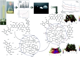
Reusing wastewater can enormously reduce environmental pollution and save water. One of the key obstacles to reuse is the scaling caused by calcium (Ca) ions and humic matter (HM). The interactions among HM, Ca ions and cation-exchange resins (CERs) during the water softening process were explored via lab-scale and pilot-scale tests. Surface topography, pore structure, and surface molecular structure of the CERs were determined using an atomic-force microscope, an automatic mercury porosimeter, a laser-Raman spectrum and X-ray photoelectron spectroscopy, respectively. The surface charge of CERs was assessed by measuring zeta potential. Experimental results showed that HM can react with Ca-ions to form HM–Ca+ and Ca ions can act as a bridge for HM and CER, suggesting that Ca ions and HM can be simultaneously removed by CERs. Kinetic analyses indicate that a pseudo-first order equation was able to accurately describe the overall adsorption kinetics and diffusion of Ca ions through a liquid film of CER was the rate-determining step for the overall adsorption process. Further investigation revealed that during the first 60 minutes of adsorption, the effect of HM on the CER adsorption kinetics can be described well by pseudo-first and pseudo-second order equations. This indicates that diffusions of Ca ions, HM, and HM–Ca+ and their interactions were equivalent for controlling the initial stage of adsorption. In addition, 99% of Ca ions and 30–60% of HM in wastewater were removed in the pilot-scale test, suggesting that CERs have significant potential to remove Ca ions and HM simultaneously in wastewater.


1. Introduction

Severe water scarcity is an increasing problem in China, which has the world's fastest growing economy and a growing industrial water demand.1 Large amounts of industrial wastewater are produced in China, causing serious environmental pollution. As industrial facilities require large quantities of water and produce considerable amounts of wastewater, it is important to reduce fresh-water consumption and wastewater generation by reusing wastewater.2,3

The largest single use of water in China's industrial sector is as boiler feedwater in steam generators.4,5 It is thus necessary to reuse wastewater as boiler feedwater during industrial processes to minimize fresh-water requirements and protect the environment. However, steam generators often require large volumes of soft water. Humic matter (HM, as a significant portion of dissolved organic matter (DOM)) and hardness cations (i.e., calcium and magnesium), which can cause reversible and irreversible scaling, are common constituents of wastewater, especially the increasing volumes of water produced as a byproduct of oil extraction from subsurface geological formations in oilfields.6 It is well known that scale can detrimentally affect the thermal efficiency and safety of boilers.7 Therefore, it is vital to develop an effective strategy for removing HM and hardness cations to enable wastewater to be reused as boiler feedwater.

The universally practiced processes for removing DOM and hardness cations are coagulation, lime softening, and ion exchange.8–10 However, coagulation and lime softening produce a large amount of sludge that requires safe disposal and it will generate serious secondary pollution if it is not disposed of appropriately. According to previous reports,11–13 ion exchange is another effective means of removing DOM and hardness cations. Cation-exchange resins (CERs), which can release positively charged ions in exchange for cationic impurities, have long been used to soften water, with proven effectiveness.14,15 Similarly, anion-exchange resins (AERs) have been used to remove anions. Recently, numerous studies on the removal of DOM by AERs have been reported.16–19 However, the retention phenomenon of DOM on the AERs is a complex process, integrating several mechanisms.16 Researchers have made considerable effort to investigate the properties of DOM and the AERs. A series of interaction mechanisms between DOM and the AERs, such as ion exchange, size exclusion, adsorption, electrostatic interaction, hydrophobic/hydrophilic repulsion, and entropy-assisted interaction, have been proposed.18–21 The properties of DOM, such as the distribution of carbon (C) between aromatic and aliphatic domains, charge density, and molecular weight, have been found to influence interactions between DOM and the AERs.19,21,22 The effects of resin properties on these interactions have also been discussed. Bolto and Wang23,24 observed an increase in DOM removal by AERs with a more open structure, enhanced exchange capacity, and higher water content. Resin structure, exchange capacity, and water content are affected by polar groups and the degree of crosslinking. In a study of nine AERs, Cornelissen et al.25 reported that the rate of removal of DOM increased as the water content of the resins increased and resin size decreased. Researchers have also reported that DOM is most effectively removed by macroporous resins with quaternary ammonium functional groups and polyacrylic skeletons.26–29 However, these findings indicate that the interaction between DOM and ion-exchange resins is not always predominantly electrostatic. It implies that the DOM with negatively charge also may be adsorbed on the surface of CERs which are also with negatively charge. Lots of studies have reported that Ca ions can promote the bonding between DOM and membrane by acting as a bridge.30–33 In addition, both of the CERs and membrane have large amounts of carboxyl groups. Therefore, we envisioned that the retention phenomenon of HM onto CERs also can be found in the process of water softening, suggesting that simultaneous removal of HM and Ca ions by CERs may be achieved. Then, how does HM interact with Ca ions and CERs? How does HM influence the removal of Ca ions in the ion-change system? Little research in this area has been conducted.

The goal of this research was to increase understanding of the interactions between HM, Ca ions, and CER with model water in the process of water softening. And to a certain extent simultaneous removal of HM and Ca ions by CERs can be achieved. The specific objectives of the research were as follows: (1) to characterize the physical/chemical properties of CERs in a mixed solution of HM and Ca ions, (2) to investigate the effect of HM on adsorption/ion-exchange kinetics of CERs for Ca ions, and (3) to determine the mechanism of the influence of HM on Ca ions removal by CERs. Insights into these interactions can provide guidelines for approaches to the simultaneous removal of DOM and hardness cations and development of a new kind of CER.

2. Experimental

2.1 Materials

CERs with a macroporous structure, polyacrylic composition, and carboxylic-Na functional groups (D113) were provided by Zhengguan Resin Chemical Co., Ltd. (Hangzhou, China). The basic performance parameters of CERs are summarized in the Table S1 in ESI. Prior to use, the resins were washed sequentially in hydrochloric acid, sodium hydroxide, and methanol to remove residual impurities (Text S1, ESI).34 After pretreatment, the CERs were divided into three groups. The resins in one group were dried in an oven at a controlled temperature of 50 °C for 24 h and subsequently stored in a desiccator before use. The analytical-grade CaCl2 was purchased from Sinopharm Chemical Reagent Co., Ltd., China. The concentration of Ca ions in the model water was kept constant at 3.5 mmol L−1 (350 mg L−1 as calcium carbonate, CaCO3) to approximate the general composition of reused oilfield wastewater in China. The analytical-grade HM (humic acid, sodium salt) was purchased from Aladdin Reagents Co., Ltd. The powdered HM was dissolved in water without further purification and the concentration of 20 mg L−1 stock solution was made. The stock solution was stored in a refrigerator at 4 °C before use.32 The other two groups of resins, named CER–Na and CER–HM, were soaked in deionized (DI) water and a 20 mg L−1 HM solution, respectively, for 48 h. The model water without the precipitate was prepared by adding anhydrous calcium chloride (CaCl2) and HM to DI water with no additional pH adjustment. The model water was used immediately after preparation.

2.2 Laboratory-scale experimental procedure

Two sets of experiments were conducted sequentially. The batch tests were conducted using a shaker (SHZ-82A, Ruihua) at ambient temperature (25 ± 1 °C). A sample comprising 1 g of CERs was distributed to each of seven 1000 mL Erlenmeyer flasks, which were numbered 0, 1, 2, 3, 4, 5, and 6. Subsequently, seven 800 mL samples of model water (350 mg L−1 as CaCO3) dosed with varying amounts of HM (0, 4, 8, 12, 16, 20, and 24 mg) were added to the seven Erlenmeyer flasks, respectively. Next, the flasks were shaken at 100 rpm for 48 h to attain equilibrium. Samples 0, 2, and 4 were used to investigate the mass transfer rate (MTR) of the CERs by calculating their adsorption capacity in a specified time interval. During each set of adsorption calculations, 100 uL of model water was removed from the Erlenmeyer flasks for analysis.

The fixed bed column runs were carried out using a glass column (13 mm in diameter and 450 mm in length). Each of seven fixed-bed columns was loaded with 10 g of dried CERs, and all of the column runs were performed at the same superficial liquid velocity (SLV) (1.0 ± 0.2 m h−1) for 6 h. The source water comprised seven 1000 mL samples of model water (350 mg L−1 as CaCO3) dosed with varying amounts of HM (0, 5, 10, 15, 20, 25, and 30 mg). The concentrations of Ca ions in the effluent water from the column were analyzed every hour.

All of the experiments were conducted in duplicate, and the means and standard deviations (SDs) for the duplicate samples are reported here.

2.3 Characterization of CERs

After the experiments, the CERs from samples 0 and 4 in the batch tests were collected and named CER–Ca and CER–HM/Ca, respectively. Next, all of the CER–Na, CER–Ca, CER–HM/Ca, and CER–HM resins were freeze-dried to remove residual water. The surface roughness and microstructure of CER–Na, CER–Ca, and CER–HM/Ca were assessed by scanning probe microscopy (SPM-9500J3, Shimadzu). To get a better dispersity an agate mortar was used to grind some of these resins into individual powders capable of passing through a grid with a 0.075 mm pore rating. 0.1 g samples of CER–Na powder, CER–HM powder, CER–Ca powder, and CER–HM/Ca powder, respectively, were weighed out and loaded into four 100 mL beakers. After 100 mL of ultrapure water had been added to each beaker, the beakers were closed. After 24 h, the zeta potential (ZP) of the resins was measured using a Malvern Zetasizer Nano ZS90 (U.K., the Smoluchowski conversion was used) to characterize their surface charge.35 In addition, the surface area and pore-size distribution of the CER–Na, CER–Ca, and CER–HM/Ca were determined using an automatic mercury porosimeter (PoreMasterGT 60, Quantachrome).

X-ray photoelectron spectroscopy (XPS) was performed to investigate the chemical states of C, O, and N elements on the surface of the CER–Na, CER–Ca, and CER–HM/Ca resins. The XPS experiments were carried out on an RBD upgraded PHI-5000C electron spectroscopy for chemical analysis system (Perkin Elmer) using Mg Kα radiation ( = 1253.6 eV). The full spectra (0–1100 eV) and narrow spectra of all of the elements were recorded at high resolution using an RBD 147 interface (RBD Enterprises, U.S.A.) with AugerScan 3.21 software. Raman laser spectroscopy (Pro-TT-EZRaman-B2) was used to identify and explore the molecular structural properties of the three resin powders in the 250–2350 cm−1 region. The Raman spectra were analyzed using reported methods of spectral interpretation36 and group-frequency measurement.37,38 The results are summarized in the Fig. S1.

2.4 Pilot-scale experiment

A 6 month pilot-scale test was conducted to verify the hypothesis that the retention phenomenon of HM on CERs can be found in the softening process of oilfield-produced water. A flow diagram of the softening process and details of the softeners used in the pilot-scale test are summarized in the ESI (Fig. S2–S4). The softeners (denoted as A, B, C, and D) were filled with CERs (D113). The basic water–quality parameters of the raw oilfield-produced water and the effluent obtained from the secondary treatment are summarized in Table S2. The basic parameters of softener operation are summarized in the Table S3, along with the rates at which the CERs removed hardness cations and specific 254 nm ultraviolet absorbance (SUVA254) (Table S4).

2.5 Analytical methods

The concentrations of the tested Ca ions were quantitatively measured using inductively coupled plasma-mass spectroscopy (ICP/MS-7700, Agilent) in accordance with Standard Method 3125.39 ZP of the HM solutions (0, 5, 10, 15, 20, 25, and 30 mg L−1) and model water (HM/Ca solutions) were measured by a Malvern Zetasizer Nano ZS90 (U.K., the Smoluchowski conversion was used). Dissolved organic carbon (DOC) was measured using a total organic carbon analyzer (TOC-LCPH, Shimadzu) equipped with an ASI-V autosampler. UV254 was measured on a Hitachi U-2900 spectrophotometer using a 1 cm quartz cell. SUVA254 was calculated as follows:40 (UV254/DOC) × 100. All of the samples were measured in duplicate, and the average values (AVs) are reported here.

3. Results

3.1 Adsorption capacity of CERs in model water

Batch tests and column runs were conducted to explore the changes in the equilibrium adsorption quantity (Qe) of the CERs for Ca ions in the model water. The basic characteristics of the model water containing different proportions of HM and the CERs' average equilibrium adsorption quantity (Qe) for Ca ions are summarized in the Tables S5 and S6, respectively. To further explore the effect of HM on Qe, the instantaneous adsorption quantity (Qt) of samples 0 (0 mg L−1 HM), 2 (10 mg L−1 HM), and 4 (20 mg L−1 HM) was measured. The adsorption kinetics in these samples were also explored (as outlined in the Discussion section). Fig. 1 shows the results for Qt obtained in the batch tests of CERs with different dosages of HM (0, 10, and 20 mg L−1). Sample 0 had a noticeably shorter equilibrium time than either sample 2 or sample 4. For samples 2 and 4, two adsorption stages (0–60 min and >60 min) were observed, as shown in Fig. 1. Fig. 2 shows the rate of removal of Ca ions with different HM dosages at the same SLV in the dynamic-adsorption experiments. The corresponding rates of removal of DOC and UV254 are summarized in the Fig. S5 and S6, respectively. In contrast with the results of the static-adsorption experiments, the rate of Ca ions removal decreased as HM dosage increased with the same empty bed contact time and the same SLV. In addition, the rate of removal of Ca ions became less stable as HM dosage increased, suggesting that there are important interactions between HM and Ca ions during this process.
image file: c6ra22113k-f1.tif
Fig. 1 Instantaneous adsorption quantity (Qt) of resins with different dosages of HM (0, 10, and 20 mg L−1).

image file: c6ra22113k-f2.tif
Fig. 2 Rate of removal of Ca ions with different dosages of HM in column-run tests.

3.2 Surface topography and pore structure of CERs

The changes in the surface topography of the CER–Na, CER–Ca, and CER–HM/Ca are depicted in Fig. 3. The changes in the pore structure of the resins are documented in Table 1. Surface roughness differed noticeably between the three resins (Fig. 3). The CER–Na surface had a 130 nm height difference, and the CER–Ca and CER–HM/Ca surfaces had height differences of 210 nm and 250 nm, respectively. Unlike the CER–Na, some precipitation of Ca ions with HM may be deposed on the surface of CER–HM/Ca. These findings can be attributed to differences in the interactions between the adsorbates and the surface of the resins. As is shown in Table 1, CER–HM/Ca had a greater specific pore volume (SPV) and specific surface area (SSA) than either CER–Na or CER–Ca. It suggests that CER–HM/Ca has a potential adsorption capacity, which is mainly attributed to the adsorbed HM. Moreover, the greatest porosity, an important parameter of mass transfer performance inside CERs, was found in the CER–HM/Ca.
image file: c6ra22113k-f3.tif
Fig. 3 Surface topography of CER–Na, CER–Ca, and CER–HM/Ca by SPM.
Table 1 Basic pore-structure parameters of CER–Na, CER–Ca, and CER–HM/Caa
  CER–Na CER–Ca CER–HM/Ca
AV SD AV SD AV SD
a MPD = Median Pore Diameter; AV = Average Value; SD = Standard Deviation.
MPD (μm) 106.7 ±9.454 127.2 ±8.900 113.2 ±4.750
SPV (mL g−1) 0.19 ±0.052 0.21 ±0.070 0.37 ±0.060
SSA (m2 g−1) 6.45 ±0.575 4.71 ±1.085 8.74 ±0.775
Porosity (%) 23.04 ±1.135 23.92 ±2.030 36.03 ±5.625


3.3 Surface charge of HM, HM/Ca, and CERs

To obtain more information on the electrostatic interaction between the HM, Ca ions, and CERs, their ZP were measured. Fig. 4(a) shows the ZP of the HM and HM/Ca at different dosages of HM. The average ZP of the HM was approximately −43.1 mV, and that of the HM/Ca was approximately −12.4 mV, indicating that the electrostatic interaction between the HM and Ca ions happened. This finding is consistent with previously published results.41,42 The ZP measurements for CER–Na, CER–Ca, CER–HM, and CER–HM/Ca are presented in Fig. 4(b). After 48 h of sorption in the model water, the ZP of the CERs had fallen from −35.9 mV (CER–Na) to −10.2 mV (CER–Ca) due to ion exchange between CER–Na and the Ca ions. However, the ZP of the CERs increased from −35.9 mV (CER–Na) to −44.4 mV (CER–HM) in the HM solution (20 mg L−1) over 48 h of sorption, indicating that more negative charges are produced on the surface of CER–Na. Both of CER–Na and HM are negatively charged in the solution. Thus, it can be postulated that a force (such as hydrophobic effect) exceeding that of electrostatic repulsion enabled HM to be adsorbed on the surface of the CERs.
image file: c6ra22113k-f4.tif
Fig. 4 Zeta potential of HM, resins, and HM/Ca; (a) zeta potential of HM and HM/Ca with different dosages of HM; (b) zeta potential of different resins.

3.4 Characterization of surface molecular structure of resins

The XPS was used to survey the distribution of species on the surfaces of the CER–HM/Ca, CER–Ca, and CER–Na resins (Fig. S7). A signal at 399.92 eV corresponding to N 1s was present in the spectrum shown in Fig. S7(a), but not in the spectra presented in Fig. S7(b) and (c). As is well known to all, N is one of important elements in HM,43 while CERs in this study does not containing N element (Table S1), indicating that the HM was adsorbed on the surface of CER–HM/Ca. The results of XPS spectra analyses for C and O elements on the surface of the resins are displayed in Fig. 5. The analyses were conducted in accordance with the Standard Reference Database and appropriate literature.44,45
image file: c6ra22113k-f5.tif
Fig. 5 XPS spectrums for C, O, and Ca for different resins: (a) C spectra for CER–Na and CER–HM/Ca; (b) O spectra for CER–Na and CER–HM/Ca.

The C 1s XPS regions for CER–Na and CER–HM/Ca shown in Fig. 5(a) indicate four main C-containing species. The peaks at 283.30 eV, 285.42 eV, and 288.40 eV for CER–Na resulted from the O–C[double bond, length as m-dash]O bond of the polyacrylic. The peaks at 284.25 eV and 286.79 eV corresponded to the C in polymethyl methacrylate (PMMA). Obviously, the polyacrylic and PMMA are the main components of CERs (Table S1). However, different C-containing species were found on the surface of the CER–HM/Ca resin. Fig. 5(a) shows peaks at 283.70 eV and 284.80 eV, corresponding to the carbazole C–N bond and the aromatic C–C bond, respectively. Carbazole is well known not to be a component of resin, and it comes from HM, also suggesting that HM was adsorbed on the surface of CER–HM/Ca. Fig. 5(b) shows O 1s spectra corresponding to three O species for CER–Na and four O species for CER–HM/Ca. The peaks at 532.39 eV and 533.78 eV were caused by the O of the polyacrylic. The peak at 531.60 eV indicates the O[double bond, length as m-dash]C–O–C bond of PMMA. These peaks are consistent with the polyacrylic structure of CERs. However, the O 1s XPS spectrum for CER–HM/Ca comprises four main peaks with binding energies of 531.80 eV, 532.39 eV, 533.40 eV, and 535.10 eV. The highest peak, at 532.39 eV, is attributed to the O[double bond, length as m-dash]C–O bond in the polyacrylic. The other peaks correspond to the O of aromatics. It is well known that some functional groups (such as benzene ring) of the aromatics in HM are hydrophobic,43 and the CERs matrices are also hydrophobic.11 Thus, hydrophobic effect as one of important driving forces for the adsorption process is possible. In addition, according to the results of Raman spectrum (Fig. S1), Ca-bridge between HM and CERs was also found by the ranges 900–1000 cm−1, indicating that Ca ions can be as bridge for HM and CERs.

4. Discussion

4.1 Influence of HM on MTR of Ca ions

Fig. 1 shows the variation in Qt for CERs at different HM concentrations. The Qt of sample 0 was consistently larger than that of either 2 or 4 during the initial stage, and sample 0 had the shortest adsorption equilibration time, indicating that without the effect of HM CERs has a faster reaction rate of adsorption for Ca ions in this stage. It can be inferred that at the initial stage MTR of Ca ions decreased as HM concentration increased, mainly due to a reaction between the HM and Ca ions that reduced the content of free Ca ions. As shown in Fig. 4(a), average ZP of the HM was −43.1 mV and that of HM/Ca was −12.4 mV, indicating that HM can react with Ca ions to form HM/Ca complex. Interestingly, HM/Ca complex with negative charges (−12.4 mV) is stable and not precipitated in the solution with large amounts of Ca ions, suggesting that there must be some electrostatic repulsion between HM/Ca complex and Ca ions. In other words, some positive charges are in the HM/Ca complex. One reasonable explanation is that HM–Ca+ is objective existence in the HM/Ca complex, suggesting that the reaction (eqn (1)) can occur. Ca ions can be as the bridges between CERs and HM molecules, which is also supported by the results of Raman spectrum. In addition, previous researchers41,46 have reported that the diffusivity of HM and Ca ions is reduced by reactions between the two, indicating that the MTR of HM–Ca+ is much smaller than that of free Ca ions. This suggests that HM–Ca+ can not compete with free Ca ions for reacting with CER–Na unless there are some other driving forces of adsorption.
 
image file: c6ra22113k-t1.tif(1)

4.2 Influence of HM on physicochemical properties of CERs

Analysis of surface roughness revealed the largest height difference in the CER–HM/Ca (Fig. 3), suggesting that the precipitation of HM and HM/Ca complexes is deposed on the surface of the CERs. These also can be supported by the results of XPS surface analysis and Raman spectrum. As the precipitation of HM and HM/Ca complexes on the surface and has the largest SSA (Table 1), CER–HM/Ca can be surrounded by a thicker liquid film than that of CER–Na. This film can affect the diffusions of free Ca ions and HM–Ca+,11 which could be the main rate-determining steps in the adsorption process. To some extend, the thickness of liquid film is related to the HM dosage. And this also can explain the results that the rate of removal of Ca ions decreased with increasing HM dosage within the same interval in the column-run tests (Fig. 2). In addition, according to the results of pore structure analysis CER–HM/Ca has the greatest porosity, indicating that mass transfer performance of Ca ions inside CER–HM/Ca is better than the others. That the diffusion of ions inside the material and diffusion of ions through the liquid film are two main rate-determining steps in most of ion-exchange process has been cognized.11 Therefore, it can be concluded that the main rate-determining steps for CER–HM/Ca is the diffusion of free Ca ions and HM–Ca+ through the liquid film. That is to say, if SLV decreases, more HM and Ca ions can be removed at the same time, which was also supported by Fig. S6. It also can explain the phenomenon that conflicting results of the static-adsorption and column-run experiments.

4.3 Influence of HM on adsorption kinetics of CERs

Kinetic study is very important because it describes the interaction rate. Adsorption kinetics generally depends on three main steps comprising external diffusion, intra-particle diffusion, and interaction between absorbate and absorbent.35,47 For the ion exchange process, the ion exchange rates can be controlled by rates of actual chemical reaction, and the intra-particle diffusion is a more common case for the overall process.11,47 The first order reaction generally corresponds to diffusion dominant case, and the second order reaction indicates the existence of some other mass transfer mechanisms, such as the interaction between one absorbate and another.11 In this paper, there are more than one absorbate, and thus the interaction also could be the rate-limiting process. The raw data on the CERs kinetics are summarized in the Fig. S8. To further analyze the adsorption kinetics, a pseudo-first order equation and a pseudo-second order equation were used to model the adsorption. The models can be expressed as follows:35,47

Pseudo-first order equation:

 
Qt = Qe(1 − exp(−k1t)) (2)

Pseudo-second order equation:

 
Qt = (k2tQe2)/(1 + k2tQe) (3)

In eqn (2) and (3), Qe (mg g−1) is the amount of adsorbed Ca ions at equilibrium, and Qt (mg g−1) is the amount of adsorbed Ca ions at time t. k1 (min−1) and k2 (g (mg−1 min−1)) are the rate constants for the pseudo-first order and pseudo-second order equations, respectively. v0 (mg (g−1 min−1)) is defined as the initial adsorption rate, as in eqn (4), and t1/2 (min) is defined as the time at which the value of Qt equals half of Qe, as calculated in eqn (5).

 
v0 = k2Qe2 (4)
 
t1/2 = 1/k2Qe (5)

Table 2 provides the basic parameters for the CERs kinetics. According to the R2 values, the kinetics of samples 0, 2, and 4 are described better by the pseudo-first order equation, indicating that the diffusion process of Ca ions is the main rate-determining step for overall adsorption process. Interestingly, k1 value of sample 0 is nearly twice as big as the sample 2 or sample 4, indicating that the driving force of diffusion process for samples 0 and 2 or 4 is different. And the k1 value of sample 2 is slightly bigger than that of sample 4, indicating that resistance to mass transfer of Ca ions is increased with increasing the HM dosage. It also suggests that the thickness of liquid film surrounding CERs increased with HM dosage. For sample 0, as only Ca ions and CERs took part in the reaction the main driving force of diffusion was electrostatic interaction between them. For samples 2 and 4, two obvious adsorption stages were found and can be discussed separately. During the initial stage (0–60 min), the corresponding parameters are summarized in Table 3 and Fig. S9. As shown in Table 3, the R2 values obtained for samples 2 and 4 indicate that their kinetics data are described well by the pseudo-first order and pseudo-second order equations, suggesting that the main rate-determining step is not just the diffusion process at the initial stages of adsorption and some reactions between Ca ions, HM, and HM–Ca+ may affect the adsorption process. Actually, with the influence of HM four main reactions in this stage were assumed: (1) Ca ions react with CER–Na to form CER–Ca; (2) Ca ions react with HM to form HM–Ca+; (3) HM–Ca+ react with CER–Na to form CER–Ca–HM; (4) HM–Ca+ were adsorbed on the CERs. The reaction between Ca ions and HM have reduced the free Ca ions concentration, resulting in weakening the electrostatic interaction between Ca ions and CERs, which can be supported by the ZP of HM/Ca (Fig. 4(a)). It also can explain the result that k1 value of sample 2 or 4 was smaller than that of sample 0. At this initial stage, the HM–Ca+ surrounding CERs, with partial positive charge, is a new kind of adsorbate. And it also can compete with Ca ions for reacting with CERs due to the hydrophobic effect and electrostatic interaction. These can explain why the pseudo-second order equation can describe this stage well. After 60 min of adsorption, the kinetics of samples 0, 2, and 4 were more accurately described by the pseudo-first order equation as shown in Table 4 and Fig. S10, suggesting that the effect of the interaction between Ca ions and HM on the adsorption kinetics had weakened for samples 2 and 4. At this stage, the reaction between Ca ions and HM had reached equilibrium, and HM–Ca+ is stable. Based on the above-mentioned discussion, most of HM–Ca+ which is surrounding the CERs had been adsorbed on the surface of CERs at the initial stage. In a short time, the remainder HM–Ca+ can not be adsorbed on the surface of CERs due to a thicker film formed by HM–Ca+ and molecule of water. In addition, MTR of HM–Ca+ is much smaller than that of Ca ions.41 HM–Ca+ can not compete with Ca ions for reacting with CERs at this sage. Therefore, kinetics of samples 2 and 4 can be accurately described by the pseudo-first order equation. However, it can be concluded that the diffusion of Ca ions is the rate-determining step for overall adsorption process, and the reactions between Ca ions, HM, and HM–Ca+ were equivalent to their diffusions for controlling the initial stages of adsorption (0–60 min).

Table 2 Kinetic constants for Ca adsorption on resins with different dosages of HMa
S Pseudo-first order
R2 Coefficient
Qe SD k1 SD
a S = Sample; SD = Standard Deviation.
0 0.9822 194.1 ±4.414 0.0092 ±0.0007
2 0.9902 214.6 ±4.317 0.0057 ±0.0004
4 0.9863 230.2 ±5.739 0.0052 ±0.0004

S Pseudo-second order
R2 Coefficient
v0 SD t1/2 k2/10−5
0 0.7767 1.811 ±0.2689 136.9 2.94
2 0.8003 1.361 ±0.1231 212.8 1.62
4 0.7477 1.292 ±0.1356 250.0 1.24


Table 3 Basic parameters of fitting curves at initial stage (0–60 min)a
S Model R2 Constant Coefficient SD
a S = Sample; SD = Standard Deviation.
0 Pseudo-first order 0.9626 log[thin space (1/6-em)]Qe 2.27 0.0058
k1 0.0062 0.0005
Pseudo-second order 0.7509 1/v0 0.513 0.0547
1/Qe 0.009 0.0018
2 Pseudo-first order 0.9709 log[thin space (1/6-em)]Qe 2.31 0.0027
k1 0.0032 0.0002
Pseudo-second order 0.9042 1/v0 0.638 0.0522
1/Qe 0.015 0.0017
4 Pseudo-first order 0.9501 log[thin space (1/6-em)]Qe 2.33 0.0028
k1 0.0025 0.0002
Pseudo-second order 0.9064 1/v0 0.683 0.0617
1/Qe 0.018 0.0020


Table 4 Basic parameters of fitting curves after 60 min of adsorptiona
S Model R2 Constant Coefficient SD
a S = Sample; SD = Standard Deviation.
0 Pseudo-first order 0.9975 log[thin space (1/6-em)]Qe 2.58 0.0353
k1 0.0184 0.0005
Pseudo-second order 0.9004 1/v0 0.525 0.1008
1/Qe 0.003 0.0005
2 Pseudo-first order 0.9912 log[thin space (1/6-em)]Qe 2.34 0.0279
k1 0.0067 0.0004
Pseudo-second order 0.8470 1/v0 0.846 0.1401
1/Qe 0.003 0.0005
4 Pseudo-first order 0.9846 log[thin space (1/6-em)]Qe 2.48 0.0499
k1 0.0081 0.0006
Pseudo-second order 0.7822 1/v0 0.924 0.1740
1/Qe 0.003 0.0005


4.4 Interactions among the HM, Ca ions, and CERs and implications for environment

The results of analysis above suggest that the influence of HM on the removal of Ca ions by CERs can be described as in Fig. 6. In the samples not subject to the influence of HM (Fig. 6(a)), the main force propelling the adsorption of Ca ions was electrostatic interaction. However, under the influence of HM, as shown in Fig. 6(b), HM can react with Ca ions to form HM–Ca+, resulting in a solution containing Ca ions, HM, and HM/Ca complexes. Most of the HM and HM/Ca complexes surrounding CERs were adsorbed on the surface of the CERs to form a layer of liquid film due to the hydrophobic effect and electrostatic interaction. Diffusion of Ca ions through the liquid film is the main rate-determining step for whole exchange reactions according to the analysis of adsorption kinetics. It can be concluded that with an appropriately lengthened adsorption time, the Ca ions and HM can be removed simultaneously by the CERs. This conclusion was further verified by the results of a 6 month pilot-scale test. According to the results of lab-scale study, this test was conducted in an oilfield to simultaneously remove HM and Ca ions by shortening the SLV (0.5–0.7 m h−1) which was below the normal SLV of CERs (1.0 ± 0.2 m h−1). And the rates of removal of Ca ions and SUVA254 (Table S5) were approximately 99% and 30–60%, respectively. Although this is just a preliminary test and verification, it also can inspire scientists to develop some kind of resin that it can remove Ca ions and HM simultaneously.
image file: c6ra22113k-f6.tif
Fig. 6 Schematic diagram of mechanism of influence of HM on removal of Ca ions.

5. Conclusions

Reusing the softened wastewater by CERs as boiler feedwater is an environmentally friendly method, which can enormously reduce environmental pollution and save water. One of the key obstacles to reuse is the scaling caused by Ca ions and HM that are well known to be universally present in wastewater. In this study, the influence of HM on the removal of Ca ions by CERs was provided and the interactions among the HM, Ca ions and CER were explored. The conclusions of this research are as follows:

(1) With the effect of HM, MTR of Ca ions is decreased and Ca ions can be as a bridge for HM and CER, resulting in that CERs have a potential adsorption capacity for Ca ions and HM.

(2) The pseudo-first order equation was able to accurately describe the overall adsorption kinetics and the diffusion of Ca ions through liquid film of CER is the rate-determining step for overall adsorption process.

(3) At the initial stage of adsorption (0–60 min), Ca ions can react with HM to form HM–Ca+ and the reactions between Ca ions, HM and HM–Ca+ were equivalent to controlling the initial adsorption process.

(4) Contacting time among the HM, Ca ion, and CER is one of the key factors for removing Ca ions and HM simultaneously.

The objective of this study is to explore the interactions among the HM, Ca ions and CERs, and provide a possible reference for removing HM and Ca ions simultaneously. However, there are also several restricted problems (such as the regeneration, performance after multiple cycles, and the treatment of regenerated solutions) that need to be discussed. In fact, the relevant experiments have been underway.

Acknowledgements

This research was supported by Liaohe Oilfield Co., Ltd., Karamay Oilfield Ltd., and the National Natural Science Foundation of China (51578397). The authors are particularly grateful to the engineers at Liaohe Oilfield Co., Ltd. for their cooperation and assistance with the field experiments.

Notes and references

  1. J. G. Liu and W. Yang, Science, 2012, 337, 649–650 CrossRef CAS PubMed.
  2. W. L. Zhang, C. Wang, Y. Li, P. F. Wang, Q. Wang and D. W. Wang, Environ. Sci. Technol., 2014, 48, 1094–1102 CrossRef CAS PubMed.
  3. M. A. G. U. S. Selene, M. F. Xavier, A. Silva and A. A. U. Souza, Ind. Eng. Chem. Res., 2011, 50, 7428–7436 CrossRef.
  4. F. Macedonio, A. Brunetti, G. Barbieri and E. Drioli, Ind. Eng. Chem. Res., 2013, 52, 1160–1167 CrossRef CAS.
  5. B. Dong, Y. Xu, S. J. Jiang and X. H. Dai, Desalination, 2015, 372, 17–25 CrossRef CAS.
  6. S. E. Weschenfelder, A. C. C. Mello, C. P. Borges and J. C. Campos, Desalination, 2015, 360, 81–86 CrossRef CAS.
  7. B. Alireza and B. V. Hari, Appl. Therm. Eng., 2010, 30, 250–253 CrossRef.
  8. J. N. Apell and T. H. Boyer, Water Res., 2010, 44, 2419–2430 CrossRef CAS PubMed.
  9. E. G. John and K. S. Arup, Environ. Sci. Technol., 2006, 40, 370–376 CrossRef.
  10. K. A. Indarawis and T. H. Boyer, Sep. Purif. Technol., 2013, 118, 112–119 CrossRef CAS.
  11. A. A. Zagorodni, Ion Exchange Materials: Properties and Applications, Elsevier, UK, 2007 Search PubMed.
  12. T. H. Boyer and P. C. Singer, Water Res., 2005, 39, 1265–1276 CrossRef CAS PubMed.
  13. D. A. Fearing, J. Banks, S. Guyetand, C. M. Eroles, B. Jefferson, D. Wilson, P. Hillis, A. T. Campbell and S. A. Parsons, Water Res., 2004, 38, 2551–2558 CrossRef CAS PubMed.
  14. M. L. Inamuddin, Ion Exchange Technology: II Applictions, Springer, Dordrecht, 2012 Search PubMed.
  15. C. E. Harland, Ion Exchange Theory and Practice, Royal Society of Chemistry, UK, 1994 Search PubMed.
  16. J. P. Croue, D. Violleau, C. Bodaire and B. Legube, Water Sci. Technol., 1999, 40, 207–214 CrossRef CAS.
  17. P. H. S. Kim and J. M. Symons, J. – Am. Water Works Assoc., 1991, 83, 61–68 CAS.
  18. B. Bolto, D. Dixon and R. Eldridge, React. Funct. Polym., 2004, 60, 171–182 CrossRef CAS.
  19. H. Humbert, H. Gallard, H. Suty and J. P. Croue, Water Res., 2005, 39, 1699–1708 CrossRef CAS PubMed.
  20. N. Ates and F. B. Incetan, Ind. Eng. Chem. Res., 2013, 52, 14261–14269 CrossRef CAS.
  21. Y. R. Tan and J. E. Kilduff, Water Res., 2007, 41, 4211–4221 CrossRef CAS PubMed.
  22. T. H. Boyer, P. C. Singer and G. R. Aiken, Environ. Sci. Technol., 2008, 42, 7431–7437 CrossRef CAS PubMed.
  23. B. Bolto, D. Dixon, R. Eldridge, S. King and K. Linge, Water Res., 2002, 36, 5057–5065 CrossRef CAS PubMed.
  24. Q. J. Wang, A. M. Li, J. N. Wang and C. D. Shuang, J. Environ. Sci., 2012, 24, 1891–1899 CrossRef CAS.
  25. E. R. Cornelissen, N. Moreau, W. G. Siegers, A. J. Abrahamse, L. C. Rietveld, A. Grefte, M. Dignum, G. Amy and L. P. Wessels, Water Res., 2008, 42, 413–423 CrossRef CAS PubMed.
  26. P. C. Singer and K. Bilyk, Water Res., 2002, 36, 4009–4022 CrossRef CAS PubMed.
  27. J. Symons, P. Fu and P. Kim, Sorption and desorption behaviour of natural organic matter onto strong base anion exchangers, in Ion exchange technology, Technomic Publishing Co Inc., USA, 1995 Search PubMed.
  28. M. Gottlieb, Ultrapure Water, 1996, 11, 53–58 Search PubMed.
  29. R. Kunin and P. Yarnel, Ultrapure Water, 1997, 10, 58–64 Search PubMed.
  30. X. Jin, X. F. Huang and E. M. V. Hoek, Environ. Sci. Technol., 2009, 43, 3580–3587 CrossRef CAS PubMed.
  31. W. Y. Ahn, A. G. Kalinichev and M. M. Clark, J. Membr. Sci., 2008, 309, 128–140 CrossRef CAS.
  32. S. H. Yoon, C. H. Lee, K. J. Kim and A. G. Fane, Water Res., 1998, 32, 2180–2186 CrossRef CAS.
  33. Q. L. Li and M. Elimelech, Environ. Sci. Technol., 2004, 38, 4683–4693 CrossRef CAS PubMed.
  34. K. Indarawis and T. H. Boyer, Environ. Sci. Technol., 2012, 46, 4591–4598 CrossRef CAS PubMed.
  35. C. D. Shuang, J. Wang, H. B. Li, A. M. Li and Q. Zhou, J. Colloid Interface Sci., 2015, 437, 163–169 CrossRef CAS PubMed.
  36. J. L. Peter, Infrared and Raman Spectroscopy: Principles and Spectral Interpretation, Elsevier’s Science & Technology Rights Department, U.S.A., 2011 Search PubMed.
  37. G. Socrates, Infrared and Raman Characteristic Group Frequencies: Tables and Charts, John Wiley & Sons, Ltd. Publication, England, 3rd edn, 2001 Search PubMed.
  38. K. Nakamoto, Infrared and Raman Spectra of Inorganic and Coordination Compounds-Part B: Applications in Coordination, Organometallic, and Bioinorganic Chemistry, John Wiley & Sons, Inc. Publication, 6th edn, 2009 Search PubMed.
  39. S. C. Lenore, E. G. Arnold, D. E. Andrew and A. H. F. Mary, Standard Methods for the Examination of Water and Wastewater, American Public Health Association, Washington, US, 21st edn, 2005 Search PubMed.
  40. J. N. Apell and T. H. Boyer, Water Res., 2010, 44, 2419–2430 CrossRef CAS PubMed.
  41. N. Kloster, M. Brigante, G. Zanini and M. Avena, Colloids Surf., A, 2013, 427, 76–82 CrossRef CAS.
  42. J. A. Leenheer, G. H. Brown, P. Maccarthy and S. E. Cabaniss, Environ. Sci. Technol., 1998, 32, 2410–2416 CrossRef CAS.
  43. T. Edward, Cation binding by humic substances, Cambridge University Press, UK, 2004 Search PubMed.
  44. V. N. Alexander, K. V. Anna, W. G. Stephen and J. P. Cedric, NIST X-ray Photoelectron Spectroscopy Database, NIST Standard Reference Database 20, version 4.1, 2012, website: http://srdata.nist.gov/xps/Default.aspx Search PubMed.
  45. C. D. Wagner, Handbook of X ray photoelectron spectroscopy, Perkin-Elmer Corporation Physical Electronics Division, U.S.A., 1979 Search PubMed.
  46. A. Nebbioso and A. Piccolo, Environ. Sci. Technol., 2009, 43, 2417–2424 CrossRef CAS PubMed.
  47. C. D. Shuang, M. Q. Wang, B. H. Li, A. M. Li, Q. Zhou, F. Pan and W. W. Zhou, J. Soils Sediments, 2014, 14, 312–319 CrossRef CAS.

Footnotes

Electronic supplementary information (ESI) available. See DOI: 10.1039/c6ra22113k
Ying Xu and Bin Dong contributed to the work equally and should be regarded as co-first authors.

This journal is © The Royal Society of Chemistry 2016