Farid Hajareh Haghighiab,
Hassan Hadadzadeh*a and
Hossein Farrokhpour*a
aDepartment of Chemistry, Isfahan University of Technology, Isfahan 84156-83111, Iran. E-mail: hadad@cc.iut.ac.ir; h-farrokh@cc.iut.ac.ir
bDepartment of Molecular Biotechnology, Cell Science Research Center, Royan Institute for Biotechnology, ACECR, Isfahan, Iran
First published on 14th November 2016
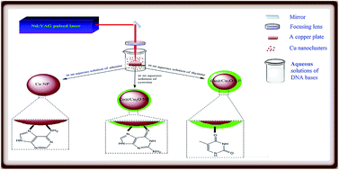
The pulsed-laser ablation (PLA) method was used as a facile and green approach to prepare oxide-free copper nanoparticles (Cu NPs), and was performed by laser ablation of a copper target in aqueous solutions of DNA bases (adenine, cytosine, and thymine). The structural analyses reveal that the in situ adsorption of the cytosine and thymine molecules on the surface of Cu NPs, although improving the stability of the colloids, cannot prevent the interaction of Cu NPs with the dissolved oxygen. However, the nanoparticles obtained in the aqueous solution of adenine are free from an oxide layer, indicating the high stabilizing effect of adenine molecules on the copper nanoparticles. FT-IR spectroscopy was used to study the mode of interaction of the DNA bases with the Cu NPs. Our results show that the participation of the nitrogen atoms of the DNA bases in the coordination to the Cu surface has a significant effect on the stabilization of the Cu NPs. In the case of adenine, the FT-IR spectra reveal that the excellent stabilizing effect originates from the ability of adenine to protect the laser-generated Cu NPs through its –NH2 group and imidazole nitrogen atoms. Thymine has no such nitrogen atoms and the NH2 group of cytosine cannot significantly participate in the coordination to the Cu surface. To the best of our knowledge, this is the first report on the preparation of oxide-free copper nanoparticles by the laser ablation of bulk copper in aqueous solutions containing small neutral biomolecules. The synthesized Cu NPs/adenine shows effective antibacterial activity against Gram-negative Escherichia coli and Gram-positive Staphylococcus aureus.
A variety of chemical and physical methods have been employed to prepare Cu NPs.1 Some of these methods include chemical and thermal reduction, pulsed sonoelectrochemical reduction, radiation methods, metal vapor synthesis, vacuum vapor deposition, microemulsion techniques, and laser ablation.1,8–10 Most of these techniques are either required hazardous chemicals and solvents or sophisticated instruments. Moreover, there are very few reports exist on the Cu NPs synthesis without oxygen-free environment.1,11–13 So, a more robust, cost-effective, and eco-friendly method for the fabrication of Cu NPs is in a rudimentary state of development relative to what has been achieved for both Au and Ag NPs.1,4 In the past decade, pulsed-laser ablation (PLA) in liquid has been exhibited to be a promising approach for the preparation of nanoparticles with different composition, including metals, semi-conductors, carbon-based nanomaterials, and alloys.14,15 In this technique, a laser pulse is focused on a target which placed in a liquid, especially water. During such a process, a microplasma is formed which rapidly expands, quenches, and decays in times on the order of microsecond. The atomized materials ablated from the target, nucleate and start to grow up to the formation of related nanoparticles.15 The PLA technique has several advantages with respect to the most used chemical methods. This method provides opportunity to produce nanoparticles of whatever composition. In the PLA technique, it is possible to control the nanoparticle properties (size, shape, and crystallinity) by tuning several experimental parameters (laser energy and wavelength, solvent composition, surfactant, etc.). Furthermore, the nanoparticles produced by the PLA approach are pure and free from chemical agents including chemical precursors, reducing agents, and (in the most cases) chemical stabilizers.15 The purity of the laser-generated nanoparticles has paramount importance on the application of them in the biomedical or biological fields. Moreover, the absence of chemical stabilizers on the surface of the obtained nanoparticles, increases the possibility of their subsequent functionalization with desired biomolecules,15 which makes them very attractive for using as biomarkers or for targeted cell therapies.16
To date, gold, silver, and copper NPs have been synthesized using PLA technique in a series of pure solvents.17 In the case of copper nanoparticles produced by PLA in pure water, literature reports show that the surface oxidation of the Cu NPs occurs immediately after generation of the metallic copper nanoparticles.17–19 Indeed, there are only a few reports devoted to the fabrication of the pure oxide-free Cu NPs using PLA method in an aqueous medium. In an excellent work, Muniz-Miranda and his coworkers20 have studied the nanosecond laser ablation of a copper plate in acetone, pure water, and aqueous solutions containing 1,10-phenanthroline (phen) and 4,4′-bipyridine (bpy). In the presence of phen and bpy, they have obtained the Cu NPs in the aqueous medium which are stable in time, without addition of any stabilizers. They have reported that the use of phen and bpy dramatically improves the long-term stability of the colloids. However, these heterocyclic ligands cannot prevent the interaction of Cu NPs with the dissolved oxygen.20 Liu et al.21 have shown that the copper nanoparticles obtained by laser ablation of a copper target in the aqueous solution of poly(vinylpyrrolidone) (PVP), are easily oxidized to copper(I) oxide (Cu2O) NPs. However, in 2015, Deo Malviya and his coworker22 have reported the synthesis of oxide-free Cu NPs through laser ablation of pure copper targets in the aqueous solutions of PVP and tuning the quality and size of NPs through the change of experimental parameters. Liu and his coworkers23 also have another good paper in this field. Recently, they have studied the feasibility of controlled synthesis of Cu@Cu2O nanoparticles by laser ablation of a copper target in the aqueous solutions of sodium dodecyl sulfate (SDS), in which SDS (as a negative capping agent) decreases the surface oxidation of Cu NPs. Also, by tuning the concentration of SDS, the relative amount of Cu and Cu2O components can be controlled in the solution.23
Recent studies on nanomaterials show that different metallic nanoparticles could have effective roles as antibacterial agents.24 According to the literature, most of the antibacterial studies involving metallic nanoparticles have been limited to the use of silver25–29 few on copper,24,30 and least on gold.31 However, the cost and the availability of silver and gold can be considered as the main limitations to their applications.25 Therefore, metallic Cu NPs can be a good candidate as an antibacterial agent in the near future.24
As mentioned above, few organic stabilizers have been introduced for the PLA synthesis of chemically stable Cu NPs in the aqueous media. Generally, these stabilizers have been limited to the polymers (such as PVP)22 or large molecules (such as SDS),23 which have high affinity for capturing the laser-generated Cu NPs as long as they are formed by laser ablation. However, the use of small biomolecules to stabilize the laser generated Cu NPs has been rarely reported.20 For biological applications, attempts have been made to stabilize metallic nanoparticles in the presence of biomolecules such as DNA, RNA, DNA bases, and proteins.32 The DNA bases are the fundamental constituent of nucleic acids containing nitrogen found within nucleotides. Thus, the interaction of metallic nanoparticles with nucleic acids is important in the bioinorganic field because of its imminent effects on the synthesis, replication, and structural integrity of DNA and RNA.33 Proteins and nucleic acids are the most studied biomolecules for stabilizing the noble metallic nanoparticles.32 To the best of our knowledge, the DNA bases have not been used for stabilizing the Cu NPs. Keeping in view the advantage of the DNA bases, the aim of this work is study of the generation of oxide-free copper nanoparticles using the laser ablation of bulk copper in the aqueous solutions of the DNA bases (adenine, cytosine, and thymine). Here, we investigate the interaction of these three bases with the laser generated Cu NPs by means of several spectroscopic and analytical techniques. In the following, we also study the antibacterial property of the oxide-free Cu NPs (Cu NPs/adenine) against Gram-negative Escherichia coli (ATCC 8739) and Gram-positive Staphylococcus aureus (ATCC 6538). Our results support the hypothesis that the Cu NPs are suitable and effective for the formulation of new types of antibacterial agents.
The spectral data were collected at the ambient temperature. The electronic absorption spectra of the DNA bases and the Cu NPs–DNA bases colloidal solutions were recorded on a JASCO 7580 UV-Vis-NIR double-beam spectrophotometer using quartz cells with a path length of 10 mm. Fourier transform infrared (FT-IR) spectra were recorded on an FT-IR JASCO 680-PLUS spectrometer in the region of 4000–400 cm−1 using KBr pellets. Steady-state luminescence measurements were performed on a SHIMADZU RF 5301PC spectrofluorophotometer. The TEM images were obtained using a Zeiss-EM10C-80 kV (Germany). The size distribution of the particles was determined from the TEM images using the SPSS program. The zeta potentials of the freshly prepared NPs were measured with a Malvern Zetasizer (Nano ZS, Malvern Instrument, UK). XRD patterns were recorded by an X-ray diffractometer (Philips EXPERTMPD) using Cu Kα (λ = 0.154 nm) radiation. The samples were scanned from 30 to 80° of 2θ.
The strong SPR band between 550 and 600 nm is well accepted to originate from the oxide-free Cu NPs.38,39 It should be mentioned that the UV-Vis spectra do not show the characteristic absorption band of the copper oxide at around 800 nm.37,40 These results indicate that the reddish-brown colloidal solution (Fig. 1) contains pure oxide-free Cu NPs. It is worthy to mention that there is no need to use an inert gas atmosphere in this method. The observed enhancement in the intensity of the SPR band can be related to the increase in concentration of the oxide-free Cu NPs generated during the ablation process.22,23 As can be seen in Fig. 1, the position of the plasmon band, which does not shift during the ablation process, is in good agreement with the theoretical prediction using Mie theory for the pure oxide-free Cu NPs.20,37 This result also indicates that the agglomeration of the Cu NPs can be avoided in the bioconjugated colloid. The reddish-brown solution of the Cu NPs is stable even in the presence of air (oxygen) for a long period of time (at least two weeks). The stability of the Cu NPs was confirmed by recording the SPR band intensity at 580 nm after 14 days. The results demonstrate no significant changes in the SPR band intensity and position after this period of time (Fig. 2).
![]() | ||
| Fig. 2 Absorption spectra of the freshly prepared Cu NPs/adenine sample (red line) and the sample after 14 days (green line). | ||
In the cytosine and thymine solutions, the position of the plasmon bands are far from that predicted using the Mie theory for small metallic Cu NPs.20 As can be seen in Fig. 3 and 4, the SPR bands of the cytosine and thymine colloidal solutions appear at 640 and 655 nm, respectively. The observed red-shift of the plasmon bands (from 580 nm) and the green color of the samples can be related to the oxidation reaction of the Cu NPs with air oxygen dissolved in the colloidal solution and formation of an oxide layer on the surface of the Cu NPs during ablation. The presence of an oxide layer switches the particles to core–shell structures, with consequent red-shift of the plasmon resonance (from that predicted using the Mie theory), depending on the dielectric constant and thickness of the shell.20
In pure water, the unprotected laser-generated Cu NPs undergo collapse in few hours due to the fast oxidation reaction of the particles.20 It should be mention that in the cytosine and thymine colloidal solutions, the protected Cu NPs do not precipitate in this period of time and the stable copper colloids are obtained in the aqueous solutions of cytosine and thymine by formation of the surface complexes, which is consistent with the previous report.20 However, these colloidal solutions lose their green color and turn clear in a couple of weeks. The complete oxidation of the Cu NPs leads to loose their metallic nature and hence plasmon resonance and related color,20 which shows that the oxidation process (formation of the oxide layer during the ablation) continues over time. This result is strongly supported by UV-Vis absorption spectroscopy (data are not shown here), which shows decay and finally disappearance of the SPR bands of the colloidal Cu/cytosine and Cu/thymine solutions as the time passes. This effect can be reasonably related to the interaction of the Cu NPs with dissolved air oxygen and water molecules in the aqueous solutions,20,23,37 and it is not totally prevented by the presence of cytosine or thymine molecules on the surface of the Cu NPs.41 The absorption spectra of the thymine solution exhibit larger red-shift and lower intensity of the SPR band (Fig. 5) with respect to the cytosine solution.
![]() | ||
| Fig. 5 The comparison of the absorption spectra of the NPs obtained in the aqueous solutions of thymine, cytosine, and adenine at the same experimental conditions. | ||
The red-shift of the plasmon band of Cu NPs is an indicator of the degree of the interaction of Cu NPs with the dissolved oxygen.23 In general, the red-shift of the plasmon band is also an indicator of the NPs aggregation.42 In the case of the Cu NPs, the observed red-shift can be originated from the degree of the interaction of Cu NPs with the dissolved oxygen and NPs aggregation, simultaneously. To compare the degree of the interaction of Cu NPs with the dissolved oxygen in the cytosine and thymine solutions (Fig. 5), the size of the Cu NPs also must be known. Thus, the absorption spectra of the Cu/cytosine and Cu/thymine colloidal solutions (Fig. 3 and 4) do not provide enough information to study the relative surface oxidation in these systems. These results show the different efficiency of the DNA bases in the formation of the pure oxide-free Cu NPs related to the different plasmon resonance intensity, as shown in Fig. 5. In addition, the different stabilizing effect of these small biomolecules can be seen in Fig. 5. On the basis of these results, it can be demonstrated that the order of capping efficiency varies as adenine > cytosine > thymine.
In the laser ablation synthesis of the noble metal nanoparticles (such as Au, Pd, and Pt) in an aqueous solution, it is well accepted that their surfaces are partially oxidized, which is an inherent characteristic of this synthesis method.44–46 This phenomenon has been confirmed by X-ray photoelectron spectroscopy (XPS) analysis.45 However, the positive surface charge can be overcompensated by the excess electrons formed within the plasma phase, which change the surface charge of the metallic particles from positive to negative.44 In the laser ablation synthesis of Cu NPs, it is reasonable to accept that the degree of surface oxidation of Cu NPs generated in the plasma phase is larger than those observed in the case of noble metal NPs because of lower oxidation potential of copper with respect to noble metals. In addition, the electron affinity of copper is very smaller than that of gold. Thus, the positively-charged Cu NPs cannot take electrons from the plasma effectively. In fact, we believe that the positive charge observed in the Cu NPs–adenine complex originates from the initial oxidation of Cu NPs in the plasma phase which is an inherent characteristic of the laser ablation synthesis of NPs.45 Recently, the oxidation mechanism and formation of a layer of Cu2O on the copper surface was studied by Fujita et al.47 For example, in a partial pressure of 10.1 kPa for oxygen, the formation of a 2 nm layer of Cu2O requires ca. 10 ks at 60 °C.47 This time (10 ks) is much larger than the lifetime (μs) for the formation of a cavity.22 Thus, the formation of a core–shell structure occurs after the collapse of the cavity in the aqueous medium. Our results show that the addition of adenine in water effectively isolates the Cu NPs by forming a capping layer at the later stages, while the nucleation and growth of the particles occur in the cavity as indicated by recent works.22 These particles, coming out from the plasma plume, are quenched and capped by adenine to prevent their interactions with oxygen. In summary, in the laser ablation synthesis, the initial oxidation of Cu NPs surface in the plasma plume is an unavoidable phenomenon, due to the inherent characteristics of the plasma.45 We suggest that the formation of an oxide layer must be taking place when the Cu NPs are engulfed by the aqueous medium. This is also consistent with the fact that the expansion cavity is primarily filled with the ablated plume, and therefore, the expected partial pressure of oxygen is extremely low.22 The presence of suitable ligand molecules such as adenine, which can capture positively-charged Cu NPs (generated in the plasma plume), impairs the interaction of dissolved oxygen and/or hydroxide anions with the positively-charged Cu NPs.
However, precipitation of the nanoparticles in the cytosine solution occurs within 10 days with a zeta potential value of −29.1 mV, which can indicate the moderate stability of the solution.32 In the Cu NPs–thymine assembly, the aggregation occurs after 14 days. The Cu NPs–thymine solution has a zeta potential of −39.5 mV, which can exhibit a good stability of the colloidal NPs.32 The surface oxidation of the Cu NPs can be a dynamic process, which means that the surface oxidation (formation of the oxide layer after ablation) continues slowly over time, thereby the Cu atoms gradually undergo an oxidation reaction and form a thicker shell of copper oxide on the surface of the related NPs. After a long time, the Cu atoms in the core oxidize and finally form the pure copper oxide nanoparticles.48 The high degree of surface oxidation leads to a high number of negative charges which stabilize the NPs. In fact, the ionic bonding of copper oxide NPs produces a high potential surface that enables the adsorption of the hydroxide ions and water molecules, which can explain the presence of a high negative charge in the surface of these particles.49 Surface hydroxylation reaction is an important process to consider, as the surface interactions of oxide NPs with water can give rise to a certain degree of hydroxylation.50 It should be mentioned that the aggregation process is driven by thermodynamic and kinetic factors.51 The classic term ‘colloid stability’ refers to kinetic stability.52 In fact, the presence of electrostatic repulsion forces between the negatively-charged copper oxide NPs provides kinetic stability for the system. However, the dispersion of nanoparticles in natural systems is often thermodynamically unfavorable, as a lower total free energy of the system may be achieved by aggregation of the particles to reduce their surface area.53 When the surface of Cu NPs is covered with the hydroxyl groups, the interaction between the nanoparticles becomes substantially stronger than the particle/water interactions.54 The increase in the interaction can be due to the direct hydrogen bonds formed between the surfaces containing hydroxide groups. It's worthy to mention that the OH⋯OH hydrogen bond is stronger than OH⋯OH2 bond. Hydrogen bonds affect the nature of the nanoparticle agglomerates, and thus influence the dynamics of the suspension.54–56 So, in the Cu NPs/cytosine and Cu NPs/thymine colloidal solutions, the direct hydrogen bonds between the hydroxyl groups of each NPs are stronger and more thermodynamically favorable than the other possible hydrogen bonds which can be formed in these two systems (such as hydrogen bonds between water and nanoparticle or even hydrogen bond between the adsorbed ligands of each NPs). The formation of stronger hydrogen bonds is more thermodynamically favorable and results in the aggregation of the copper oxide NPs. The importance of surface modification for the successful preparation of homogeneously dispersed nanocomposites becomes more evident in the case of oxide nanoparticles where the particle–particle interaction is very strong due to the hydrogen bonds.51
![]() | ||
| Fig. 6 TEM image of the adenine-stabilized Cu NPs prepared in an aqueous solution of adenine (10−4 M). | ||
The spherical copper NPs have an average particle size of 11 nm with a relatively broad range size distribution (3–30 nm). However, a majority of these particles are within the 3–12 nm range (Fig. 7).
In the PLA method, the presence of the suitable ligands at the moment of the production of NPs can effectively control the growth processes including the coalescence of the nuclei and the adsorption of free atoms in solution.57 Adenine is a small biomolecule with suitable electron donor moieties for trapping the small Cu NPs. The TEM results also show that the adenine molecules can effectively control the growth process of the Cu NPs. In addition, there is an inverse correlation between the concentration of the stabilizing ligand and the average particle size of NPs,57 so that the surface coverage rate of NPs increases in higher concentration of the stabilizers.57 Therefore, it is expected that at the higher concentration of adenine, the average size and the standard deviation of size distribution for the Cu NPs are reduced with respect to those observed in the present study.
![]() | ||
| Fig. 8 Fluorescence spectra of the adenine solution (10−4 M) in the presence of increasing amounts of the Cu NPs (0–200 μg mL−1) during the laser ablation. | ||
The free adenine solution shows two broad emission peaks around 310 and 380 nm, when excited at 232 nm. The fluorescence intensity of the adenine solution gradually decreases as the Cu NPs concentration increases (Fig. 8), which indicates that the Cu NPs act as quenchers for the adenine fluorescence. When an appropriate fluorophore is directly adsorbed on a metal surface, its fluorescence is quenched.58 However, at a distance of a few nanometers from the nanoparticle surface, the fluorescence can be strongly enhanced.58 Quenching of the intensity is mostly due to the energy transfer processes, which involve the surface energy transfer from the fluorophore molecules to the nanoparticles. Quenching is not only caused by an increase in the non-radiative rate but also, by a drastic decrease in the fluorophore radiative rate.59 This result can indicate that there is no oxide layer between the Cu NPs surface and the adenine molecules in the Cu NPs–adenine conjugates and the adenine molecules directly attach to the metal surface.
As can be seen in Fig. 9, the free cytosine solution has an emission peak around 370 nm, when excited at 226 nm, and emission of the free thymine appears at 352 nm (λex = 255 nm) (Fig. 10).
In the presence of Cu NPs, the cytosine and thymine solutions show emission peaks at 365 and 363 nm, respectively (Fig. 9 and 10). The fluorescence intensity of the cytosine and thymine solutions increases as the Cu NPs are generated in the solution. As mentioned above, metallic nanoparticles cause quenching of the fluorescence intensity when they directly attach to the fluorophore molecule, whereas the fluorescence intensity can be strongly enhanced at a specific distance of a few nanometers from the surface of NPs.58 The observed enhancement of the fluorescence intensity of these colloidal solutions can be attributed to the metal-enhanced fluorescence (MEF) effect of the Cu NPs due to the presence of a spacer layer on the Cu NPs surface, which makes the distance between the fluorophore molecules and NPs.60,61 The MEF phenomenon occurs when a fluorophore interacts with the enhanced localized electric fields around the metallic nanoparticles that are induced by the incident light.62 In this case, the presence of metallic NPs held in a specific distance (either by a desired spacer layer or by a naturally oxide layer) from the fluorophores, can result in decreasing the fluorescence lifetime of the fluorophores, which leads to the increasing of their emission intensity.63 Decreasing the lifetime, often provides photostability for the fluorophores, because they spend less time “on average”, in an excited state, before they return to their ground state, thus the possibility of photodestruction significantly decreases.63 The specific distance for the appearance of the MEF effect should be typically less than 10 nm from the metal surface.63,64 In our studies, however, no additional spacer layer was used. It is expected that the native oxide layer on the Cu NPs acts as a natural spacer layer. Other metals have also reported to show MEF effect, including gold,58,65 silver,66–69 zinc,70 and aluminum.62,71 In this regard, copper, silver, and gold nanoparticles were employed for the applications of MEF with the fluorophores which have emission wavelength in the Vis-NIR region. However, aluminum and zinc nanoparticles were demonstrated to increase the fluorescence emission of fluorophores in the UV and blue spectral regions.70 These observations are in good agreement with the previously described MEF phenomenon of metallic NPs.58,67,71 The interactions of the Cu NPs with cytosine and thymine molecules can result in an increase in their quantum yield (i.e., emission intensity). It is worth nothing that the most nucleic acids and nucleotides have low intrinsic fluorescence quantum yield (Q0), for example, the quantum yield of the DNA bases is often reported to be on the order of 10−4.72–74 Therefore, using the metal NPs with appropriate geometry and compositions for emission enhancement is a useful approach and can increase the detection sensitivity of these low quantum yield fluorophores. This strategy is of great importance in the DNA sequencing detection and genetic analysis.58
The observed diffraction peaks can be indexed according to Joint Committee for Powder Diffraction Studies (JCPDS), File No. 04-0836.75 The results clearly confirm that the adenine molecules play the key role in the production of the oxide-free Cu NPs and protect them from the interaction of Cu NPs with the dissolved oxygen. The crystalline nature of the sample is reflected in the sharp XRD peaks. The size of the metallic Cu NPs can be calculated using the Debye–Scherrer equation,76 given as follows (eqn (1)):
D = 0.89λ/β cos θ
| (1) |
The XRD results obtained from the Cu NPs/cytosine and Cu NPs/thymine samples, confirm the presence of both copper and cuprous oxide (Cu2O) phases in the samples (Fig. 12).
As can be seen in Fig. 12, the XRD patterns show six distinguishable peaks that can be assigned to the crystalline metallic Cu and Cu2O planes. These XRD patterns show the aerobic oxidation of the Cu NPs produced in the aqueous solutions of cytosine and thymine. It should be mentioned that there are not any other peaks related to the CuO phase.23 These results are in agreement with the UV-visible absorption spectra and clearly show the core–shell structure of the Cu NPs.23 In addition, the average crystalline sizes of the nanoparticles obtained in the cytosine and thymine solutions are found to be 19 and 22 nm, respectively, and various degrees of the interaction of Cu NPs with the dissolved oxygen can also be seen in these patterns. The ratios of Cu and Cu2O components of the nanoparticles for the Cu/cytosine and Cu/thymine samples are depicted in Fig. 12. In the cytosine and thymine solutions, the position of the plasmon bands are far from that predicted using the Mie theory for small metallic Cu NPs. As can be seen in Fig. 3 and 4, the SPR bands of the cytosine and thymine solutions appear at 640 and 655 nm, respectively. The average crystalline sizes of the nanoparticles obtained in the cytosine and thymine solutions were found to be 19 and 22 nm, respectively. According to these data, the difference between SPR bands of the thymine and cytosine samples (655 − 640 = 15 nm) is mainly due to the difference in the degree of the interaction of Cu NPs with the dissolved oxygen for these two samples. It is hard to accept that the increasing in the NPs size to about 3 nm (22 − 19 = 3 nm) can have a significant contribution in the 15 nm red-shift (640 to 655 nm) in the SPR bands.
In the Cu NPs–cytosine assembly, a change in the frequency and intensity of the carbonyl group of cytosine is observed. The position of carbonyl band shifts from 1657 (for the free molecule) to 1617 cm−1 (for the adsorbed molecule), which demonstrates that the cytosine base interacts with the Cu NPs through its carbonyl group (Fig. S2, S4; and Table S2, ESI†). The N(3)
C(4) stretching frequency shows a red-shift from 1543 (free) to 1495 cm−1 (adsorbed), which suggests that the cytosine base can also bind to the Cu NPs via its N(3) atom (Fig. S2, S4 and Table S2, ESI†). In the case of Cu NPs–thymine interaction, the changes in the position and intensity of the carbonyl stretching vibration are significant (Fig. S3 and Table S3, ESI†). It should be mentioned that the thymine molecule has the two different carbonyl groups, C(2)
O and C(4)
O,78 which show two IR bands at 1739 and 1674 cm−1, respectively. The frequency of the C(4)
O group shifts from 1674 (free) to 1630 cm−1 (adsorbed), which confirms that the thymine molecules bind to the Cu NPs through its C(4)
O carbonyl group (Fig. S4, ESI†). It is worth noting that the FT-IR spectrum of the Cu NPs/cytosine sample shows a new band at around 625 cm−1 (in comparison to the free cytosine), which can be attributed to the Cu(I)–O vibration of the Cu2O layer.82 As can be seen in Fig. S3, (ESI†), the FT-IR spectrum of the free thymine base shows a vibrational band at around 625 cm−1, and it may overlap with the Cu(I)–O vibrational band in the Cu NPs/thymine spectrum.
The difference in the stabilizing efficiency of the DNA bases is due to the varying ability of the bases for the coordination to the Cu surface as a result of the different available coordination sites. Thus, it can be inferred that the number and the type of binding sites, which participate in the coordination process, play the key roles in the stabilizing efficiency of these DNA bases. As mentioned previously, the surface oxidation of the Cu NPs occurs immediately after their generation by laser ablation in pure water. To obtain the oxide-free Cu NPs sample in the aqueous solution, the appropriate ligands with suitable electron donor moieties (such as nitrogen- and oxygen-donor atoms) are necessary to trap and protect the laser generated Cu NPs. Thymine has no exocyclic nitrogen atom and the ring nitrogen cannot actively participate in the coordination process, thus the entire bonding takes place through the O atoms (Fig. S4, ESI†). The FT-IR spectra show that the NH2 group of cytosine cannot effectively coordinate to the copper surface. We suggest that the excellent stabilizing effect of adenine originates from its ability to trap the Cu NPs through two suitable coordination sites. The FT-IR spectra reveal that both NH2 and nitrogen of the imidazole ring can participate in the Cu NPs–adenine interaction (Fig. S4, ESI†). The type and the number of the atoms (nitrogen and oxygen) which participate in the coordination process are the two main parameters which can affect the stabilizing ability of these DNA bases. Fig. S4 (ESI†) shows the structures of the DNA bases (cytosine, thymine, and adenine) and their binding interactions with Cu NPs. Generally, the Cu–N bond is stronger than the Cu–O bond,78,83,84 therefore, it is reasonable to accept that the weakest stabilizing ability of thymine is due to the absence of appropriate nitrogen atoms (which can participate in the coordination process) in its structure (Fig. S4, ESI†). The interaction of the Cu NP with the adenine base results in the formation of stronger chemical bonds (i.e., two Cu–N bonds) (Fig. S4, ESI†). In the pulsed laser ablation of a copper target in the presence of small biomolecules as ligands, it seems that the presence of the suitable nitrogen atoms in the ligand structure and participation of these nitrogen atoms in the coordination have significant effect on the stabilizing ability of the small biomolecules.
Footnote |
| † Electronic supplementary information (ESI) available: (Scheme S1), the general schematic experimental setup used for the preparation of the Cu NPs in aqueous solutions of the DNA bases (adenine, cytosine, and thymine) (Fig. S1), FT-IR spectra of adenine (red line) and the adsorbed adenine (Cu NPs/adenine) (green line) (Fig. S2), FT-IR spectra of cytosine (red line) and the adsorbed cytosine (Cu NPs–cytosine) (green line) (Fig. S3), FT-IR spectra of thymine (red line) and the adsorbed thymine (Cu NPs–thymine) (green line) (Table S1), main vibrational frequencies of the free and adsorbed adenine (Table S2), main vibrational frequencies of the free and adsorbed cytosine (Table S3), main vibrational frequencies of the free and adsorbed thymine. See DOI: 10.1039/c6ra22038j |
| This journal is © The Royal Society of Chemistry 2016 |